Can you generate image
Generate easy microscopic slide diagram of following questions according to textbook of pathology by Harsh Mohan 1.Septic thrombus in lung 2.apostematous nephritis, 3.cryptococcal meningitis (HIV) 4.cytomegaloviral pneumonia (HIV)
Medical education microscopic slide diagram labeled "1. SEPTIC THROMBUS IN LUNG" - H&E stained histology teaching diagram. Show a pulmonary blood vessel cross-section (medium-sized artery/vein) filled with a thrombus containing: a central zone of necrotic debris and fibrin meshwork, dense infiltration of neutrophils (polymorphonuclear cells) throughout the thrombus, colonies of bacteria (dark cocci/bacilli clusters) embedded within the fibrin, surrounding the vessel show hemorrhagic infarction of lung parenchyma with alveolar wall necrosis, fibrin exudate in alveolar spaces, and abundant neutrophil infiltration. Label key structures: "Infected thrombus", "Neutrophil infiltration", "Bacterial colonies", "Fibrin meshwork", "Alveolar hemorrhage", "Necrotic lung tissue", "Vessel wall". Pink and purple H&E color scheme. Include a scale bar and magnification (x100). Clean educational style with a white background title box at top reading "SEPTIC THROMBUS - LUNG (H&E x100)"
Medical education microscopic slide diagram labeled "2. APOSTEMATOUS NEPHRITIS (Renal Cortical Abscess)" - H&E stained histology teaching diagram. Show kidney cortex with multiple small abscesses (miliary abscesses): each abscess is a rounded focus filled with liquefied pus and dense neutrophils, surrounded by a rim of necrotic renal tubular cells, glomeruli nearby appear relatively preserved but compressed, renal tubules between abscesses show acute tubular injury with flattened epithelium, interstitium shows heavy neutrophilic infiltrate, prominent edema of interstitium. Label key structures: "Cortical microabscess", "Neutrophil-filled abscess cavity", "Necrotic tubular cells", "Preserved glomerulus", "Edematous interstitium", "Damaged renal tubules", "Pus". Pink and purple H&E color scheme. Include scale bar and magnification (x100). Clean educational style with title box reading "APOSTEMATOUS NEPHRITIS - KIDNEY (H&E x100)"
Medical education microscopic slide diagram labeled "3. CRYPTOCOCCAL MENINGITIS (HIV/AIDS)" - H&E and Mucicarmine stained histology teaching diagram. Show meninges and subarachnoid space: abundant Cryptococcus neoformans yeast cells - round to oval yeasts (5-10 microns) with characteristic thick mucoid capsule appearing as a clear halo around each cell, narrow-based budding visible on some yeasts, minimal inflammatory reaction (characteristic of HIV - no neutrophils, sparse lymphocytes) - "gelatinous" appearance of the subarachnoid space, some yeasts show mucicarmine-positive (pink/red) capsule staining, occasional giant cells trying to engulf organisms, "soap bubble" lesions in brain tissue (dilated Virchow-Robin spaces filled with yeasts). Label key structures: "Cryptococcus neoformans yeast", "Thick polysaccharide capsule (clear halo)", "Narrow-based budding", "Gelatinous matrix", "Minimal inflammation (HIV)", "Meningeal tissue", "India ink-like appearance". Pink and purple H&E with mucicarmine pink capsule highlights. Title box: "CRYPTOCOCCAL MENINGITIS - HIV (H&E + Mucicarmine x400)"
Medical education microscopic slide diagram labeled "4. CYTOMEGALOVIRAL (CMV) PNEUMONIA (HIV/AIDS)" - H&E stained histology teaching diagram. Show lung tissue: alveolar walls thickened with interstitial pneumonitis, scattered enlarged infected cells (cytomegaly) - cells are 2-4x normal size, each infected cell shows the pathognomonic "owl's eye" intranuclear inclusion body (large, dark purple/basophilic central inclusion surrounded by a clear halo/chromatin rim), also show multiple small basophilic cytoplasmic inclusion granules, alveolar spaces contain fibrin, proteinaceous exudate and macrophages, some hyaline membranes lining alveoli, minimal neutrophilic inflammation (HIV immunosuppression), interstitial lymphocyte infiltration. Label key structures: "CMV-infected enlarged cell (cytomegaly)", "Owl's eye intranuclear inclusion (basophilic)", "Clear halo around inclusion", "Cytoplasmic inclusions", "Thickened alveolar wall", "Hyaline membrane", "Alveolar macrophages", "Interstitial pneumonitis". Pink and purple H&E color scheme. Title box: "CMV PNEUMONIA - HIV (H&E x400)"
1. Septic Thrombus in Lung (H&E x100)
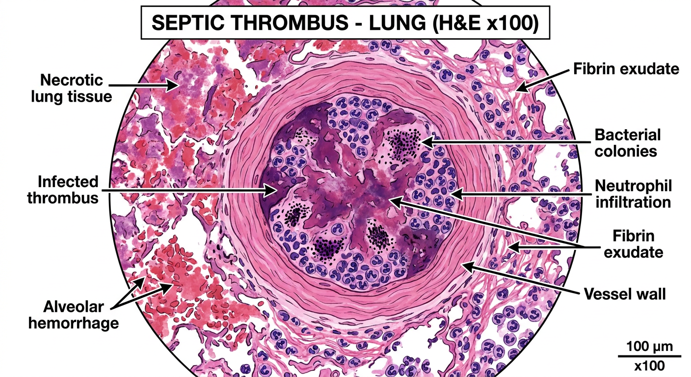
- Pulmonary vessel filled with infected thrombus (fibrin + neutrophils)
- Bacterial colonies embedded within the thrombus
- Surrounding hemorrhagic infarction and necrosis of lung parenchyma
- Dense PMN (neutrophil) infiltration throughout
2. Apostematous Nephritis (H&E x100)
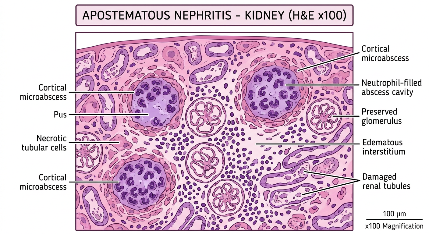
- Multiple miliary cortical abscesses filled with neutrophils/pus
- Necrotic renal tubular cells surrounding abscess cavities
- Relatively preserved glomeruli (compressed)
- Edematous, neutrophil-infiltrated interstitium
3. Cryptococcal Meningitis - HIV (H&E + Mucicarmine x400)
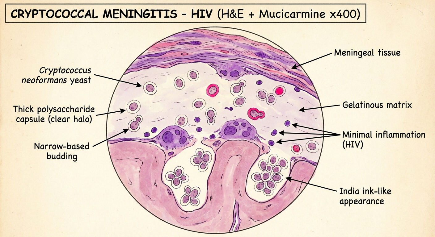
- C. neoformans yeasts with characteristic thick polysaccharide capsule (clear halo)
- Narrow-based budding
- Minimal/absent inflammatory reaction (hallmark in HIV/AIDS)
- "Soap bubble" / gelatinous appearance in subarachnoid space
- Mucicarmine-positive (pink) capsule staining
4. CMV Pneumonia - HIV (H&E x400)
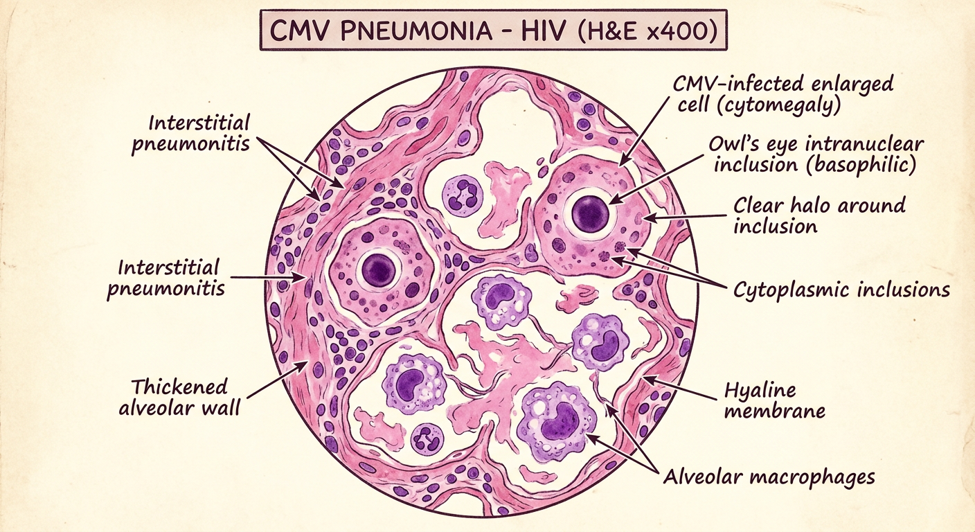
- Massively enlarged infected cells (cytomegaly)
- Pathognomonic "owl's eye" intranuclear inclusion - large basophilic inclusion with clear halo
- Small cytoplasmic inclusions
- Thickened alveolar walls (interstitial pneumonitis)
- Hyaline membranes + alveolar macrophages
- Minimal neutrophilic response (HIV immunosuppression)
Note: The library does not contain Harsh Mohan's textbook directly, but these diagrams reflect the classic microscopic descriptions as taught in Harsh Mohan's Textbook of Pathology and corroborated by Robbins Pathology. The "owl's eye" inclusion of CMV and the minimal inflammation in cryptococcal meningitis (HIV) are especially emphasized in Harsh Mohan.