
External carotid artery with carotid sheath
Reading File
The external carotid artery (ECA) is a major branch of the common carotid artery that supplies the face, scalp, and neck. The carotid sheath is a fascial sleeve that encloses the common and internal carotid arteries, internal jugular vein, and vagus nerve.
Searching sources...
Finding Sources
Finding Sources
Reading File
Reading File
Reading File
Reading File
Reading File
Reading File
Reading File
Reading File
Loading Image
Here is a detailed anatomical overview of the External Carotid Artery (ECA) and the Carotid Sheath, drawn from Gray's Anatomy for Students, Scott-Brown's Otorhinolaryngology, and K.J. Lee's Essential Otolaryngology.
External Carotid Artery (ECA)
Origin and Course
The ECA arises from the common carotid artery (CCA) at its bifurcation, typically at the level of the upper border of the thyroid cartilage (C3-C4). After the bifurcation, the ECA lies anteromedial to the internal carotid artery (ICA). It ascends, passes deep to (or enters) the parotid gland, and divides into its two terminal branches (superficial temporal and maxillary arteries) near the lower border of the ear, within the substance of the parotid gland.
Distinguishing ECA from ICA
The ECA is identified by the fact that it gives off branches in the neck, whereas the ICA has no branches in the neck.
Branches of the ECA
The ECA gives off 6 branches before its 2 terminal branches - a useful mnemonic is "Some Anatomists Like Freaking Out Poor Students" (Superior thyroid, Ascending pharyngeal, Lingual, Facial, Occipital, Posterior auricular, then Superficial temporal, Maxillary as terminals).
| Branch | Origin (surface) | Supplies |
|---|---|---|
| Superior thyroid artery | Anterior (1st branch) | Thyroid gland, larynx (internal structures), sternocleidomastoid, cricothyroid muscles |
| Ascending pharyngeal artery | Deep/posterior (smallest, may actually arise 1st) | Pharyngeal constrictors, palate, palatine tonsil, meninges of posterior cranial fossa, skull base, dura |
| Lingual artery | Anterior, at level of hyoid bone | Tongue, sublingual gland, floor of mouth, epiglottis, tonsil |
| Facial artery | Anterior (3rd anterior branch, just above lingual) | Face from mandible to medial canthus, soft palate, tonsil, submandibular gland |
| Occipital artery | Posterior (near facial origin) | Sternocleidomastoid, posterior scalp, deep back muscles, meninges of posterior cranial fossa, mastoid cells |
| Posterior auricular artery | Posterior (small, just above digastric) | Parotid gland, muscles of ear, external ear and scalp posterior to ear, middle and inner ear |
| Superficial temporal artery | Terminal (upward continuation) | Parotid gland and duct, masseter, lateral face, anterior external ear, temporalis muscle, parietal and temporal scalp |
| Maxillary artery | Terminal (passes deep to mandible) | External acoustic meatus, TMJ, meninges, teeth, nasal cavity, palate, infratemporal fossa, sinuses |
Note: When ligating the ECA for epistaxis, the ascending pharyngeal artery was traditionally preserved due to its supply to the skull base and dura. - Scott-Brown's Otorhinolaryngology Head & Neck Surgery, Vol. 1
Relation to the Parotid Gland
The ECA enters/passes deep to the inferior border of the parotid gland and gives off the posterior auricular artery before dividing into its terminal branches within the gland. The retromandibular vein (formed by junction of superficial temporal and maxillary veins) also lies within the parotid. - Gray's Anatomy for Students
Carotid Sheath
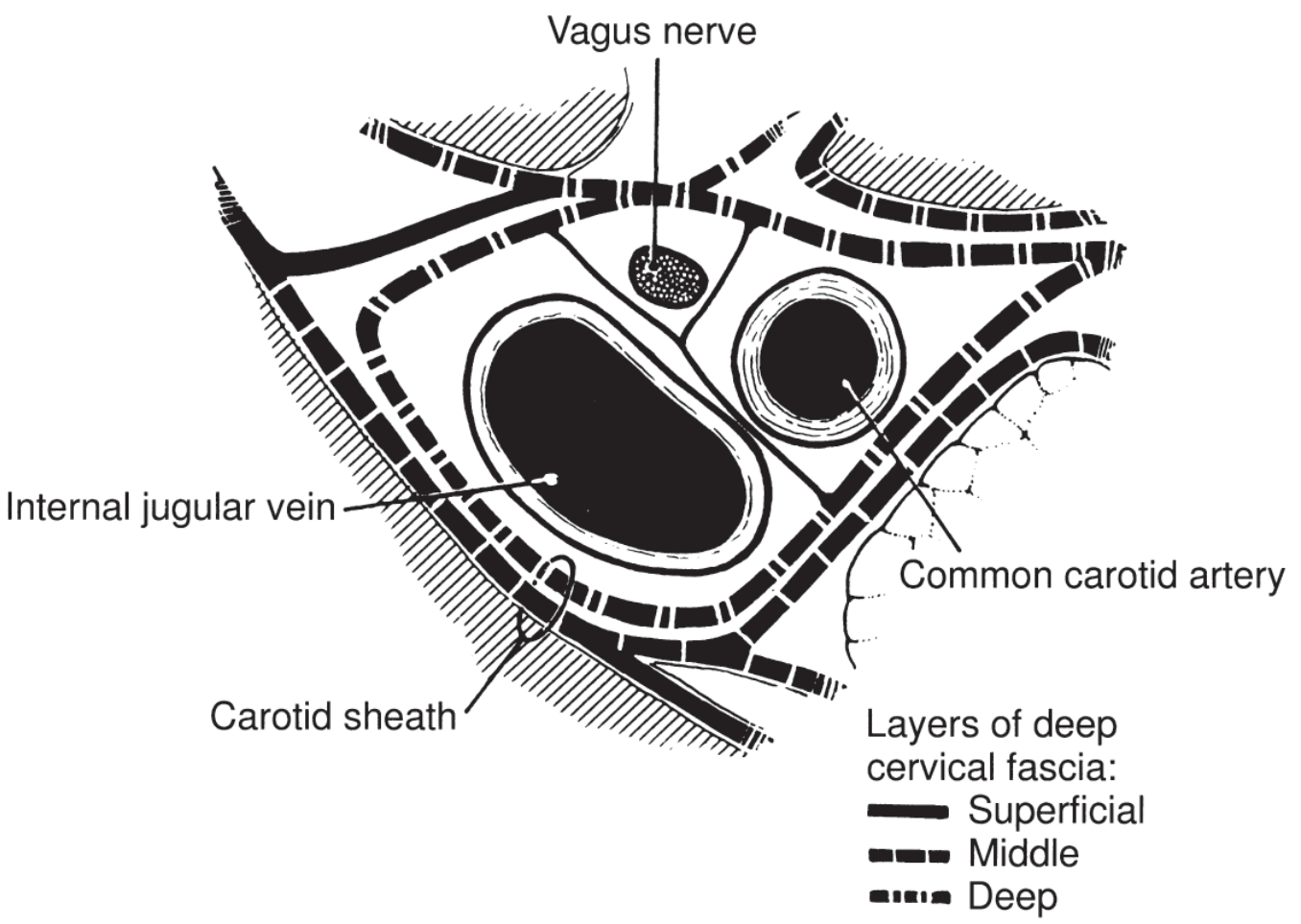
Figure: Fascial layers of the carotid sheath - K.J. Lee's Essential Otolaryngology
Structure and Composition
The carotid sheath is a fascial tube derived primarily from the investing (superficial) layer of deep cervical fascia, medial to the sternocleidomastoid. It receives contributions from all three layers of deep cervical fascia:
- Investing layer
- Pretracheal (middle) layer
- Prevertebral (deep) layer
The sheath is thicker around the arteries but loose around the internal jugular vein, which allows the vein to expand during increased venous flow. The sheath connects to surrounding fascial layers via loose areolar tissue. - Scott-Brown's Otorhinolaryngology; Gray's Anatomy for Students
Contents
| Structure | Position |
|---|---|
| Common carotid artery (below bifurcation) / Internal + External carotid above | Medial |
| Internal jugular vein (IJV) | Lateral/anterolateral |
| Vagus nerve [CN X] | Posterior (between artery and vein) |
| Ansa cervicalis | On anterior surface |
Extent
- Superior: Base of skull
- Inferior: Thorax (continues into the thoracic cavity)
Clinical Significance
The carotid sheath is known as "The Lincoln Highway of the Neck" because it is a potential avenue for rapid spread of infection from the neck down into the mediastinum. - K.J. Lee's Essential Otolaryngology
The carotid sheath space has specific boundaries:
- Anterior: Sternocleidomastoid muscle
- Posterior: Prevertebral space
- Medial: Visceral space
- Lateral: Sternocleidomastoid
Fascial Compartments of the Neck (Context)
The carotid sheath is the 4th longitudinal compartment of the neck created by cervical fascia:
- Outer compartment - investing layer
- Prevertebral compartment - deep muscles and vertebral column
- Visceral compartment - pharynx, trachea, esophagus, thyroid
- Carotid sheath - neurovascular structures
Infections spreading via these fascial spaces can pass from the neck to:
- Anterior mediastinum (via pretracheal space)
- Posterior mediastinum (via retropharyngeal space or prevertebral space)
Sources: Gray's Anatomy for Students | Scott-Brown's Otorhinolaryngology Head & Neck Surgery (Vol. 1 & 2) | K.J. Lee's Essential Otolaryngology
Ansa cervical
The ansa cervicalis is a nerve loop in the neck formed by branches of the cervical plexus (C1-C3) that innervates the infrahyoid (strap) muscles. It has a superior root from C1 fibers carried on the hypoglossal nerve, and an inferior root from C2-C3.
Searching sources...
Finding Sources
Reading File
Reading File
Reading File
Loading Image
Ansa Cervicalis
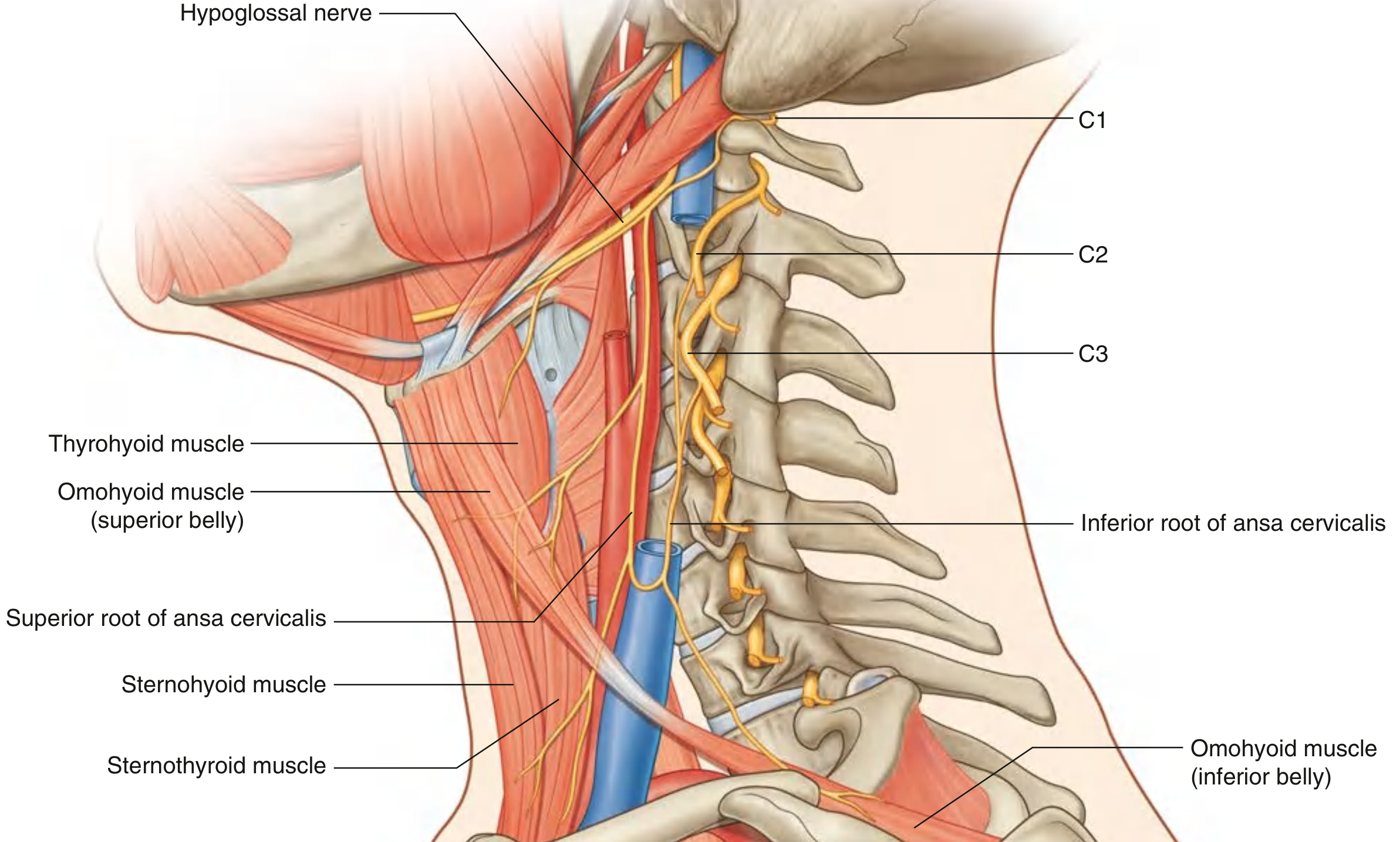
Fig. 8.180 Ansa Cervicalis - Gray's Anatomy for Students
Definition
The ansa cervicalis (meaning "handle/loop of the neck") is a nerve loop formed by fibers from cervical nerves C1, C2, and C3 that innervates three of the four infrahyoid (strap) muscles in the anterior triangle of the neck. It lies anterior to the carotid sheath.
Formation: Two Roots
1. Superior Root (Descendens Hypoglossi) - C1 (± C2)
- C1 fibers (sometimes also C2) join the hypoglossal nerve [CN XII] soon after it exits the skull via the hypoglossal canal
- They "hitch a ride" on CN XII as it descends and arches forward across the internal and external carotid arteries
- As CN XII turns forward, these fibers leave it and descend as the superior root, running between the internal jugular vein and the internal/common carotid arteries
- The superior root innervates:
- Superior belly of omohyoid
- Upper parts of sternohyoid
- Upper parts of sternothyroid
2. Inferior Root (Descendens Cervicalis) - C2, C3 (± C4 per Fischer's)
- Arises directly from the cervical plexus (C2 and C3)
- Descends either medial or lateral to the internal jugular vein before turning medially to join the superior root
- At the junction, it gives branches to innervate:
- Inferior belly of omohyoid
- Lower parts of sternohyoid
- Lower parts of sternothyroid
Muscles Supplied (Summary)
| Muscle | Innervated by |
|---|---|
| Sternohyoid | Ansa cervicalis (C1, C2, C3) |
| Sternothyroid | Ansa cervicalis (C1, C2, C3) |
| Omohyoid (both bellies) | Ansa cervicalis (C1, C2, C3) |
| Thyrohyoid | NOT ansa cervicalis - directly by C1 fibers via CN XII (nerve to thyrohyoid) |
Key point: Thyrohyoid is the exception - it is innervated independently by C1 fibers that remain on the hypoglossal nerve and never join the ansa loop. - Fischer's Mastery of Surgery
Position / Relations
- The ansa cervicalis loop forms on the anterior surface of the carotid sheath (anterior to the common carotid artery)
- It is closely related to the internal jugular vein - the inferior root can pass either medial or lateral to it
- The superior root travels closely with (and is often visible during) carotid endarterectomy and neck dissection
Clinical Significance
Ansa Cervicalis - Recurrent Laryngeal Nerve (RLN) Transfer
The ansa cervicalis has an important reinnervation role in laryngeal surgery:
- Used for nerve transfer to the RLN in cases of unilateral vocal fold paralysis
- First described by Frazier et al. (1924); current technique reported in 1986
- An end-to-end anastomosis of the proximal ansa cervicalis to the distal stump of the RLN restores muscle tone, bulk, and position to the hemilarynx
- No active movement of the vocal fold is expected - the goal is bulk and tone restoration
- Ansa cervicalis is the preferred donor nerve over the hypoglossal nerve because sacrifice of the ansa results in significantly lower morbidity (the infrahyoid muscles are not critical for phonation or swallowing) - Cummings Otolaryngology
Neck Dissection
The ansa cervicalis is at risk during neck dissections, particularly when clearing the carotid sheath region. Identifying and preserving it helps maintain infrahyoid muscle tone and avoids postoperative neck contour deformity.
Sources: Gray's Anatomy for Students | Fischer's Mastery of Surgery, 8th ed. | Cummings Otolaryngology Head and Neck Surgery
This is a shared conversation. Sign in to Orris to start your own chat.