
Atrial flutter
Reading File
Atrial flutter is a supraventricular tachyarrhythmia caused by a macro-reentrant circuit in the right atrium, classically producing a sawtooth flutter wave pattern on ECG at ~300 bpm, with 2:1 AV conduction giving a ventricular rate of ~150 bpm.
Searching sources...
Finding Sources
Searching PubMed
"atrial flutter"[MeSH Terms] AND management
Good - I have several strong sources. Let me read the main sections in parallel.
Reading File
Reading File
Reading File
Reading File
Good content from multiple sources. Let me fetch key images and read the remaining truncated treatment section.
Reading File
Reading File
Loading Image
Loading Image
Excellent - I now have comprehensive content and two key images. Let me compile the full answer.
Atrial Flutter
Definition & Mechanism
Atrial flutter is a macro-reentrant supraventricular tachyarrhythmia with an atrial rate of approximately 300 beats/min and a ventricular response that depends on the degree of AV block - typically 150 bpm (2:1), 100 bpm (3:1), or slower multiples.
Typical (CTI-dependent) atrial flutter involves a reentrant circuit confined to the right atrium. The circuit travels:
- Counterclockwise (more common): caudocranial up the interatrial septum, craniocaudal down the right atrial free wall
- Clockwise (less common): reverse direction
The cavotricuspid isthmus (CTI) - the narrow strip of tissue between the inferior vena cava and tricuspid annulus - acts as a critical slow-conduction zone and is the target of ablation.
Atypical atrial flutter involves macro-reentrant circuits that do NOT use the CTI - these often occur in the left atrium after mitral valve surgery or catheter ablation for AF, or are scar-mediated. - Braunwald's Heart Disease
ECG Findings
The classic ECG shows a sawtooth flutter wave pattern:
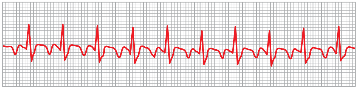
Atrial flutter at 250 bpm with 2:1 AV block giving a ventricular rate of 125 bpm - Guyton & Hall Textbook of Medical Physiology
| Feature | Counterclockwise (Typical) | Clockwise (Reverse Typical) |
|---|---|---|
| Leads II, III, aVF | Negative (sawtooth) | Positive |
| Lead V1 | Positive | Negative |
| Atrial rate | ~300 bpm | ~300 bpm |
Clinical Presentation
- Asymptomatic if AV block results in a physiologic rate
- Palpitations, light-headedness, syncope, or shortness of breath with faster ventricular rates
- Can precipitate heart failure or myocardial ischemia in patients with underlying heart disease
- If high-grade AV block develops: bradycardia, light-headedness, syncope
Aetiology / Associations
Typical atrial flutter almost always occurs with underlying cardiovascular or pulmonary disease. It can also be triggered by antiarrhythmic drugs (e.g., sodium-channel blockers used for AF). Common precipitants include hypoxia, fever, and electrolyte abnormalities. - Goldman-Cecil Medicine
Management
The approach follows three parallel tracks:
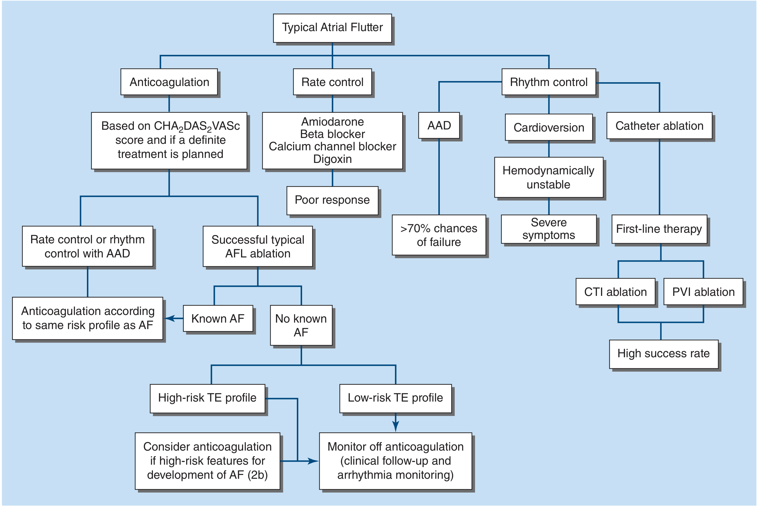
1. Anticoagulation
- Risk-stratify using CHA₂DS₂-VASc score
- Required before cardioversion unless episode is confirmed <48 hours, TEE has excluded left atrial appendage thrombus, or the patient is hemodynamically unstable requiring emergency cardioversion
- If cardioversion is not urgent: therapeutic anticoagulation (DOAC or warfarin with INR 2-3) for 4 weeks prior
- 3% per year thromboembolic risk - long-term anticoagulation similar to AF guidelines for patients at risk of recurrence
- After successful CTI ablation with no known AF and low-risk TE profile: can monitor off anticoagulation
2. Rate Control
- AV nodal blocking agents: beta-blockers (e.g., metoprolol 25-50 mg daily), calcium channel blockers (e.g., diltiazem 180-240 mg daily), amiodarone, or digoxin
- Rate control in atrial flutter is harder to achieve compared to AF - the AV node tends to conduct at fixed ratios (e.g., 2:1, 3:1), leading to abrupt rate changes
- Temporary measure while planning definitive rhythm control
3. Rhythm Control
Acute cardioversion:
- DC cardioversion is preferred and >95% effective
- IV ibutilide (1 mg over 10 min, repeat once if needed) - ~60% effective for chemical cardioversion
- Other antiarrhythmics (sotalol, dofetilide, disopyramide, amiodarone) - but >70% recurrence rate
Definitive therapy - Catheter ablation:
- Radiofrequency ablation of the CTI is >95% curative for typical flutter and is the first-line, preferred option for most patients
- Low risk of complications and low recurrence rates
- Antiarrhythmic drugs are a secondary option or for atypical flutter where ablation success is lower
Important caveat: Up to 50% of patients undergoing CTI ablation subsequently develop atrial arrhythmias, most commonly atrial fibrillation. This is an ongoing area of research.
Post-Ablation Follow-Up
- If ablation is successful and no known AF: risk of recurrence is very small - long-term anticoagulation is not necessary in low-risk patients
- If known AF coexists: continue anticoagulation based on AF risk profile
- Monitor for AF development with clinical follow-up and arrhythmia monitoring
Prognosis
Prognosis is generally determined by underlying comorbid conditions (heart disease, pulmonary disease) rather than the arrhythmia itself. Successful CTI ablation offers near-curative rates for typical flutter.
Recent evidence note: A 2025 systematic review (PMID 40434140) assessed pulsed field ablation (PFA) as an emerging modality for CTI-dependent flutter, showing favorable safety and efficacy - though this remains under investigation. A 2024 meta-analysis (PMID 39784135) found dapagliflozin (SGLT2 inhibitor) may have a protective effect against AF/flutter and reduce all-cause mortality.
Sources: Braunwald's Heart Disease; Harrison's Principles of Internal Medicine 22E; Goldman-Cecil Medicine; Guyton & Hall Textbook of Medical Physiology
This is a shared conversation. Sign in to Orris to start your own chat.