
What is vasectomy
vasectomy procedure male sterilization overview
Vasectomy
What Is It?
Anatomy
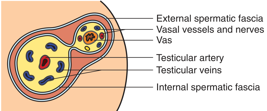
The Two Key Surgical Steps
- Vas isolation - accessing and isolating the vas deferens
- Vas occlusion - cutting, ligating, cauterizing, or blocking the vas
Methods of Vas Isolation
| Method | Description |
|---|---|
| Conventional Vasectomy (CV) | A 1.5-3 cm scrotal incision is made; the vas is grasped with a towel clamp or Allis forceps. Used when anatomy is challenging (prior surgery, thick scrotal skin, varicocele). |
| No-Scalpel Vasectomy (NSV) | A minimally invasive technique using a small puncture instead of an incision. Associated with less discomfort, fewer complications, and faster recovery. Preferred technique. |
Methods of Vas Occlusion
- Ligation (tying) with or without excision of a small segment
- Electrocautery of the lumen
- Metal or plastic clips
- Fascial interposition (placing a tissue layer between the two cut ends) - reduces risk of recanalization
- Mucosal fulguration - destroying the inner lining
Anesthesia
Pre-Procedure Counseling
- The procedure is intended to be permanent - reversal is possible but not guaranteed
- Need for a post-vasectomy semen analysis (PVSA) to confirm success
- A 1-2% risk of needing a repeat vasectomy if PVSA shows persistent sperm
- A small risk of spontaneous recanalization (the two ends reconnecting), which can result in pregnancy
- 1-2% risk of chronic scrotal pain
Effectiveness
Complications
| Complication | Notes |
|---|---|
| Hematoma | Most common early complication |
| Infection | Uncommon; scrotal skin heals rapidly |
| Chronic scrotal pain | ~1-2% of men |
| Recanalization / failure | Rare; requires PVSA to detect |
| Sperm granuloma | Local reaction to leaked sperm |
Vasectomy vs. Tubal Ligation
Reversal
Primary amminuria
Primary Aminoaciduria
Note: "Amminuria" is not a standard medical term. You likely mean aminoaciduria (amino acids in urine). This is the established term used in all major nephrology and physiology textbooks.
Definition
Types of Aminoaciduria
| Type | Mechanism | Examples |
|---|---|---|
| Renal (primary) aminoaciduria | Defective tubular transporter - plasma amino acid levels are normal | Cystinuria, Hartnup disease, iminoglycinuria |
| Overflow aminoaciduria | Plasma amino acid level is elevated, exceeding the reabsorptive threshold | Phenylketonuria, maple syrup urine disease |
Classification by Amino Acid Group
- Neutral and cyclic amino acids
- Glycine and imino acids
- Cystine and dibasic amino acids
- Dicarboxylic amino acids
- Beta-amino acids
Major Primary Aminoacidurias
1. Cystinuria (Most Clinically Significant)
- Prevalence: ~1 in 7,000 worldwide (varies by region)
- Inheritance: Autosomal recessive
- Defect: Mutations in genes encoding the two subunits of transport system b(0,+) - the rBAT (SLC3A1) and b(0,+)AT1 (SLC7A9) heterodimer
- Amino acids affected: Cystine, ornithine, lysine, arginine (dibasic group)
- Clinical consequence: Cystine stones in the urinary tract - cystine is the least soluble naturally occurring amino acid, especially at low pH
- Key point: This is the most clinically important renal aminoaciduria because of its stone-forming tendency
2. Hartnup Disease (Neutral Aminoaciduria)
- Prevalence: ~1 in 26,000
- Inheritance: Autosomal recessive
- Defect: Mutations in the neutral amino acid transporter B0AT1 (SLC6A19), found in kidney and intestine
- Amino acids affected: All monoamino-monocarboxylic (neutral) amino acids - alanine, asparagine, glutamine, histidine, isoleucine, leucine, methionine, phenylalanine, serine, threonine, tryptophan, tyrosine, valine
- Clinical consequence: Nicotinamide deficiency (tryptophan is a precursor of nicotinamide), causing:
- Photosensitive pellagra-like rash
- Intermittent cerebellar ataxia
- Rarely, mental retardation, emotional instability, psychosis
- Important: Most newborns identified by screening are asymptomatic
- Diagnosis: Urine aminogram showing elevated neutral amino acids with normal glycine, cystine, dibasic, dicarboxylic, and imino acids
- Treatment: Nicotinamide 50-300 mg/day for symptomatic cases
3. Iminoglycinuria
- Inheritance: Autosomal recessive (benign)
- Defect: Defective shared carrier for imino acids (proline, hydroxyproline) and glycine
- Amino acids affected: Proline, hydroxyproline, glycine
- Clinical consequence: None - clinically benign; mainly of scientific interest for demonstrating shared transport mechanisms
4. Dicarboxylic Aminoaciduria
- Defect: Defective SLC1A1 (EAAT3) transporter
- Amino acids affected: Glutamate (Glu) and Aspartate (Asp)
- Inheritance: Autosomal recessive
5. Generalized Aminoaciduria (Fanconi Syndrome)
Diagnosis
- Urine aminogram using chromatographic methods (ion exchange or HPLC) at specialized centers
- The pattern of amino acid excretion (not total amount) is the key diagnostic factor
- Detected in most countries through newborn urine screening programs
Summary Table
| Condition | Gene | Amino Acids Lost | Key Complication |
|---|---|---|---|
| Cystinuria | SLC3A1 / SLC7A9 | Cystine, Arg, Lys, Orn | Renal stones |
| Hartnup disease | SLC6A19 | All neutral amino acids | Pellagra-like rash, ataxia |
| Iminoglycinuria | SLC36A2 / SLC6A18 | Pro, HydroxyPro, Gly | None (benign) |
| Dicarboxylic aminoaciduria | SLC1A1 | Glu, Asp | Usually benign |
| Fanconi syndrome | Multiple | All amino acids | Rickets, acidosis, phosphaturia |
Amenorrhea means and clinical significance
Amenorrhea - Definition and Clinical Significance
Definition
The hypothalamus secretes GnRH in a pulsatile fashion → stimulates pituitary FSH and LH → promotes ovarian follicular development and ovulation → estrogen and progesterone cause endometrial development → withdrawal of these hormones causes menstrual bleeding. If any component (hypothalamus, pituitary, ovary, uterus, outflow tract) is nonfunctional, bleeding cannot occur.
- Berek & Novak's Gynecology
Types of Amenorrhea
1. Primary Amenorrhea
- Absence of menses by age 13 when there is no visible development of secondary sexual characteristics, OR
- Absence of menses by age 15 in the presence of normal secondary sexual characteristics
2. Secondary Amenorrhea
- A woman with regular cycles and a delay of even 1 week warrants a pregnancy test
- Women with fewer than 9 cycles/year or cycle length >35 days should be evaluated
It is important not to overemphasize the distinction between primary and secondary amenorrhea - their causes largely overlap.
- Berek & Novak's Gynecology
WHO Classification
| WHO Group | Hormonal Profile | Description |
|---|---|---|
| I | Hypogonadotropic hypogonadism | No endogenous estrogen, normal/low FSH, normal prolactin, no pituitary lesion |
| II | Normogonadotropic anovulation | Evidence of estrogen production, normal prolactin and FSH (e.g., PCOS) |
| III | Hypergonadotropic hypogonadism | Elevated FSH indicating gonadal insufficiency/failure |
| IV | Hyperprolactinemic anovulation | Anovulation specifically due to elevated prolactin |
Causes
Primary Amenorrhea - Major Causes
- Imperforate hymen
- Vaginal aplasia or congenital vaginal atresia
- Mullerian agenesis (Mayer-Rokitansky-Kuster-Hauser syndrome) - congenital absence of uterus
- Turner syndrome (45,XO) - gonadal dysgenesis
- XX gonadal dysgenesis
- 17-hydroxylase deficiency
- Autoimmune oophoritis
- Resistant ovary syndrome
- Constitutional delay of puberty (physiologic - most common)
- Kallmann syndrome (deficient pulsatile GnRH secretion + anosmia)
- Hypopituitarism
- Congenital adrenal hyperplasia
- Hypothyroidism
- Polycystic ovary syndrome (PCOS)
When gonadal failure occurs with primary amenorrhea, there is a high incidence of abnormal karyotype.
Secondary Amenorrhea - Major Causes
| Category | Examples |
|---|---|
| Physiologic | Pregnancy, lactation |
| Uterine | Asherman syndrome (post-traumatic synechiae), progestational agents |
| Ovarian | PCOS, primary ovarian insufficiency (idiopathic, autoimmune, post-chemo/radiation), ovarian tumors |
| Adrenal | Cushing syndrome, late-onset CAH, virilizing adrenal tumors |
| Thyroid | Hypothyroidism, hyperthyroidism |
| Pituitary | Hyperprolactinemia (prolactinoma), Sheehan syndrome, Lymphocytic hypophysitis |
| Hypothalamic | Functional hypothalamic amenorrhea (stress, excessive exercise, weight loss, eating disorders) |
| Iatrogenic/Drug | Antipsychotics, antidepressants, chemotherapy agents (busulfan, cisplatin), oral contraceptives |
Clinical Significance
1. Sign of Serious Underlying Disease
2. Estrogen Deficiency Consequences
- Osteoporosis - decreased bone mineral density
- Cardiovascular risk - loss of estrogen's cardioprotective effect
- Vaginal atrophy and genitourinary symptoms
3. Infertility
4. Endometrial Cancer Risk (Unopposed Estrogen)
5. Psychological and Quality of Life Impact
6. Karyotypic Abnormalities
Diagnostic Approach (Key Points)
- Physical examination - assess secondary sexual characteristics and pelvic anatomy
- hCG (pregnancy test) - rule out pregnancy first in secondary amenorrhea
- Serum prolactin and TSH - rule out hyperprolactinemia and thyroid disease
- FSH + estradiol - differentiate hypergonadotropic vs. hypogonadotropic hypogonadism
- Anti-Mullerian hormone (AMH) - useful for ovarian reserve assessment
- Karyotype - when gonadal failure is suspected in primary amenorrhea
Treatment Principles
- Correct the underlying cause where possible (medical or surgical)
- Hormone therapy (estrogen + progesterone) to establish/maintain secondary sexual characteristics and protect bone
- Calcium + Vitamin D for bone protection in estrogen-deficient states
- Ovulation induction (clomiphene, gonadotropins, GnRH pulsatile therapy) for women desiring pregnancy
primary hemangioma
hemangioma vascular tumor skin infantile
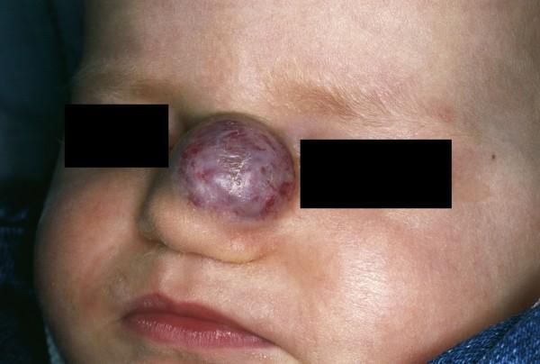
This clinical photograph displays a close-up view of an infant's face featuring a prominent vascular lesion on the nose. The lesion is identified as an infantile hemangioma, a common benign vascular tumor of childhood. Located on the nasal dorsum and extending toward the nasal tip, the tumor is a well-circumscribed, globular, and exophytic mass that significantly distorts the underlying nasal contour. Its color is variegated, showing shades of deep red and violet with surface telangiectasia, indicative of a high density of blood vessels. The surface texture is largely smooth and slightly translucent, with subtle lobulations visible under the skin. The surrounding facial skin on the cheeks, forehead, and bridge of the nose appears fair and unremarkable, without secondary signs of ulceration or additional satellite lesions. This visual represents a 'Cyrano nose' deformity, highlighting the clinical presentation of focal-type infantile hemangiomas which, if left untreated during the proliferative phase, can lead to permanent structural distortion of nasal cartilage or airway obstruction.
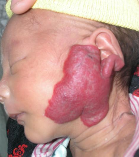
This clinical photograph displays a 40-day-old infant in a lateral view, showcasing a large infantile hemangioma in the left parotid and auricular region. The lesion is a prominent, irregularly shaped, raised plaque characterized by a vibrant crimson-to-purple hue and a lobulated, strawberry-like surface texture. The vascular mass extends from the preauricular cheek area posteriorly, partially involving and distorting the left pinna and external ear structures. The margins are well-defined against the surrounding light-toned skin. This image serves as a clinical example of a proliferative phase infantile hemangioma, a common benign vascular tumor of childhood, specifically located in the parotid area which requires monitoring for potential impact on deeper structures or functional impairment of the ear canal.
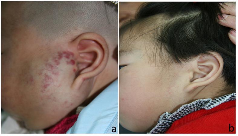
This clinical photograph represents a comparison chart of an infantile hemangioma before and after treatment. Image (a) depicts a 3-month-old male with a large, segmental infantile hemangioma located in the left parotid and preauricular region. The lesion is characterized by a cluster of raised, bright red to purple papules and plaques with a mottled distribution. Image (b) shows the same patient at 10 months of age following a course of oral propranolol therapy. There is a significant clinical resolution of the vascular tumor, showing near-complete involution of the hemangioma with minimal residual telangiectasia and healthy skin texture. This comparison illustrates the efficacy of beta-blocker therapy in managing proliferative infantile hemangiomas, particularly in high-risk areas like the parotid region where deep growth could affect underlying structures. This content is suitable for dermatology and pediatric education regarding vascular anomalies and pharmacological intervention outcomes.
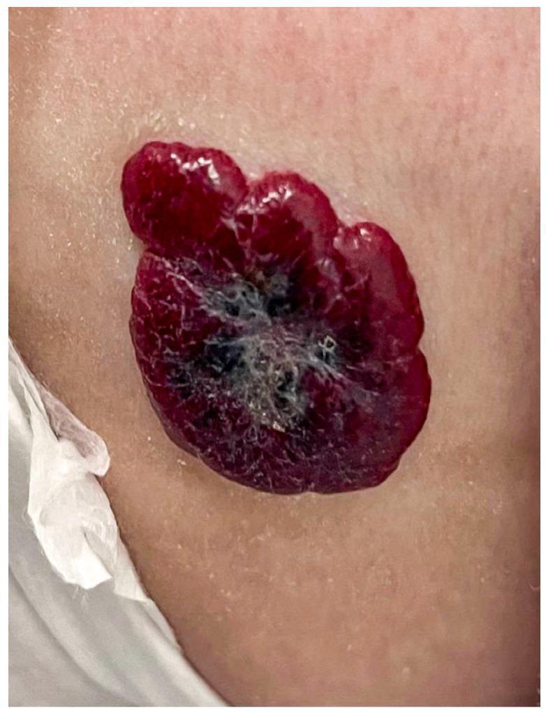
This clinical photograph displays a superficial, plaque-like skin lesion on the fair-toned skin of an infant, characteristic of an infantile hemangioma. The lesion is primarily a vibrant 'strawberry-red' color with a well-circumscribed but irregular, oval border. Its texture is notably lobulated and nodular, with a raised profile above the surrounding skin surface. The central portion of the lesion exhibits areas of deeper bluish-purple discoloration interspersed with fine grayish-white patches, which may indicate early regression or variations in vascular density. The surrounding skin appears healthy with normal texture, and small water droplets are visible nearby. This image serves as a classic pediatric dermatology example of a common benign vascular tumor during its growth phase, illustrating the typical morphology, color, and lobular architecture used for clinical diagnosis and monitoring of infantile hemangiomas.
Hemangioma
What Is a Hemangioma?
Note: "Primary" is most meaningful when distinguishing from secondary vascular changes. The most important subtypes are infantile hemangioma (skin/soft tissue), hepatic hemangioma (liver), and vertebral hemangioma (bone).
Types and Classification
By Histology
| Type | Description |
|---|---|
| Capillary | Fine, closely packed newly formed capillaries; most common type in GI tract |
| Cavernous | Large, dilated, irregular blood-filled spaces; common in liver and rectum |
| Mixed | Combination of both |
By Age of Presentation
| Type | Key Feature |
|---|---|
| Infantile hemangioma (IH) | Not present at birth; appears within 4 weeks and proliferates rapidly |
| Congenital hemangioma | Fully formed at birth; two subtypes: RICH (rapidly involuting) and NICH (non-involuting) |
1. Infantile Hemangioma (Most Common Overall)
Natural History - Phases
| Phase | Timing | Features |
|---|---|---|
| Early proliferation | 1-3 months | Rapid increase in size; lesion becomes warmer, firmer |
| Late proliferation | 3-5 months | Continued slower growth; 80% of final size by 3 months |
| Plateau | ~6 months | Stabilization |
| Involution | 6-12 months onward | Spontaneous regression; 50-70% resolve completely by age 4 |
Clinical Presentation
- Newborns may first show pale macules with thread-like telangiectasias
- Develop into raised, bright red or purple-red, lobulated ("strawberry") plaques
- May be superficial (bright red, well-defined), deep (bluish, subcutaneous), or mixed
- Pattern: focal (round, localized) or segmental (plaque-like, following embryologic segments)
Clinical Images
Complications - Indications for Early Treatment
-
Life-threatening complications:
- Airway hemangiomas (obstruction)
- Hepatic hemangiomas (high-output heart failure, severe hypothyroidism)
- Profuse bleeding from ulcerated hemangioma
-
Functional impairment:
- Near the eye - may impair visual development
- Near the mouth - may interfere with feeding
-
Ulceration: Most common complication (5-21%) - painful, usually scars; highest risk in diaper area and large lesions
-
Associated syndromes:
- PHACES syndrome - Posterior cranial fossa malformations, large facial Hemangiomas, Arterial lesions, Cardiovascular anomalies, Eye anomalies, Sternal cleft
- LUMBAR syndrome - Lower body hemangioma, Urogenital anomalies, Ulceration, Myelopathy, Bony deformities, Anorectal malformations, Arterial/Renal anomalies
-
Disfigurement: Permanent scarring or distortion of anatomic landmarks
Treatment
| Approach | Details |
|---|---|
| Watchful waiting | For uncomplicated lesions; photo documentation to track growth/regression |
| Propranolol (first-line) | Oral beta-blocker; first-line for complicated IH; initiated under supervision; minimum 6 months duration |
| Topical timolol | For small superficial lesions |
| Corticosteroids | When propranolol is contraindicated |
| Laser | Pulsed-dye laser for superficial lesions |
| Surgery | For residual deformity after involution |
2. Hepatic Hemangioma
- Most common benign tumor of the liver
- Female:male ratio 3:1; mean age ~45 years
- Lesions <5 cm = hemangiomas; lesions >5 cm = "giant hemangiomas"
- Growth occurs by ectasia (dilation), not neoplasia - no malignant potential reported
Microscopy
Clinical Features
- Most are asymptomatic - found incidentally on imaging
- Large lesions may cause vague upper abdominal discomfort
- Spontaneous rupture is exceedingly rare
- Kasabach-Merritt syndrome (rare): thrombocytopenia + consumptive coagulopathy associated with giant hemangiomas
Diagnosis
- CT and MRI: classic peripheral nodular enhancement pattern is diagnostic
- Biopsy is NOT recommended - dangerous and inaccurate for vascular lesions
Treatment
- Asymptomatic: observation only
- Symptomatic / enlarging / rupture risk: surgical enucleation (preferred over resection)
3. Vertebral Hemangioma (Bone)
- Most common benign vascular tumor of bone - found in 10-12% of autopsy specimens
- Most are clinically silent - incidental finding
- Commonest location: vertebral body, especially lumbar and lower thoracic spine
Radiological Features
- X-ray/CT: "corduroy" pattern (vertical striations, coarse trabeculae) on sagittal views; "polka dot" pattern on axial CT
- MRI: increased signal on both T1 and T2 (fat content) in typical cases
- Aggressive hemangiomas: T1 hypointense (vascular, not fatty) - red flag
Clinical Subtypes
| Type | Description |
|---|---|
| Asymptomatic | Most common; incidental finding; no treatment needed |
| Compressive | Compresses spinal cord or cauda equina |
| Symptomatic | Pain (60%), neurologic deficit (30%), fracture (10%) |
Treatment (Symptomatic/Aggressive)
- Radiation (50-80% success for symptomatic)
- Vertebroplasty (bone cement injection) - stabilizes pathologic bone
- Embolization - preoperative or for neurologic deficits
- Ethanol injection - obliterates vascular spaces
- Surgery - for neurologic deficit + fracture; embolization done first to minimize intraoperative bleeding
4. Hemangiomas of Other Sites
| Site | Key Points |
|---|---|
| Oral cavity/Head & Neck | Lip is most frequent oral site; soft, painless, red/blue mass; tends to regress spontaneously; may be associated with Sturge-Weber or von Hippel-Lindau syndrome |
| GI tract (colon/rectum) | 2nd most common vascular lesion of colon; cavernous rectal hemangiomas may cause massive hemorrhage; diagnosed by endoscopy; phleboliths on plain film suggest rectal cavernous hemangioma |
| Urologic | Can occur in kidney and bladder; usually incidental |
Summary
| Feature | Infantile (Skin) | Hepatic | Vertebral |
|---|---|---|---|
| Most common in | Infants, female | Adult females | Adults (incidental) |
| Behavior | Proliferates then involutes | Stable, ectatic growth | Usually static |
| Malignant potential | None | None | None |
| Treatment | Propranolol if complicated | Observation / enucleation | Observation / radiation / vertebroplasty |
How this is related with brain
Sturge-Weber syndrome brain MRI leptomeningeal angioma
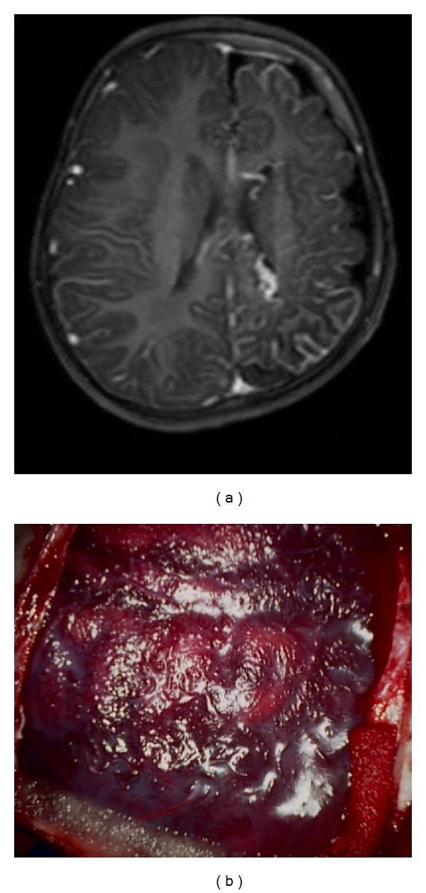
This composite image illustrates diagnostic and surgical findings of Sturge-Weber syndrome (SWS). (a) T1-weighted axial gadolinium-enhanced MRI of the brain: The left hemisphere demonstrates hallmark features of leptomeningeal angiomatosis, characterized by irregular, intense contrast enhancement and thickening of the leptomeninges. This is accompanied by ipsilateral sulcal prominence and hemispheric atrophy compared to the right side. (b) Intraoperative photograph: A direct view of the cortical surface revealing a dense, hypervascular pial angioma. The brain surface exhibits a deep reddish-purple discoloration due to engorged, tortuous abnormal vessels. The presence of these lesions is clinically significant in pediatric neurosurgery for managing refractory epilepsy, often requiring procedures like peri-insular functional hemispherectomy to disconnect the affected cortical tissue. The images provide a correlation between radiological evidence of pial enhancement and the physical presentation of vascular malformations on the cerebral cortex.
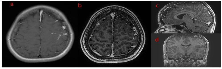
This diagnostic image composite presents brain Magnetic Resonance Imaging (MRI) from a patient with Sturge-Weber syndrome. The series includes T1-weighted sequences following gadolinium contrast administration across four views: (a) axial, (b) thin-slice axial, (c) sagittal reconstruction, and (d) coronal reconstruction. The primary finding is intense, pathological leptomeningeal enhancement localized to the left hemisphere, specifically following the gyral patterns of the frontal lobe. This enhancement represents a meningeal angioma or pial angiomatosis. In the axial views (a, b), there is a clear asymmetry compared to the right hemisphere, with contrast highlighting the deep sulci and cortical surfaces on the left. The thin-slice axial view (b) particularly emphasizes the increased vascularity and potential intracranial lipomatosis within the subarachnoid space. The sagittal (c) and coronal (d) multiplanar reconstructions further demonstrate the extent of this leptomeningeal involvement along the cortical mantle. These findings are characteristic of neurocutaneous syndromes involving vascular malformations of the pia mater.
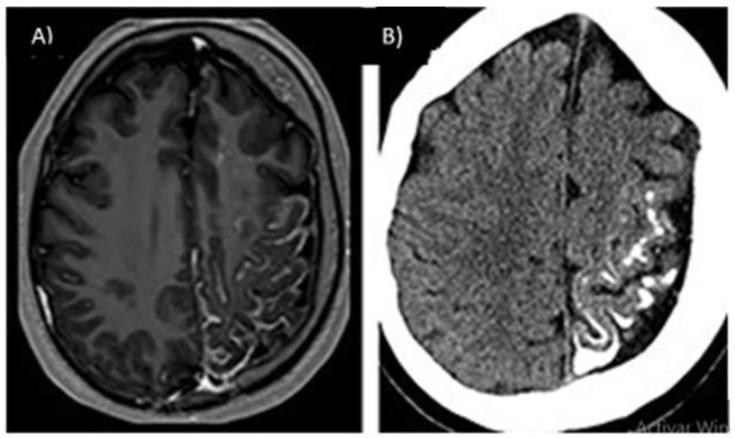
This dual-panel diagnostic image displays characteristic findings of Sturge-Weber syndrome (SWS) in the brain. Image A is a contrast-enhanced axial T1-weighted MRI showing intense, curvilinear leptomeningeal enhancement over the left cerebral hemisphere. The enhancement follows the pial surface of the gyri and sulci, consistent with a leptomeningeal angioma. Image B is a non-contrast axial CT scan of the same patient, revealing extensive gyriform calcifications (the 'tram-track' sign) within the left cortex, appearing as hyperdense, serpentine lines. Both modalities demonstrate secondary structural changes, including ipsilateral cortical atrophy and volume loss of the left hemisphere compared to the right. These findings illustrate the classic neuroimaging triad of SWS: leptomeningeal vascular malformation, intracranial calcification, and associated cerebral atrophy. This material is highly relevant for medical education in neurology, radiology, and pediatrics for the identification of phakomatoses and neurocutaneous syndromes.
Hemangioma and the Brain
- Hemangiomas can occur directly inside the brain (cavernous hemangioma / cavernoma)
- Hemangiomas on the skin/face can signal brain vascular anomalies through neurocutaneous syndromes
- Systemic/multifocal hemangiomas can have associated brain malformations (PHACES syndrome)
1. Intracranial (Brain) Hemangiomas - Cavernous Malformations
Pathology
- Composed of large, endothelium-lined, blood-filled cavities with no intervening normal brain tissue
- Prone to repeated small hemorrhages - hemosiderin deposits surround the lesion giving a characteristic "popcorn" appearance on MRI
- No arterial feeding vessels (unlike arteriovenous malformations)
Clinical Presentation
- May be asymptomatic (incidental finding)
- Seizures - most common symptomatic presentation
- Focal neurological deficits - depend on location (weakness, sensory loss, visual disturbance)
- Headache
- Intracerebral hemorrhage - can be sudden and severe
- Most common in supratentorial locations (cerebral hemispheres)
Diagnosis
- MRI is the gold standard - shows the classic "popcorn" or "mulberry" appearance with a hemosiderin rim (hypointense ring) on T2/susceptibility-weighted imaging (SWI)
- CT may show calcification or hyperdensity from hemorrhage
- Angiography is typically negative (no feeding artery)
Treatment
- Asymptomatic: observation
- Symptomatic (seizures, hemorrhage, progressive deficits): surgical resection is curative when accessible
- Radiosurgery (Gamma Knife) for deep/inoperable lesions
2. Sturge-Weber Syndrome - Facial Hemangioma + Brain Angioma
What Is It?
Key Features
| Feature | Detail |
|---|---|
| Port-wine stain | Facial capillary hemangioma/malformation, especially upper face and periorbital area |
| Leptomeningeal angioma | Abnormal vascular proliferation over the cerebral cortex (pial angiomatosis) |
| Brain calcification | Classic "tram-track" or "trolley-track" gyriform calcification on CT |
| Cerebral atrophy | Ipsilateral (same side as skin lesion) progressive atrophy |
| Glaucoma | Due to vascular anomaly involving the eye |
Brain Complications
- Focal-onset epilepsy (most common neurological manifestation) - often refractory
- Hemiparesis - contralateral to the brain lesion
- Hemianopia (visual field loss)
- Cognitive impairment (variable severity)
- Only ~15% of patients with an upper facial port-wine stain actually develop brain involvement
MRI Findings in Sturge-Weber Syndrome
Treatment
- Antiepileptic drugs for seizure control
- Aspirin (low-dose) - reduces thrombotic episodes and stroke-like events
- Hemispherectomy/disconnection surgery for drug-resistant epilepsy
- Regular ophthalmological monitoring for glaucoma
3. PHACES Syndrome - Large Facial Hemangioma + Posterior Fossa Brain Malformation
| Letter | Feature |
|---|---|
| P | Posterior cranial fossa malformations (e.g., Dandy-Walker) |
| H | Large segmental facial Hemangiomas |
| A | Arterial lesions (intracranial/cervical arteries) |
| C | Cardiac defects (aortic coarctation) |
| E | Eye anomalies |
| S | Sternal cleft / supraumbilical raphe |
Brain Connection
- Children with a large segmental facial hemangioma (especially in the "beard" distribution, V1-V3 dermatomes) must be screened for PHACES syndrome
- Brain MRI should be obtained to evaluate for posterior fossa malformations and intracranial arterial anomalies (which can cause stroke)
- The posterior fossa malformations in PHACES include hypoplasia of the cerebellum, Dandy-Walker variants, and arachnoid cysts
4. Multifocal Hemangiomas and Hepatic / CNS Involvement
- Hepatic hemangiomas (high-output cardiac failure)
- CNS involvement - screening with brain MRI is indicated
Summary: How Hemangioma Relates to the Brain
| Relationship | Condition | Key Brain Feature |
|---|---|---|
| Direct brain tumor | Cavernous hemangioma (cavernoma) | Seizures, hemorrhage, focal deficits |
| Neurocutaneous syndrome | Sturge-Weber syndrome | Leptomeningeal angioma, epilepsy, "tram-track" calcification |
| Associated syndrome | PHACES syndrome | Posterior fossa malformations, arterial anomalies |
| Multifocal disease | ≥5 cutaneous hemangiomas | Risk of CNS hemangioma involvement |
All case means
What do you mean by 'all case means'?
- Are you asking about all acronyms/abbreviations used (like PHACES, LUMBAR, RICH, NICH, SWS)?
- Are you asking about all syndromes related to hemangioma?
- Are you asking about something in a different language (e.g., did you mean "all causes" or "all cases")?
- Or something completely different?
Some common sedatives for cold and fever for 20yr old female
common medications cold fever 20 year old adult paracetamol antihistamine decongestant dosage
Important note: The term "sedatives" is not the right word here. Sedatives are drugs that induce sleep/calm (e.g., benzodiazepines). What you likely mean are symptomatic medications for cold and fever - some of which do cause drowsiness as a side effect. This answer covers exactly that.This is for general educational information only. Always consult a doctor or pharmacist before taking any medication.
Common Medications for Cold and Fever in a 20-Year-Old Female
1. Antipyretics (Fever Reducers)
Paracetamol / Acetaminophen (First Choice)
- How it works: Reduces fever by acting on the hypothalamic heat-regulating center; mild analgesic
- Dose (adult): 500 mg - 1000 mg every 4-6 hours as needed
- Max dose: 4000 mg (4 g) per 24 hours - do NOT exceed
- Safe for: Adults, pregnancy (if needed)
- Caution: Many cold/flu combination tablets already contain paracetamol - avoid double-dosing
- Side effects: Very safe at recommended doses; overdose causes serious liver damage
Ibuprofen (Alternative / Also Anti-inflammatory)
- How it works: NSAID - reduces fever, pain, and inflammation
- Dose (adult): 200-400 mg every 6-8 hours with food
- Max dose: 1200 mg/day (OTC); up to 2400 mg/day under medical supervision
- Advantages: Also relieves body aches and throat pain better than paracetamol
- Caution: Take with food; avoid on empty stomach; avoid if history of gastric ulcers or kidney disease
- Important for females: Avoid during pregnancy (especially 3rd trimester)
2. Antihistamines (For Runny Nose, Sneezing, Watery Eyes)
First-Generation (Sedating) - Cause Drowsiness
| Drug | Dose | Notes |
|---|---|---|
| Chlorphenamine / Chlorpheniramine | 4 mg every 4-6 hrs | Very commonly used in cold combos; causes drowsiness |
| Diphenhydramine (Benadryl) | 25-50 mg every 6-8 hrs | Strong sedation; often used at night |
| Promethazine | 10-25 mg at night | Stronger sedation; also antiemetic |
These are the drugs that make you "sleepy" - commonly found in nighttime cold products like NyQuil, Benadryl, Calpol Night.
Second-Generation (Non-Sedating) - No/Minimal Drowsiness
| Drug | Dose | Notes |
|---|---|---|
| Cetirizine (Zyrtec) | 10 mg once daily | Mild drowsiness in some; good for daytime use |
| Loratadine (Claritin) | 10 mg once daily | Essentially non-sedating; good for daytime |
| Fexofenadine (Allegra) | 120-180 mg once daily | Truly non-sedating; best for daytime |
Second-generation antihistamines are better if you need to study, drive, or work.
3. Decongestants (For Blocked/Stuffy Nose)
| Drug | Route | Dose | Notes |
|---|---|---|---|
| Pseudoephedrine | Oral | 60 mg every 4-6 hrs | Effective; can raise blood pressure; may cause insomnia |
| Phenylephrine | Oral | 10 mg every 4 hrs | Milder than pseudoephedrine |
| Xylometazoline / Oxymetazoline | Nasal spray | 1-2 sprays each nostril, 2-3x/day | Fast relief; use max 3-5 days only (risk of rebound congestion) |
4. Cough Suppressants / Expectorants
| Drug | Use | Dose |
|---|---|---|
| Dextromethorphan (DXM) | Dry, non-productive cough | 10-30 mg every 4-6 hrs |
| Guaifenesin | Productive cough - loosens mucus | 200-400 mg every 4 hrs; drink plenty of water |
| Honey + warm water | Mild soothing effect | Evidence supports use in adults |
5. Common Combination Cold Products (OTC)
| Product Type | Typical Contents |
|---|---|
| Day/Cold & Flu Tablets | Paracetamol + Phenylephrine (decongestant) |
| Night Cold Tablets | Paracetamol + Diphenhydramine or Chlorphenamine (sedating) |
| All-in-one Cold Capsules | Paracetamol + Decongestant + Antihistamine + Cough suppressant |
Caution: When using combination products, always check the ingredients to avoid taking duplicate medications (e.g., paracetamol in both a combo tablet and a standalone tablet).
6. Non-Drug Measures (Equally Important)
- Rest and sleep - essential for recovery
- Warm fluids - warm water, soup, herbal teas - thin secretions
- Saline nasal drops/spray - safe, no side effects, helps relieve congestion
- Steam inhalation (warm humidified air ~45°C) - evidence supports reduction in cold symptoms
- Honey - soothing for throat and cough
- Vitamin C - regular supplementation reduces cold duration by ~8% in adults
- Zinc lozenges - some evidence for shortening cold duration in adults
Summary Table
| Symptom | Recommended Drug | Causes Drowsiness? |
|---|---|---|
| Fever | Paracetamol / Ibuprofen | No |
| Body aches | Ibuprofen / Paracetamol | No |
| Runny nose / sneezing | Cetirizine (day) / Chlorphenamine (night) | Night option: Yes |
| Blocked nose | Xylometazoline spray / Pseudoephedrine | No |
| Dry cough | Dextromethorphan | No |
| Productive cough | Guaifenesin | No |
| Sleep difficulty | Diphenhydramine / Promethazine | Yes (intended) |
When to see a doctor:
- Fever above 39.5°C (103°F) not responding to medication
- Symptoms lasting more than 10 days
- Difficulty breathing or chest pain
- Severe headache or stiff neck
- Ear pain or facial pain (may indicate bacterial infection)
As a medical practitioner what tablet i can give someone who is suffering from sleepless ness