
Penile carcinoma
"penile cancer" AND treatment
penile squamous cell carcinoma histology clinical
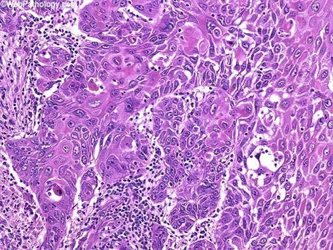
High-power histology image of penile squamous cell carcinoma with dermal invasion. Malignant squamous cells form irregular nests and cords that extend from the overlying epithelium into the dermis, producing finger-like projections and disruption of normal dermal architecture. Cells exhibit squamous differentiation with hyperchromatic pleomorphic nuclei, increased nuclear-to-cytoplasmic ratio, conspicuous nucleoli, and keratinizing features. Rare dyskeratotic cells are present within tumor nests, reflecting abnormal keratinization. Focal parakeratosis and occasional keratin pearls may be observed in places, along with a peritumoral inflammatory infiltrate in the adjacent dermis. Depth of invasion should be measured from the basement membrane of the overlying squamous epithelium to the deepest invasive cancer cells; this parameter has established prognostic significance in penile SCC. Vascular invasion, when present, further increases risk of metastasis and warrants more aggressive management. The cited Mod Pathol 2001 study emphasizes depth of invasion and lymphovascular invasion as predictors of cancer progression, underscoring the clinical relevance of accurate histologic assessment for staging and treatment planning. Clinically, these histopathologic features inform surgical margins, nodal assessment, and adjuvant therapy considerations. Overall, the image depicts an infiltrative penile SCC with dyskeratosis, an aggressive tumor subset requiring careful correlation with clinical and radiologic data. For accurate prognosis and management.
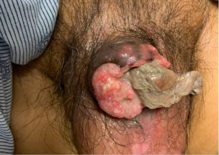
Clinical photograph of the male pelvic region demonstrating an advanced stage of penile squamous cell carcinoma. The image shows a large, exophytic mass that has extensively destroyed and replaced the normal penile architecture, including the glans and distal shaft. The lesion is multi-lobulated and exhibits heterogenous surface features, characterized by erythematous, granular tissue and significant areas of gray-white tissue necrosis (slough). There is visible foul, purulent discharge emanating from the central portion of the mass. The surrounding skin of the pubic area and proximal shaft shows signs of secondary inflammation, erythema, and anatomical distortion. This image serves as a clinical example of a T4 stage penile malignancy, illustrating the aggressive locally invasive nature of the disease and the resulting complete obliteration of normal external genitalia structures.
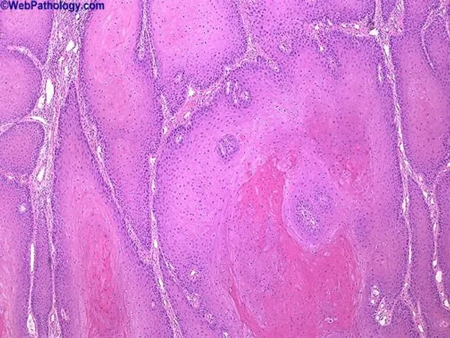
Imaging modality: Light microscopy of hematoxylin and eosin stained penile tissue biopsy. The histology shows a verruciform, exophytic growth pattern arising from penile skin and involving the glans region. The surface demonstrates marked hyperkeratosis with pronounced acanthosis. Beneath this, large, well-differentiated nests of keratinizing squamous epithelium extend into the dermis with broad, pushing margins rather than infiltrative invasion. Cellular atypia is minimal, with orderly maturation and abundant keratin production; mitotic figures are scarce. The nests are composed of mature squamous cells exhibiting extensive keratinization, sometimes forming keratin-filled cores or keratin pearls; the overlying epithelium remains thickened and papillomatous. Inflammation may be present at the periphery. Overall, this pattern is characteristic of verrucous carcinoma of the penis (penile carcinoma, verruciform variant), a slow-growing, locally aggressive squamous cell carcinoma with low metastatic potential. Clinically, it presents as a large exophytic mass in middle-aged men and requires wide local excision with clear margins; differentiation from condyloma acuminatum is essential because of treatment implications. The image underscores the diagnostic importance of architectural pushing borders, minimal cytologic atypia, and pronounced keratinization in establishing a verrucous carcinoma diagnosis and guiding surgical management. No lymphovascular invasion identified; margins critical. Immunohistochemistry may aid in ruling out analogous lesions.
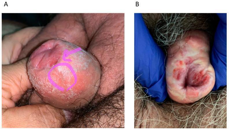
Clinical photograph comparison of two cases of local recurrence of penile squamous cell carcinoma following previous penis-sparing surgery. Image A displays a recurrence at the neoglans and foreskin interface characterized by a raised, hyperkeratotic, and scaly plaque, highlighted by a pink circle and arrow, suggesting a superficial verrucous or squamous growth. Image B demonstrates a more aggressive multifocal recurrence on the neoglans surface, presenting with deeply erythematous, friable, and ulcerated lesions, indicating increased vascularity and potential tissue erosion. Both images highlight the importance of post-glansectomy surveillance. These clinical signs are critical for identifying penile intraepithelial neoplasia (PeIN) or invasive squamous cell carcinoma recurrences in patients who have undergone glansectomy or partial penectomy.
Penile Carcinoma
Epidemiology
- Smith and Tanagho's General Urology, 19th Ed.
Risk Factors
| Risk Factor | Notes |
|---|---|
| Phimosis / poor genital hygiene | Smegma accumulation under phimotic foreskin causes chronic inflammation |
| Lack of neonatal circumcision | Disease is virtually absent in males circumcised at birth |
| HPV infection | Implicated in ~50% of penile cancers (especially types 16 and 18) |
| Smoking | Independent risk factor |
| Chronic inflammatory conditions | Balanitis, lichen sclerosus |
| Low socioeconomic status | Associated factor |
Pathology
A. Precancerous Lesions
- Leukoplakia - white plaque usually at the meatus; histology shows acanthosis, hyperkeratosis, parakeratosis; most common in diabetics. May precede or co-exist with frank carcinoma.
- Balanitis xerotica obliterans (lichen sclerosus) - white patch on prepuce or glans involving the meatus; associated with atrophic epidermis and collagen deposition abnormalities; common in middle-aged diabetics.
- Giant condylomata acuminata (Buschke-Lowenstein tumor) - cauliflower-like lesions from prepuce or glans; HPV-driven; may be difficult to distinguish from well-differentiated squamous cell carcinoma.
B. Penile Intraepithelial Neoplasia (PeIN / Carcinoma In Situ)
- Erythroplasia of Queyrat - CIS involving the glans; red, velvety plaque with ulcerations
- Bowen's disease - CIS involving the shaft; similar red plaque appearance
- Histology: hyperplastic cells in disordered array with vacuolated cytoplasm and mitotic figures
- Can be subdivided into differentiated (HPV-negative) or undifferentiated (HPV-positive) subtypes
C. Invasive Carcinoma
- Squamous cell carcinoma (SCC): >98% of all penile cancers
- Most commonly originates on the glans (48%), followed by prepuce, then shaft
- Appearance: papillary (exophytic/verrucous) or ulcerative (endophytic/infiltrative)
- Endophytic/ulcerative tumors carry worse prognosis than papillary tumors
- Verrucous carcinoma: 5-16% of cases; well-demarcated deep margin; locally aggressive but low metastatic potential; HPV-driven
Patterns of Spread
- Local extension: Carcinoma begins as ulcerative or papillary lesion, gradually invading glans, then shaft. Buck's fascia acts as a temporary barrier to deeper invasion - once penetrated, the tumor can invade the corpora and urethra.
- Lymphatic spread: Drains to the inguinal lymph nodes first (superficial then deep), then to pelvic (iliac) lymph nodes. Lymph node metastasis is the most important single predictor of survival.
- Hematogenous spread: Occurs late; liver, lung, and bone are the most common sites.
Key point: ~25% of patients with clinically nonpalpable inguinal nodes will still harbor lymphatic micrometastases.
- Campbell Walsh Wein Urology, 3-Volume Set
TNM Staging (AJCC 8th Edition)
Primary Tumor (T)
| Stage | Description |
|---|---|
| Tis | Carcinoma in situ (PeIN) |
| Ta | Noninvasive localized SCC |
| T1a | Invades lamina propria/dermis; no LVI, no PNI, not high-grade |
| T1b | Invades lamina propria/dermis; with LVI and/or PNI or high-grade (G3/sarcomatoid) |
| T2 | Invades corpus spongiosum (glans or ventral shaft) ± urethral invasion |
| T3 | Invades corpora cavernosa (including tunica albuginea) ± urethral invasion |
| T4 | Invades adjacent structures (scrotum, prostate, pubic bone) |
Regional Lymph Nodes (N)
| Stage | Description |
|---|---|
| cN0 | No palpable/visually enlarged inguinal lymph nodes |
| cN1 | Palpable mobile unilateral inguinal lymph node |
| cN2 | Palpable mobile ≥2 unilateral or bilateral inguinal lymph nodes |
| cN3 | Fixed inguinal nodal mass or pelvic lymphadenopathy |
| pN0 | No regional node metastasis |
| pN1 | ≤2 unilateral inguinal metastases, no extranodal extension |
| pN2 | ≥3 unilateral inguinal or bilateral inguinal metastases |
| pN3 | Extranodal extension or pelvic lymph node metastasis |
Distant Metastasis (M)
| Stage | Description |
|---|---|
| M0 | No distant metastasis |
| M1 | Distant metastasis |
- Campbell Walsh Wein Urology
Diagnosis and Workup
- Biopsy (incisional or punch): mandatory to confirm histology and grade before definitive management
- MRI of penis: best for local staging - especially useful with artificial erection to assess corpora cavernosa invasion
- Ultrasound: to assess depth of invasion
- CT abdomen/pelvis: evaluate inguinal and pelvic lymph nodes (especially in obese patients or those with prior inguinal surgery)
- PET/CT: higher sensitivity (96%) for enlarged/palpable nodes; not routinely recommended for staging in current practice; useful when inguinal metastases are confirmed to evaluate pelvic/distant disease
Treatment
Primary Tumor
| Approach | Indication |
|---|---|
| Topical therapy (5-FU, imiquimod cream) | Tis only |
| Laser ablation (CO₂ or Nd:YAG) | Tis, Ta, T1a (small, superficial) |
| Mohs surgery / wide local excision | Tis, Ta, T1 |
| Glansectomy with resurfacing | T1-T2 confined to glans |
| Partial penectomy | T1b-T3; requires 5-10 mm clear margin (grade-dependent) |
| Total penectomy with perineal urethrostomy | Bulky T3-T4 at penile base |
| Radiation (brachytherapy or EBRT) | T1-T2, glans lesions <4 cm in centers with expertise |
Important: The traditional 2 cm margin rule has been challenged. Grade 1-2 tumors spread maximally 5 mm proximally; grade 3 tumors spread up to 10 mm. Intraoperative frozen sections can guide organ-sparing surgery.
Regional Lymph Nodes - Inguinal Lymph Node Dissection (ILND)
- Nonpalpable nodes: Intermediate risk (T1b) and high-risk tumors (T2, T3)
- Palpable nodes <4 cm: Positive on FNA, or high-risk primary (T1b+)
- Delay in treating inguinal nodes >12 weeks significantly worsens survival (5-year DSS 64% vs 40%)
- 1-3 positive inguinal nodes: ~75%
- 4-5 positive nodes: ~8%
- Neoadjuvant chemotherapy (cisplatin + 5-fluorouracil) first
- If response occurs, consolidative surgery with superficial and deep inguinal node dissection
Systemic Disease / Metastatic Penile Cancer
- BMP (bleomycin, methotrexate, cisplatin)
- TIP (paclitaxel, ifosfamide, cisplatin) - widely used in neoadjuvant setting
- Cisplatin + 5-FU - standard for locally advanced/palpable nodal disease
Prognosis
| Nodal Status | 5-Year Survival |
|---|---|
| Node-negative | 65-90% |
| Positive inguinal nodes | 30-50% |
| Positive iliac/pelvic nodes | <20% |
| Soft tissue or bone metastases | Virtually 0% |
- Depth of invasion (most important)
- Lymphovascular invasion (LVI)
- Perineural invasion (PNI)
- Grade (G3 / sarcomatoid = worst)
- Positive surgical margins
Key Teaching Points
- HPV (types 16, 18) is implicated in ~50% of cases; neonatal circumcision is nearly 100% protective.
- SCC accounts for >98% of cases; glans is the most common site.
- Lymph node status is the single most important prognostic factor.
- Buck's fascia acts as a barrier to deep invasion; once breached, prognosis worsens significantly.
- T1b vs T1a distinction (LVI, PNI, high grade) drives the surgical decision to perform ILND even with nonpalpable nodes.
- Organ preservation is the modern standard for early-stage disease.
- Delays in inguinal lymph node management beyond 12 weeks worsen survival.
- Campbell Walsh Wein Urology, 3-Volume Set (AJCC 8th Ed. staging, surgical management, ILND protocols)
- Smith and Tanagho's General Urology, 19th Ed. (epidemiology, pathology, spread, prognosis)
- Suartz et al., BJU Int 2025 (PMID 39856798) - open vs. endoscopic lymphadenectomy meta-analysis
- Fadigas et al., Arch Esp Urol 2024 (PMID 39763248) - immunotherapy in penile cancer systematic review