
Essay questions with neat labelled diagram and flowchart 1. Describe Ascending track 2. Explain corticospinal track 3. Functions and connections of thalamus 4. Structure connections and functions of cerebral cortex 5. Explain Visual pathway with the lesions 6. Organ of corti
Reading File
Finding Sources
Finding Sources
Finding Sources
Finding Sources
Finding Sources
Finding Sources
Finding Sources
Finding Sources
Finding Sources
Finding Sources
Reading File
Reading File
Reading File
Reading File
Reading File
Reading File
Reading File
Reading File
Reading File
Loading Image
Loading Image
Loading Image
Loading Image
Loading Image
Loading Image
Reading File
Reading File
Loading Image
Loading Image
Loading Image
Here are comprehensive essay answers for all 6 topics, complete with labelled diagrams and flowcharts drawn from authoritative textbooks.
1. ASCENDING TRACTS
Introduction
Ascending tracts are white matter pathways that carry sensory information from the body to the brain. They are classified by the modality they convey.
Two Major Ascending Systems
A. Dorsal Column - Medial Lemniscal (DCML) Pathway
Carries: Fine touch, vibration, proprioception, 2-point discrimination
B. Spinothalamic (Anterolateral) Pathway
Carries: Pain, temperature, crude touch, pressure
FLOWCHART: Dorsal Column - Medial Lemniscal Pathway
PERIPHERAL RECEPTOR
(Meissner's corpuscle, Pacinian corpuscle, muscle spindle)
|
▼
1st ORDER NEURON (Cell body in Dorsal Root Ganglion)
|
▼ Enters ipsilateral DORSAL COLUMN
┌──────────────────────┐
│ Gracile fasciculus │ → Leg/lower body (medial)
│ (below T6) │
│ Cuneate fasciculus │ → Arm/upper body (lateral)
│ (above T6) │
└──────────────────────┘
|
▼ Ascends IPSILATERALLY to MEDULLA
2nd ORDER NEURON
(Nucleus gracilis / Nucleus cuneatus)
|
▼ CROSSES → MEDIAL LEMNISCUS (contralateral)
|
▼ Travels through Pons → Midbrain
3rd ORDER NEURON
(Ventral Posterolateral nucleus [VPL] of THALAMUS)
|
▼ Internal capsule (posterior limb)
|
▼ PRIMARY SOMATOSENSORY CORTEX
(Postcentral gyrus, Brodmann areas 3, 1, 2)
FLOWCHART: Spinothalamic (Anterolateral) Pathway
The diagram below from Neuroanatomy through Clinical Cases shows the full spinothalamic pathway:
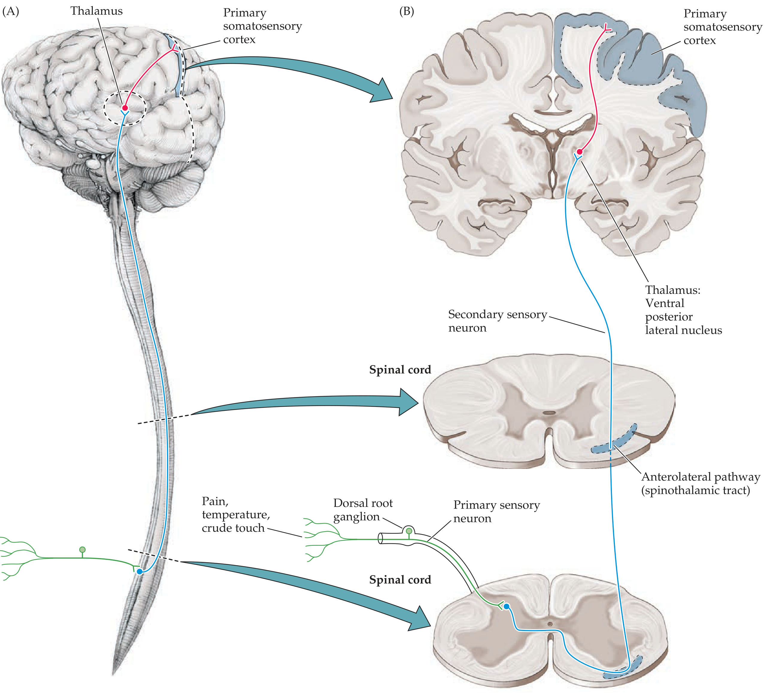
Figure: Spinothalamic Sensory Pathway - Neuroanatomy through Clinical Cases, 3rd Ed.
PERIPHERAL RECEPTOR
(Free nerve endings - Aδ and C fibers)
|
▼
1st ORDER NEURON (Cell body in Dorsal Root Ganglion)
|
▼ Enters dorsal horn (Rexed laminae I, II, V)
|
▼ CROSSES immediately in ANTERIOR WHITE COMMISSURE
2nd ORDER NEURON
Ascends in CONTRALATERAL ANTEROLATERAL COLUMN
┌──────────────────────────┐
│ Lateral spinothalamic │ → Pain & Temperature
│ Anterior spinothalamic │ → Crude touch & Pressure
└──────────────────────────┘
|
▼ Passes through Medulla → Pons → Midbrain
3rd ORDER NEURON
(VPL of THALAMUS)
|
▼
PRIMARY SOMATOSENSORY CORTEX (Postcentral gyrus)
Key Comparison Table
| Feature | DCML | Spinothalamic |
|---|---|---|
| Modality | Fine touch, vibration, proprioception | Pain, temperature, crude touch |
| 1st neuron | Dorsal root ganglion | Dorsal root ganglion |
| Where it crosses | Medulla | Spinal cord (same level) |
| Location in cord | Ipsilateral dorsal column | Contralateral anterolateral |
| 2nd neuron | Nucleus gracilis/cuneatus | Dorsal horn (laminae I, V) |
| 3rd neuron | VPL thalamus | VPL thalamus |
| Cortex | Postcentral gyrus | Postcentral gyrus |
Clinical Significance
- Hemicord lesion (Brown-Séquard syndrome): ipsilateral DCML loss + contralateral spinothalamic loss below the lesion
- Syringomyelia: bilateral spinothalamic loss at the level of the syrinx (cape-like distribution) with preserved DCML
2. CORTICOSPINAL TRACT
Introduction
The corticospinal (pyramidal) tract is the most clinically important descending motor pathway in the nervous system. It controls voluntary movements, especially of the extremities, and originates primarily in the motor cortex.
Origin
-
50% from primary motor cortex (Brodmann area 4) - precentral gyrus
- Remainder from premotor/supplementary motor areas (area 6) and parietal lobe (areas 3,1,2,5,7)
- Giant Betz cells (~3% of corticospinal neurons) are the largest neurons in the nervous system
LABELLED DIAGRAM
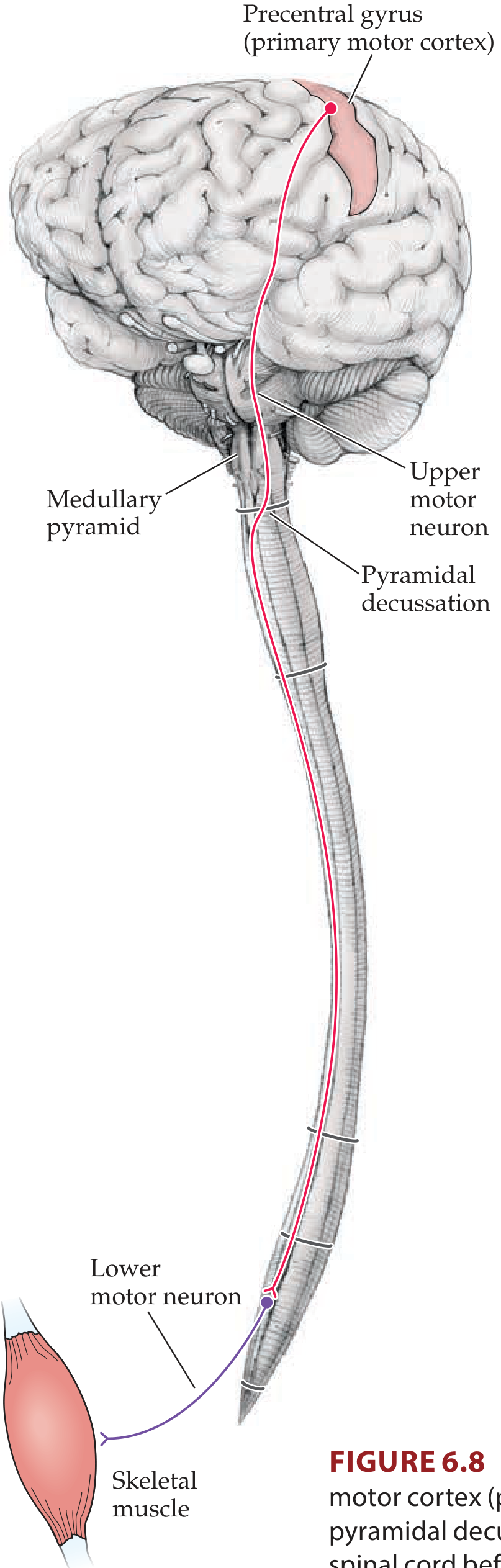
Figure 6.8: Lateral Corticospinal Tract - Neuroanatomy through Clinical Cases, 3rd Ed.
FLOWCHART
PRIMARY MOTOR CORTEX (Precentral gyrus, area 4)
+ Premotor (area 6) + Parietal cortex
|
▼
CORONA RADIATA (cerebral white matter)
|
▼
INTERNAL CAPSULE - Posterior limb
(Somatotopy: Face → Arm → Leg, anterior to posterior)
|
▼
CEREBRAL PEDUNCLE (Basis pedunculi)
Middle 1/3 → corticobulbar + corticospinal fibers
|
▼
PONS - scattered fascicles (ventral)
|
▼
MEDULLARY PYRAMIDS (ventral surface of medulla)
|
▼ ── PYRAMIDAL DECUSSATION ──
┌─────────────────────────────────┐
│ 85% cross │ 15% stay
▼ ▼
LATERAL CORTICOSPINAL ANTERIOR CORTICOSPINAL
TRACT (contralateral) TRACT (ipsilateral)
(in dorsolateral funiculus) (in ventral funiculus)
| |
▼ ▼ Crosses at level
LOWER MOTOR NEURON LOWER MOTOR NEURON
(Anterior horn, ventral horn) (Axial muscles)
|
▼
SKELETAL MUSCLE (voluntary movement)
Key Features
| Structure | Relevance |
|---|---|
| Internal capsule lesion | Contralateral hemiplegia (face + arm + leg) |
| Cerebral peduncle lesion | Weber syndrome (CN III palsy + contralateral hemiplegia) |
| Pyramidal decussation | 85% cross → contralateral control |
| Lateral CST in cord | Somatotopy: arm medial, leg lateral |
Upper vs Lower Motor Neuron Signs
| UMN Lesion (above anterior horn) | LMN Lesion (anterior horn/below) |
|---|---|
| Spastic paralysis | Flaccid paralysis |
| Hyperreflexia | Hyporeflexia/areflexia |
| Babinski sign positive | No Babinski |
| No muscle wasting | Muscle wasting |
| Clasp-knife rigidity | Fasciculations |
3. THALAMUS - FUNCTIONS AND CONNECTIONS
Introduction
The thalamus is an egg-shaped diencephalic structure sitting above the brainstem. It is the main relay center of the brain - nearly all pathways projecting to the cerebral cortex do so after synapsing in the thalamus.
LABELLED DIAGRAMS
Thalamic Connections (inputs and outputs):
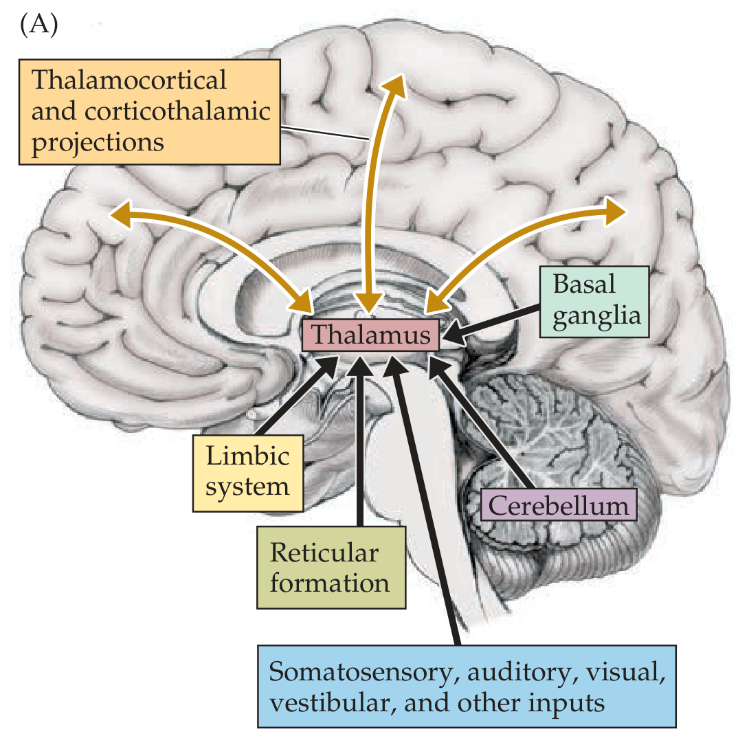
Figure 2.20A: Thalamic inputs and reciprocal connections - Neuroanatomy through Clinical Cases, 3rd Ed.
Thalamic Nuclear Divisions:
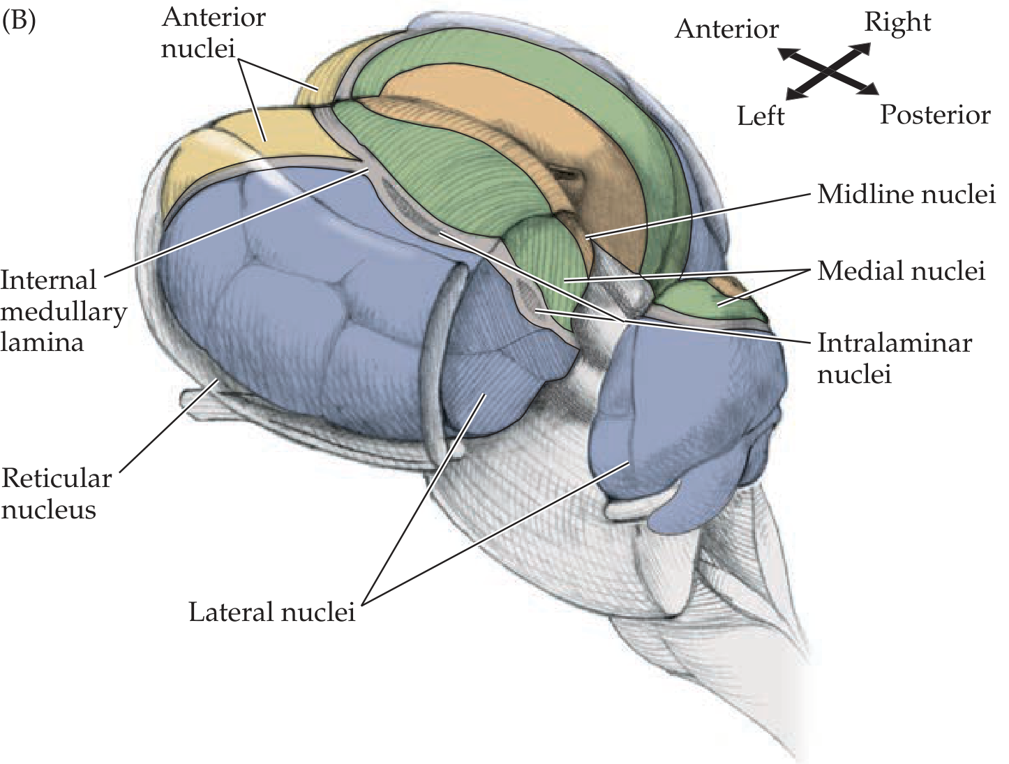
Figure 2.20B: Major thalamic nuclear divisions - Neuroanatomy through Clinical Cases, 3rd Ed.
Structure: Thalamic Nuclei and Their Connections
THALAMIC NUCLEI
├── ANTERIOR NUCLEI
│ Input: Mammillary bodies (via mammillothalamic tract), hippocampus
│ Output: Cingulate gyrus
│ Function: Limbic system, memory, emotion
│
├── MEDIAL NUCLEI (Dorsomedial)
│ Input: Amygdala, prefrontal cortex, basal ganglia
│ Output: Prefrontal cortex
│ Function: Emotion, memory, cognitive integration
│
├── LATERAL NUCLEI
│ ├── Ventral anterior (VA)
│ │ Input: Basal ganglia (globus pallidus)
│ │ Output: Motor cortex (area 6)
│ │ Function: Motor planning
│ ├── Ventral lateral (VL)
│ │ Input: Cerebellum (dentate nucleus)
│ │ Output: Motor cortex (area 4)
│ │ Function: Coordination, voluntary movement
│ ├── Ventral posterolateral (VPL)
│ │ Input: Medial lemniscus, spinothalamic tract (body)
│ │ Output: Somatosensory cortex (areas 3,1,2)
│ │ Function: Somatic sensation - body
│ ├── Ventral posteromedial (VPM)
│ │ Input: Trigeminal lemniscus (face), taste
│ │ Output: Somatosensory cortex
│ │ Function: Somatic sensation - face
│ ├── Lateral geniculate nucleus (LGN)
│ │ Input: Retina (via optic tract)
│ │ Output: Primary visual cortex (area 17)
│ │ Function: Vision
│ └── Medial geniculate nucleus (MGN)
│ Input: Inferior colliculus
│ Output: Primary auditory cortex (area 41,42)
│ Function: Hearing
│
├── PULVINAR
│ Input: Superior colliculus, visual cortex
│ Output: Association cortex (parietal, temporal, occipital)
│ Function: Visual attention, multimodal integration
│
├── INTRALAMINAR NUCLEI (within internal medullary lamina)
│ Input: Reticular formation, spinothalamic tract, basal ganglia
│ Output: Widespread cortex + striatum
│ Function: Arousal, consciousness, pain
│
└── RETICULAR NUCLEUS
Input: Cortex + other thalamic nuclei (collateral branches)
Output: Inhibits other thalamic nuclei (GABAergic)
Function: Gating/filtering of thalamic signals
Functions of the Thalamus (Summary)
- Relay center - relays all sensory input (except olfaction) to cortex
- Motor integration - VA/VL nuclei link cerebellum and basal ganglia to motor cortex
- Arousal & consciousness - intralaminar nuclei via ascending reticular activating system
- Limbic function - anterior nucleus in Papez circuit
- Reciprocal modulation - cortex projects back to thalamus via layer VI neurons, enabling feedback control
Thalamic Syndrome (Dejerine-Roussy)
Caused by VPL infarct: initial contralateral hemianesthesia followed by severe burning, intractable pain (thalamic pain).
4. CEREBRAL CORTEX - STRUCTURE, CONNECTIONS, AND FUNCTIONS
Introduction
The cerebral cortex is the outermost layer of gray matter. It contains >14 billion neurons. About 70% of all CNS neurons are in the cortex, and 75% of those are in association cortex.
Structure
Layers of Neocortex (6 layers):
SURFACE ──────────────────────────────────
Layer I │ Molecular layer │ Mainly dendrites and horizontal axons
Layer II │ External granular │ Small pyramidal + stellate cells
Layer III │ External pyramidal │ Pyramidal cells → cortico-cortical
Layer IV │ Internal granular │ Stellate cells → INPUT layer (receives thalamocortical)
Layer V │ Internal pyramidal │ Large pyramidal cells → LONG projections
│ │ (CST, corticobulbar, corticostriate)
Layer VI │ Multiform/fusiform │ → Corticothalamic projections
DEEP ──────────────────────────────────────
Types of Cortex:
- Neocortex (isocortex): 6 layers, ~90% of cortex, most of cerebrum
- Allocortex: ancient cortex - hippocampus (3 layers), olfactory cortex
- Mesocortex: transitional - cingulate gyrus
Lobes and Key Areas
CEREBRAL CORTEX
│
├── FRONTAL LOBE
│ ├── Primary motor cortex (area 4) - precentral gyrus
│ │ Homunculus: Face lateral → Arm → Leg medial
│ ├── Premotor area (area 6) - motor planning
│ ├── Supplementary motor area (area 6 medial)
│ ├── Frontal eye fields (area 8)
│ ├── Broca's area (areas 44, 45) - motor speech (left dominant)
│ └── Prefrontal cortex - executive function, working memory
│
├── PARIETAL LOBE
│ ├── Primary somatosensory cortex (areas 3,1,2) - postcentral gyrus
│ ├── Superior parietal lobule (areas 5,7) - spatial awareness
│ └── Inferior parietal lobule
│ ├── Supramarginal gyrus (area 40)
│ └── Angular gyrus (area 39) - reading, calculation
│
├── TEMPORAL LOBE
│ ├── Primary auditory cortex (areas 41,42) - Heschl's gyrus
│ ├── Auditory association cortex (area 22)
│ ├── Wernicke's area (area 22, posterior) - language comprehension
│ └── Inferior temporal (areas 20,21) - object recognition
│
└── OCCIPITAL LOBE
├── Primary visual cortex (area 17) - calcarine sulcus
├── Visual association (areas 18,19)
└── "What" pathway (ventral) + "Where" pathway (dorsal)
Connections
Cortico-cortical:
- Association fibers - connect areas within same hemisphere
- Short U-fibers (adjacent gyri)
- Long fasciculi (arcuate, uncinate, cingulum)
- Commissural fibers - connect hemispheres via corpus callosum, anterior commissure
Cortical efferents (Layer V):
- Corticospinal tract → spinal cord
- Corticobulbar tract → brainstem motor nuclei
- Corticopontine → pons → cerebellum
- Corticostriate → basal ganglia
Cortical afferents (Layer IV):
- From thalamus (all sensory relay nuclei)
Functions by Lobe
| Lobe | Key Functions |
|---|---|
| Frontal | Voluntary motor, speech production, executive function, personality |
| Parietal | Somatosensory, spatial awareness, reading, calculation |
| Temporal | Auditory, memory (hippocampus), language comprehension |
| Occipital | Vision, visual processing |
Association Cortex
- Unimodal: processes one modality (e.g., visual association cortex areas 18,19)
- Heteromodal: integrates multiple modalities (inferior parietal lobule, prefrontal)
- 75% of cortical neurons are in association cortex - underlies all higher cognitive function
5. VISUAL PATHWAY WITH LESIONS
Introduction
The visual pathway transmits information from the retina to the occipital cortex. Precise knowledge of this pathway allows clinicians to localize lesions from the visual field defect.
Visual Field Representation
- Each retina is divided into nasal and temporal halves
- The nasal retina receives light from the temporal visual field
- The temporal retina receives light from the nasal visual field
- The macula (central vision) is represented at the posterior pole of the visual cortex
FLOWCHART: Visual Pathway
VISUAL STIMULUS (light)
|
▼
RETINA (photoreceptors → bipolar cells → ganglion cells)
|
▼
OPTIC NERVE (CN II)
- Carries fibers from entire ipsilateral retina
|
▼
OPTIC CHIASM
┌────────────────────────────────────┐
│ Nasal retina fibers CROSS │
│ Temporal retina fibers STAY │
│ Result: each optic tract carries │
│ info from CONTRALATERAL visual field│
└────────────────────────────────────┘
|
▼
OPTIC TRACT
- Ipsilateral temporal + contralateral nasal retinal fibers
|
├──→ Small branch → PRETECTUM + SUPERIOR COLLICULUS
│ (pupillary light reflex, eye movements)
|
▼
LATERAL GENICULATE NUCLEUS (LGN) of THALAMUS
|
▼
OPTIC RADIATIONS (Geniculocalcarine tract)
┌──────────────────────────────────────┐
│ UPPER fibers → parietal lobe │
│ → Superior calcarine cortex │
│ → Inferior visual field │
│ │
│ LOWER fibers → temporal lobe │
│ (Meyer's loop around inf. horn │
│ of lateral ventricle) │
│ → Inferior calcarine cortex │
│ → Superior visual field │
└──────────────────────────────────────┘
|
▼
PRIMARY VISUAL CORTEX (Area 17)
Calcarine sulcus, occipital lobe
(Macula → posterior; Periphery → anterior)
VISUAL FIELD DEFECTS WITH LESIONS
The diagram below shows the visual pathway and lesions at numbered sites:
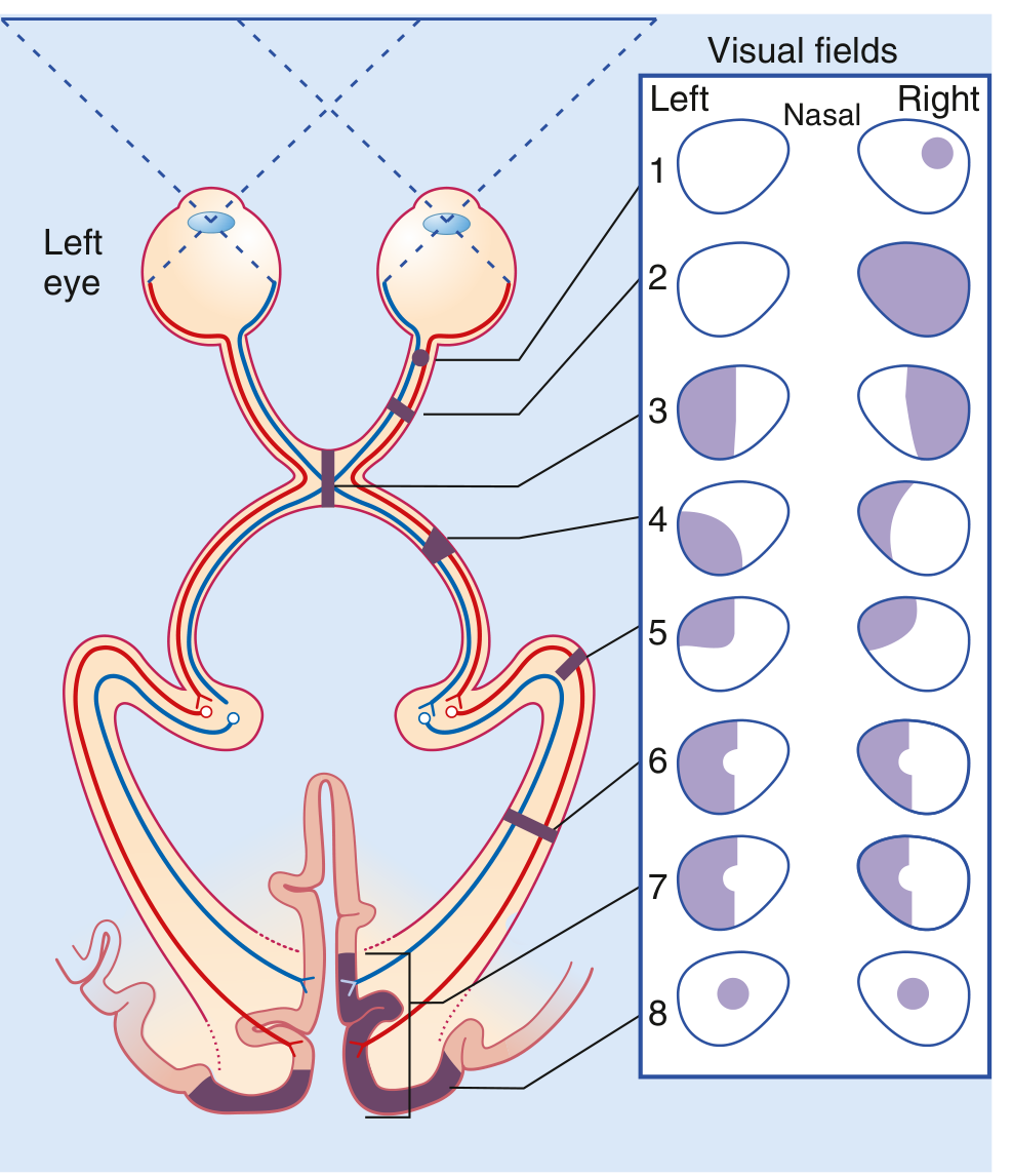
Figure: Visual Field Deficits with Lesions Along the Visual Pathway - Gray's Anatomy for Students
Visual field representation:
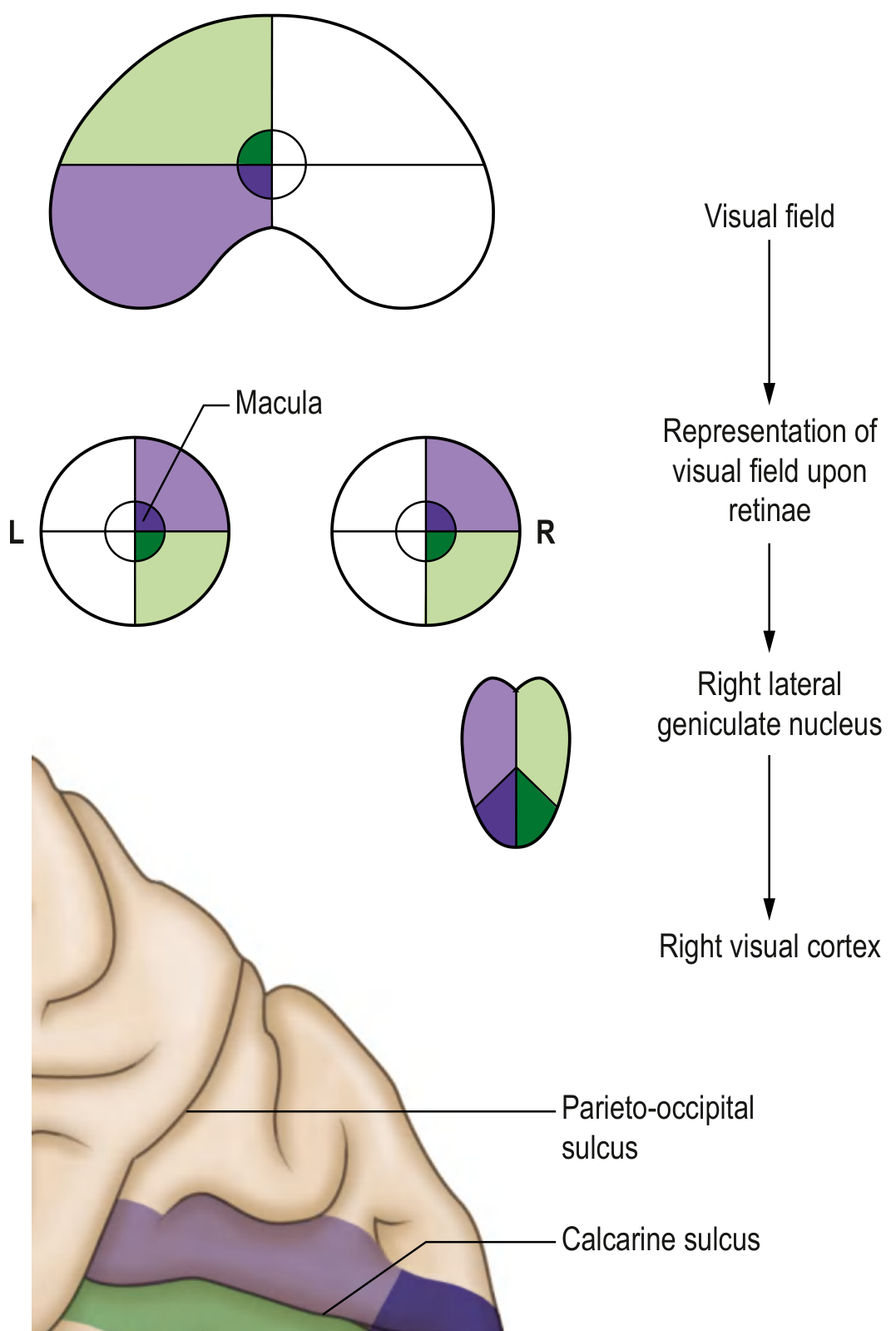
Figure: Left visual field representation to right visual cortex via right LGN - Gray's Anatomy for Students
Lesion Summary Table
| Site | Lesion | Visual Field Defect |
|---|---|---|
| 1. Optic nerve (unilateral, central) | Central scotoma | Ipsilateral central scotoma |
| 2. Optic nerve (complete) | e.g., optic neuritis | Monocular blindness - ipsilateral eye |
| 3. Optic chiasm (medial crossing fibers) | Pituitary adenoma | Bitemporal hemianopia |
| 4. Optic tract | Stroke | Contralateral homonymous hemianopia |
| 5. Meyer's loop (temporal radiation) | Temporal lobe lesion | Contralateral homonymous superior quadrantanopia ("pie in the sky") |
| 6. Upper optic radiation (parietal) | Parietal lobe lesion | Contralateral homonymous inferior quadrantanopia |
| 7. Complete optic radiation | Lesion near occipital pole | Contralateral homonymous hemianopia WITH macular sparing |
| 8. Bilateral occipital cortex | Bilateral lesions | Bilateral central scotoma |
Macular Sparing
- Occurs with posterior cerebral artery territory strokes involving visual cortex
- The macular region has dual blood supply (MCA + PCA) so may be preserved
6. ORGAN OF CORTI
Introduction
The organ of Corti is the specialized sensory receptor organ for hearing. It sits on the basilar membrane within the cochlea (scala media) and converts mechanical vibrations into electrical nerve impulses.
Structure of the Cochlea (Context)
COCHLEA (2.75 turns)
│
├── Scala vestibuli (perilymph) - above vestibular membrane
├── Scala media / Cochlear duct (endolymph) ← contains Organ of Corti
└── Scala tympani (perilymph) - below basilar membrane
(communicates with scala vestibuli at helicotrema, apex)
LABELLED DIAGRAM: Organ of Corti
TECTORIAL MEMBRANE
↕ (stereocilia in contact with / embedded in tectorial membrane)
━━━━━━━━━━━━━━━━━━━━━━━━━━━━━━━━━━━━━━━━━━━━━
RETICULAR LAMINA (stiff plate)
│ │
│ INNER HAIR CELLS (IHC) │ OUTER HAIR CELLS (OHC)
│ • Single row │ • 3-4 rows
│ • ~3,500 cells │ • ~12,000 cells
│ • 12 μm diameter │ • 8 μm diameter
│ • 90-95% of afferent │ • 5-10% afferent
│ nerve fibers │ • Receive efferent fibers
│ │ from brainstem (tuning)
│ TUNNEL OF CORTI │
│ (space between IHC │ SPACES OF NUEL
│ and OHC regions) │ (around OHCs)
└────────── RODS OF CORTI ───────┘
(inner and outer pillar cells - triangular support)
━━━━━━━━━━━━━━━━━━━━━━━━━━━━━━━━━━━━━━━━━━━━━
BASILAR MEMBRANE
FLOWCHART: Mechanism of Sound Transduction
SOUND WAVE ENTERS EAR
|
▼
Tympanic membrane vibrates
|
▼
Ossicles (malleus → incus → stapes) AMPLIFY vibration
|
▼
Oval window → pressure waves in perilymph (scala vestibuli)
|
▼
BASILAR MEMBRANE vibrates
|
▼ TONOTOPIC ORGANIZATION
┌──────────────────────────────────────┐
│ BASE of cochlea → HIGH frequency │
│ APEX of cochlea → LOW frequency │
└──────────────────────────────────────┘
|
▼
Reticular lamina + rods of Corti move as RIGID UNIT
|
▼
Stereocilia of hair cells SHEAR against tectorial membrane
Basilar membrane UP → stereocilia deflect toward modiolus
Basilar membrane DOWN → stereocilia deflect outward
|
▼
K⁺ enters hair cell through mechanosensitive channels (MET)
→ DEPOLARIZATION
|
▼
Ca²⁺ influx → neurotransmitter (glutamate) release at base
|
▼
SPIRAL GANGLION OF CORTI (bipolar neurons, in modiolus)
~30,000 afferent fibers
|
▼
COCHLEAR NERVE (CN VIII)
|
▼
COCHLEAR NUCLEI (dorsal + ventral) - pontomedullary junction
|
▼
Superior olivary complex → Lateral lemniscus
|
▼
INFERIOR COLLICULUS (midbrain) - sound localization
|
▼
MEDIAL GENICULATE NUCLEUS (thalamus)
|
▼
PRIMARY AUDITORY CORTEX (Heschl's gyrus, areas 41, 42)
Temporal lobe - tonotopic organization preserved
Key Points on Hair Cells
| Feature | Inner Hair Cells | Outer Hair Cells |
|---|---|---|
| Number | ~3,500 (1 row) | ~12,000 (3-4 rows) |
| Afferent innervation | 90-95% of cochlear nerve | 5-10% |
| Efferent innervation | Sparse | Dense (from olivocochlear bundle) |
| Primary role | Detection/transduction of sound | "Tuning" - amplify and sharpen frequency selectivity |
| Damage effect | Severe hearing loss | Reduced frequency discrimination |
Endocochlear Potential
- Scala media (endolymph): high K⁺, low Na⁺, +80 mV potential (produced by stria vascularis)
- Hair cell interior: -70 mV
- Therefore: ~150 mV driving force for K⁺ entry when MET channels open → very sensitive transduction
Basilar Membrane Tonotopy
BASE (narrow, stiff) ──────────────→ APEX (wide, compliant)
HIGH FREQUENCY (20,000 Hz) LOW FREQUENCY (20 Hz)
This tonotopic organization is preserved throughout the auditory pathway all the way to the cortex.
Sources
- Neuroanatomy through Clinical Cases, 3rd Edition (Blumenfeld) - Corticospinal tract, Thalamus, Spinothalamic pathway
- Guyton and Hall Textbook of Medical Physiology - Motor cortex, Organ of Corti
- Gray's Anatomy for Students - Visual pathway and lesions
- Scott-Brown's Otorhinolaryngology Head & Neck Surgery - Organ of Corti structure
- Bradley and Daroff's Neurology in Clinical Practice - Cerebral cortex organization
More elaborate as for 20 marks
Reading File
Reading File
Reading File
Reading File
Reading File
Reading File
Reading File
Reading File
Reading File
Reading File
Reading File
Reading File
Loading Image
Loading Image
Reading File
Reading File
Loading Image
Loading Image
I now have comprehensive material from multiple authoritative textbooks. Let me compile the full 20-mark essays.---
20-MARK ESSAY ANSWERS IN NEUROANATOMY & NEUROPHYSIOLOGY
Q1. DESCRIBE THE ASCENDING TRACTS (20 Marks)
Definition and Classification
Ascending tracts (sensory tracts) are bundles of nerve fibers within the white matter of the spinal cord and brainstem that convey sensory information from the periphery to higher brain centers. They are organized somatotopically and modality-specifically.
General rule of somatotopy: Arms are medial to legs, with two exceptions - primary sensorimotor cortex and posterior columns, where arms are lateral to legs. - Neuroanatomy through Clinical Cases, 3rd Ed.
THE TWO PRINCIPAL ASCENDING SYSTEMS
A. DORSAL COLUMN - MEDIAL LEMNISCAL (DCML) PATHWAY
Modalities Carried
- Fine (discriminative) touch
- Vibration sense
- Conscious proprioception (position sense)
- Two-point discrimination
- Stereognosis (object recognition by touch)
Receptors
| Receptor | Modality |
|---|---|
| Meissner's corpuscles | Fine touch, texture |
| Pacinian corpuscles | Vibration, deep pressure |
| Merkel's discs | Sustained pressure, fine touch |
| Ruffini endings | Skin stretch, joint movement |
| Muscle spindles / Golgi tendon organs | Proprioception |
Three-Neuron Chain
1st Order Neuron:
- Cell body in dorsal root ganglion (DRG)
- Peripheral process carries impulse from receptor
- Central process enters the spinal cord via the dorsal root and ascends ipsilaterally in the dorsal (posterior) column
Somatotopic Arrangement in Dorsal Column:
MEDIAL LATERAL
│ │
Gracile fasciculus Cuneate fasciculus
(T7 and below - lower limbs) (T6 and above - upper limbs, trunk)
│ │
Nucleus gracilis Nucleus cuneatus
(in medulla) (in medulla)
2nd Order Neuron:
- Cell body in nucleus gracilis (leg fibers) or nucleus cuneatus (arm fibers) in the medulla
- Axons arch ventrally, cross the midline as internal arcuate fibers = sensory decussation
- Form the medial lemniscus on the contralateral side
- Ascend through the brainstem (medulla → pons → midbrain) to the thalamus
3rd Order Neuron:
- Cell body in the Ventral Posterolateral (VPL) nucleus of the thalamus
- Axons pass through the posterior limb of the internal capsule
- Terminate in the primary somatosensory cortex (postcentral gyrus, Brodmann areas 3, 1, 2)
FLOWCHART: DCML Pathway
RECEPTOR (Meissner, Pacinian, muscle spindles)
|
▼
1st ORDER NEURON
Cell body → DORSAL ROOT GANGLION
Central axon enters DORSAL COLUMN (ipsilateral)
┌─────────────────────────────────────┐
│ GRACILE FASCICULUS (medial) │ ← T7 and below (leg/lower trunk)
│ CUNEATE FASCICULUS (lateral) │ ← T6 and above (arm/upper trunk)
└─────────────────────────────────────┘
| ascends ipsilaterally
▼
MEDULLA OBLONGATA
┌──────────────────────────────────────────┐
│ Nucleus gracilis → leg fibers │
│ Nucleus cuneatus → arm fibers │
└──────────────────────────────────────────┘
|
▼ 2nd ORDER NEURON
INTERNAL ARCUATE FIBERS ──► CROSS MIDLINE
|
▼
MEDIAL LEMNISCUS (contralateral)
Ascends through PONS and MIDBRAIN
|
▼ 3rd ORDER NEURON
VPL NUCLEUS OF THALAMUS
|
▼ Posterior limb of internal capsule
PRIMARY SOMATOSENSORY CORTEX
(Postcentral gyrus, areas 3,1,2)
Homuncular representation
B. SPINOTHALAMIC TRACT (Anterolateral System)
Modalities Carried
- Lateral spinothalamic tract: Pain and temperature
- Anterior spinothalamic tract: Crude touch and pressure
Receptors
- Free nerve endings (nociceptors, thermoreceptors) via:
- Aδ fibers (fast, sharp, well-localized pain; cold)
- C fibers (slow, dull, burning, poorly localized pain; warm)
Three-Neuron Chain
1st Order Neuron:
- Cell body in dorsal root ganglion
- Enters spinal cord via dorsal root
- Ascends/descends 1-2 levels in Lissauer's tract (dorsolateral fasciculus)
- Synapses in dorsal horn of spinal cord gray matter:
- Rexed Lamina I (marginal zone) - sharp pain
- Rexed Lamina II (substantia gelatinosa) - modulation of pain
- Rexed Lamina V - multimodal input
2nd Order Neuron:
- Cell body in dorsal horn
- Axon crosses the midline immediately through the anterior white commissure (within 1-2 spinal cord levels)
- Ascends in the contralateral anterolateral white matter column
3rd Order Neuron:
- Cell body in VPL nucleus of thalamus
- Projects to primary somatosensory cortex
The diagram below shows this pathway from Neuroanatomy through Clinical Cases:
Figure: Spinothalamic Sensory Pathway - Neuroanatomy through Clinical Cases, 3rd Ed.
FLOWCHART: Spinothalamic Pathway
RECEPTOR (Free nerve endings: Aδ and C fibers)
|
▼
1st ORDER NEURON
Cell body → DORSAL ROOT GANGLION
Enters dorsal horn → ascends/descends in LISSAUER'S TRACT
Synapses in REXED LAMINAE I, II, V
|
▼ 2nd ORDER NEURON
CROSSES IMMEDIATELY via ANTERIOR WHITE COMMISSURE
(1-2 spinal segments above entry)
|
▼
CONTRALATERAL ANTEROLATERAL COLUMN
┌──────────────────────────────────────┐
│ Lateral spinothalamic → Pain, Temp │
│ Anterior spinothalamic → Crude touch │
└──────────────────────────────────────┘
| ascends through medulla, pons, midbrain
▼ 3rd ORDER NEURON
VPL NUCLEUS OF THALAMUS
|
▼
PRIMARY SOMATOSENSORY CORTEX (Postcentral gyrus)
COMPARISON TABLE: DCML vs SPINOTHALAMIC
| Feature | DCML | Spinothalamic |
|---|---|---|
| Modality | Fine touch, vibration, proprioception | Pain, temperature, crude touch |
| 1st neuron | Dorsal root ganglion | Dorsal root ganglion |
| Spinal cord position | Ipsilateral dorsal column | Contralateral anterolateral |
| Where it crosses | Medulla (internal arcuate fibers) | Spinal cord (ant. white commissure) |
| 2nd neuron nucleus | Nucleus gracilis / cuneatus | Dorsal horn (laminae I, II, V) |
| Ascent | Medial lemniscus | Spinothalamic tract |
| 3rd neuron | VPL of thalamus | VPL of thalamus |
| Cortical area | Postcentral gyrus (areas 3,1,2) | Postcentral gyrus (areas 3,1,2) |
| Fiber size | Large, myelinated (Aβ) | Small myelinated (Aδ), unmyelinated (C) |
OTHER ASCENDING TRACTS
| Tract | Modality | Pathway |
|---|---|---|
| Spinocerebellar (dorsal) | Ipsilateral proprioception (lower limb) | Clarke's column → inferior cerebellar peduncle |
| Spinocerebellar (ventral) | Bilateral proprioception | Crosses twice → superior cerebellar peduncle |
| Spinoreticular | Diffuse pain, arousal | Anterolateral → reticular formation → intralaminar thalamus |
| Spinomesencephalic | Pain modulation | Anterolateral → PAG → endogenous analgesia |
CLINICAL CORRELATIONS
Brown-Séquard Syndrome (Hemisection of spinal cord)
Ipsilateral below lesion:
- Loss of fine touch, vibration, proprioception (DCML - uncrossed in cord)
- Upper motor neuron signs (corticospinal tract - uncrossed in cord)
Contralateral below lesion (1-2 levels below lesion):
- Loss of pain and temperature (spinothalamic - crossed in cord)
Syringomyelia
- Cyst in central spinal cord
- Bilateral loss of pain and temperature at that spinal level
- Cape-like distribution (shoulders, arms)
- DCML preserved (lies peripherally, away from central canal)
Posterior Column Disease (e.g., Tabes dorsalis, Vitamin B12 deficiency)
- Loss of vibration and proprioception
- Romberg sign positive
- Sensory ataxia
Q2. EXPLAIN THE CORTICOSPINAL TRACT (20 Marks)
Introduction
The corticospinal tract (CST), also called the pyramidal tract, is the most clinically important descending motor pathway in the human nervous system. It controls voluntary movements, particularly skilled movements of the distal extremities. The pathway runs from the cerebral cortex to the spinal cord, with a single synapse at the lower motor neuron in the ventral horn.
ORIGIN
The corticospinal tract does NOT arise exclusively from the motor cortex. Its origin is distributed:
- ~30% from the primary motor cortex (area 4, precentral gyrus) - including Betz cells
- ~30% from premotor and supplementary motor areas (area 6)
- ~40% from somatosensory areas (areas 3, 1, 2, 5, 7) in the parietal lobe - these fibers modulate sensory processing rather than drive movement directly
The giant Betz cells (layer V of primary motor cortex) are the largest neurons in the human nervous system (~60 μm diameter). Their axons constitute only ~3% of the total 1 million fibers in the CST but conduct at the fastest velocity (~70 m/sec). The remaining 97% are small fibers (<4 μm) conducting tonic signals.
COURSE (Detailed Level by Level)
1. CEREBRAL CORTEX → CORONA RADIATA
- Axons from cortical layer V converge into the corona radiata (fan-shaped white matter)
- Multiple fiber systems mix here including corticobulbar, corticopontine, and CST
2. INTERNAL CAPSULE
The internal capsule is the most compact segment. In horizontal section, it appears as an arrowhead or inverted "V":
- Anterior limb: separates head of caudate from lenticular nucleus
- Genu: transition zone (level of foramen of Monro)
- Posterior limb: separates thalamus from lenticular nucleus - CST lies here
Somatotopic arrangement in internal capsule posterior limb (anterior → posterior):
FACE → ARM → TRUNK → LEG
(anterior) (posterior)
The diagram below shows somatotopic organization through internal capsule, midbrain, and spinal cord:
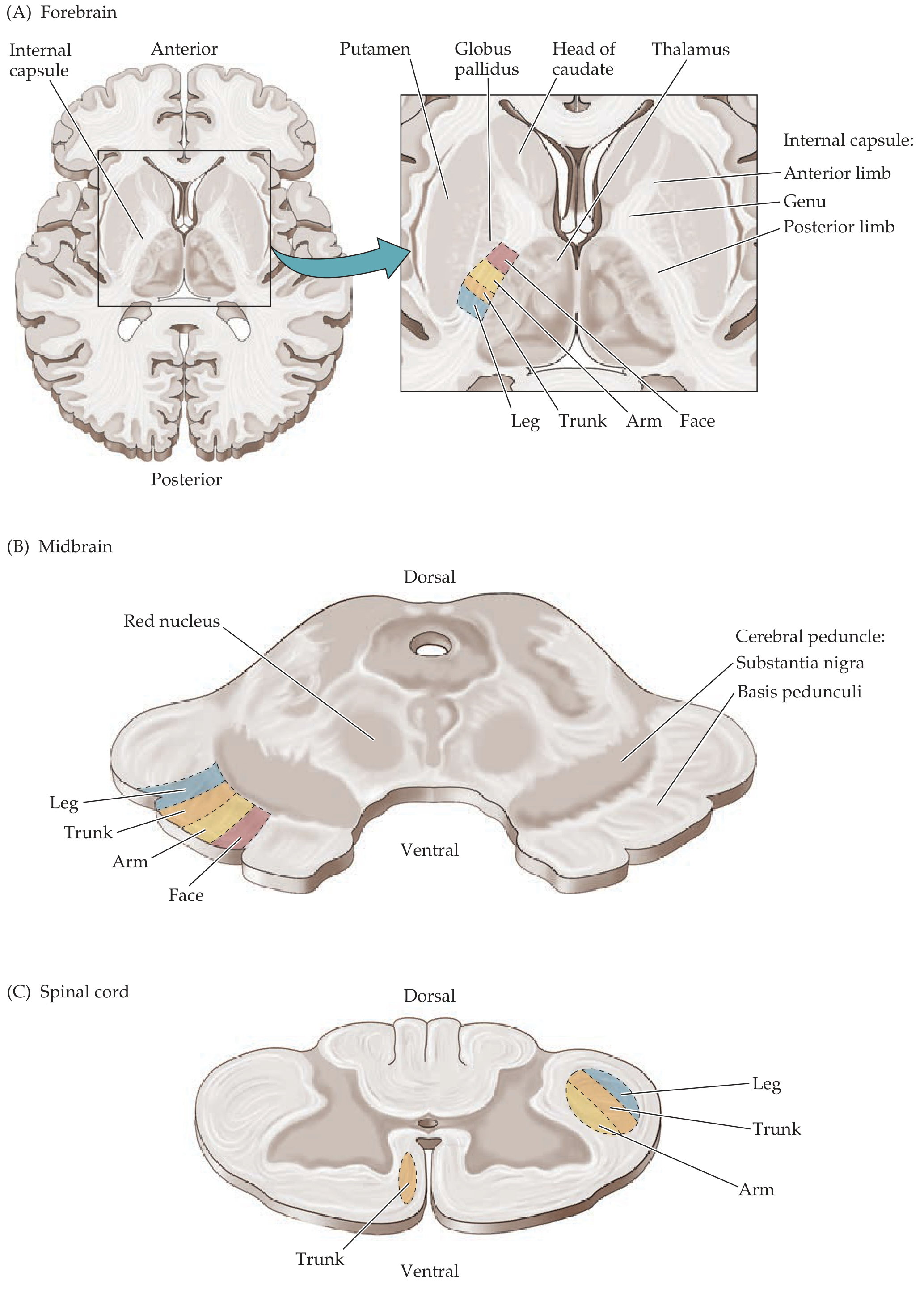
Figure 6.10: Somatotopic Organization of Corticobulbar and Corticospinal Tracts - Neuroanatomy through Clinical Cases, 3rd Ed.
3. CEREBRAL PEDUNCLES (Midbrain)
- Internal capsule continues as the basis pedunculi (ventral cerebral peduncle)
- CST and corticobulbar fibers occupy the middle 1/3 of the basis pedunculi
- Somatotopy: Face medial → Arm → Leg lateral
- Corticopontine fibers occupy the medial and lateral thirds
4. PONS
- CST fibers become scattered into fascicles as they are separated by transversely running pontine nuclei and transverse pontocerebellar fibers
- They travel through the ventral (basilar) pons
5. MEDULLARY PYRAMIDS
- Fibers re-collect on the ventral surface of the medulla forming the pyramids
- This is why the CST is also called the pyramidal tract
- A small % of fibers leave as corticobulbar fibers to cranial nerve nuclei (VII, IX, X, XI, XII)
6. PYRAMIDAL DECUSSATION (Cervicomedullary junction)
At the junction of the medulla and spinal cord at the foramen magnum:
- ~85% of fibers cross to form the lateral corticospinal tract (in dorsolateral funiculus)
- ~15% continue ipsilaterally as the anterior corticospinal tract (in ventral funiculus)
7. SPINAL CORD
Lateral corticospinal tract (LCST):
- In the dorsolateral funiculus
- Somatotopy: Arm fibers medial, Leg fibers lateral (opposite of internal capsule)
- Descends entire length of cord
- Synapses on anterior horn cells (lower motor neurons) and interneurons in Rexed laminae VII, VIII, IX
- Controls contralateral distal limb movements (fine, skilled)
Anterior corticospinal tract (ACST):
- In the ventral funiculus, ipsilateral
- Most fibers eventually cross via the anterior white commissure at their spinal level
- Controls bilateral axial/postural muscles
LABELLED DIAGRAM
Figure 6.8: Lateral Corticospinal Tract - Neuroanatomy through Clinical Cases, 3rd Ed.
FLOWCHART
PRIMARY MOTOR CORTEX (area 4) + PREMOTOR (area 6) + PARIETAL (3,1,2,5,7)
Layer V neurons (Betz cells + pyramidal neurons)
|
▼ CORONA RADIATA (white matter fan)
|
▼ INTERNAL CAPSULE - Posterior limb
[Somatotopy: Face-Arm-Trunk-Leg, A→P]
|
▼ CEREBRAL PEDUNCLE - Basis pedunculi
Middle 1/3 [Face medial, Leg lateral]
+ CORTICOBULBAR fibers diverge to CN nuclei
|
▼ PONS - scattered fascicles (ventral/basilar)
|
▼ MEDULLARY PYRAMIDS (ventral medulla)
|
═══ PYRAMIDAL DECUSSATION ═══
↙ 85% cross 15% stay ↘
LATERAL CST ANTERIOR CST
(dorsolateral funiculus) (ventral funiculus)
Contralateral cord Ipsilateral cord
| |
▼ ▼
Lower Motor Neuron Crosses at spinal level
(Anterior horn, → bilateral axial muscles
Rexed laminae VII-IX)
|
▼
SKELETAL MUSCLE (voluntary contraction)
CORTICOBULBAR TRACT
A closely related pathway - fibers leave the CST at brainstem levels to supply cranial nerve motor nuclei:
- Facial nucleus (CN VII): Bilateral supply to upper face; contralateral supply to lower face only. (This explains why UMN facial palsy spares the forehead)
- Hypoglossal nucleus (CN XII): Primarily contralateral supply
- Nucleus ambiguus (CN IX, X): Bilateral supply (pharynx, larynx)
- Trigeminal motor nucleus (CN V): Bilateral supply
UMN vs LMN SYNDROME
| Feature | UMN Lesion | LMN Lesion |
|---|---|---|
| Site | Cortex, internal capsule, brainstem, spinal cord (above ant. horn) | Anterior horn cells, ventral root, peripheral nerve, NMJ |
| Weakness | Yes, contralateral | Yes, ipsilateral |
| Tone | Spasticity (increased) | Flaccidity (decreased) |
| Reflexes | Hyperreflexia (DTRs increased) | Hyporeflexia/areflexia |
| Plantar response | Extensor (Babinski +ve) | Flexor or absent |
| Wasting | Mild (disuse) | Severe |
| Fasciculations | Absent | Present |
| Type of paralysis | Spastic/clasp-knife | Flaccid |
| Clonus | Present | Absent |
CLINICAL CORRELATION: LEVEL OF LESION
| Level | Clinical Finding |
|---|---|
| Motor cortex | Contralateral monoparesis (arm or leg, depends on area) |
| Internal capsule | Contralateral hemiplegia (face + arm + leg) - compact fibers |
| Midbrain (cerebral peduncle) | Weber syndrome: CN III palsy (ipsilateral) + contralateral hemiplegia |
| Pons | Millard-Gubler syndrome: CN VI, VII palsy (ipsilateral) + contralateral hemiplegia |
| Medulla | Medial medullary syndrome: CN XII palsy (ipsilateral) + contralateral hemiplegia |
| Lateral medulla | Spares pyramidal tract (only lateral structures affected) |
| Above pyramidal decussation | Contralateral weakness |
| Below pyramidal decussation (spinal cord) | Ipsilateral weakness |
Q3. FUNCTIONS AND CONNECTIONS OF THE THALAMUS (20 Marks)
Introduction
The thalamus is a paired, egg-shaped gray matter structure forming the dorsal part of the diencephalon. Together with the hypothalamus and epithalamus, it constitutes the diencephalon. It is the gateway to consciousness - virtually all sensory information (except olfaction) must synapse in the thalamus before reaching the cortex. Its name comes from the Greek "thalamos" (inner room/bridal chamber).
Key anatomical facts:
- Located deep in the cerebral white matter, above the brainstem, behind the basal ganglia
- Shaped like two eggs angled with posterior ends outward, forming an inverted V on horizontal section
- Connected across the midline by the massa intermedia (interthalamic adhesion)
- Surrounded by white matter: internal capsule laterally, hypothalamic sulcus inferiorly
GROSS STRUCTURE
The thalamus is divided by a Y-shaped sheet of white matter - the internal medullary lamina - into:
- Anterior nuclear group
- Medial nuclear group
- Lateral nuclear group (includes ventral and dorsal tiers)
Additional nuclei:
- Intralaminar nuclei (within the internal medullary lamina)
- Midline nuclei (along the third ventricle wall)
- Reticular nucleus (thin shell surrounding the thalamus laterally, separated by external medullary lamina)
LABELLED DIAGRAMS
Thalamic connections with cortex and subcortical structures:
Figure 2.20A: Thalamic inputs and reciprocal connections - Neuroanatomy through Clinical Cases, 3rd Ed.
3D Thalamic nuclear divisions:
Figure 2.20B: Major thalamic nuclear divisions - Neuroanatomy through Clinical Cases, 3rd Ed.
THALAMIC NUCLEI, CONNECTIONS, AND FUNCTIONS
1. ANTERIOR NUCLEAR GROUP
INPUTS:
├── Mammillary bodies (via mammillothalamic tract)
├── Hippocampus (via fornix)
└── Cingulate cortex (reciprocal)
OUTPUTS:
└── Cingulate gyrus (area 24)
FUNCTION:
├── Limbic system - memory, emotion
├── Part of PAPEZ CIRCUIT (memory)
└── Attention and learning
Papez Circuit: Hippocampus → Fornix → Mammillary bodies → Mammillothalamic tract → Anterior thalamus → Cingulate gyrus → Cingulum → Entorhinal cortex → Hippocampus
2. DORSOMEDIAL (MEDIODORSAL) NUCLEUS
INPUTS:
├── Amygdala
├── Prefrontal cortex (reciprocal)
├── Temporal lobe (olfactory cortex, perirhinal cortex)
└── Basal ganglia (indirect via pallidum)
OUTPUTS:
└── Prefrontal association cortex (areas 9, 10, 11, 12)
FUNCTION:
├── Affective behavior and emotion
├── Judgment and decision-making
└── Working memory
Clinical: Mediodorsal lesion → personality change, memory defects (Korsakoff syndrome involves this nucleus)
3. LATERAL NUCLEAR GROUP
DORSAL TIER (lateral dorsal, lateral posterior, pulvinar)
- Lateral dorsal: connects with cingulate gyrus
- Lateral posterior: connects with parietal association cortex
- Pulvinar (largest thalamic nucleus): inputs from superior colliculus, visual cortex; outputs to parieto-temporo-occipital association cortex; function - visual attention, cross-modal integration
VENTRAL TIER:
| Nucleus | Major Inputs | Major Outputs | Function |
|---|---|---|---|
| Ventral anterior (VA) | Globus pallidus, substantia nigra | Premotor cortex (area 6), prefrontal | Motor planning, initiation |
| Ventral lateral (VL) | Cerebellum (dentate nucleus via sup. cer. peduncle), globus pallidus | Primary motor cortex (area 4) | Motor coordination, cerebellar relay |
| Ventral posterolateral (VPL) | Medial lemniscus, spinothalamic tract (body) | Primary somatosensory cortex (areas 3,1,2) | Somatic sensation - body |
| Ventral posteromedial (VPM) | Trigeminothalamic tract (face), nucleus solitarius (taste) | Primary somatosensory cortex (face area), taste cortex | Somatic sensation - face; taste |
| Lateral geniculate nucleus (LGN) | Optic tract (retinal ganglion cells) | Primary visual cortex (area 17) via optic radiation | Vision |
| Medial geniculate nucleus (MGN) | Inferior colliculus (brachium of IC) | Primary auditory cortex (areas 41, 42) | Hearing |
4. INTRALAMINAR NUCLEI
(within the internal medullary lamina: centromedian, parafascicular, central lateral, etc.)
INPUTS:
├── Spinothalamic tract (pain fibers - collaterals)
├── Ascending reticular activating system (ARAS)
└── Basal ganglia, cerebellum
OUTPUTS:
├── Widespread cortical areas
└── Striatum (caudate, putamen)
FUNCTION:
├── AROUSAL and CONSCIOUSNESS
├── Diffuse pain signaling
└── Regulating basal ganglia motor loops
Clinical: Bilateral lesions → persistent vegetative state; centromedian lesions → impaired arousal
5. MIDLINE NUCLEI
- Adjacent to the third ventricle wall
- Connections with hypothalamus, hippocampus, amygdala
- Functions: visceral, autonomic, limbic integration
6. RETICULAR NUCLEUS
UNIQUE FEATURES:
├── Does NOT project to cortex
├── Receives collaterals from BOTH thalamocortical and corticothalamic fibers
└── Outputs ONLY to other thalamic nuclei (GABAergic - INHIBITORY)
FUNCTION:
├── "Gatekeeper" - filters/gates thalamic output
├── Prevents sensory overload
└── Involved in sleep spindles (together with intralaminar nuclei)
CONNECTIONS SUMMARY FLOWCHART
SUBCORTICAL INPUTS TO THALAMUS:
Somatosensory (medial lemniscus) ──────► VPL ────► Somatosensory cortex (3,1,2)
Pain/temperature (spinothalamic) ──────► VPL ────► Somatosensory cortex
Trigeminal input (face) ───────────────► VPM ────► Somatosensory cortex (face)
Taste (NTS via solitariothalamic) ─────► VPM ────► Taste cortex
Retina (optic tract) ──────────────────► LGN ────► Visual cortex (17)
Inferior colliculus (auditory) ────────► MGN ────► Auditory cortex (41, 42)
Cerebellum (dentate nucleus) ──────────► VL ─────► Motor cortex (4)
Basal ganglia (globus pallidus, SNr) ──► VA ─────► Premotor cortex (6)
Hippocampus / Mammillary bodies ───────► AN ─────► Cingulate gyrus
Reticular formation (ARAS) ────────────► IL ─────► Widespread cortex (arousal)
Limbic system (amygdala) ──────────────► MD ─────► Prefrontal cortex
CORTICAL FEEDBACK (Corticothalamic - from Layer VI):
All cortical areas ──────────────────────────────► Corresponding thalamic nuclei
(This is RECIPROCAL - the dominant direction of thalamocortical traffic
is modulated by this feedback)
INHIBITORY MODULATION:
Corticothalamic + Thalamocortical axon collaterals ── Reticular nucleus
↓ (GABAergic)
Inhibit all thalamic nuclei
FUNCTIONS OF THE THALAMUS
- Sensory relay - all sensory modalities except olfaction relay here before reaching cortex
- Motor integration - VL and VA relay cerebellar and basal ganglia signals to motor cortex
- Consciousness and arousal - intralaminar nuclei form part of the ascending reticular activating system
- Pain processing - relay for spinothalamic pain input; also participates in affective component of pain
- Limbic/memory function - anterior nucleus in Papez circuit
- Gating and attention - reticular nucleus filters irrelevant sensory input
- Sleep regulation - oscillatory circuits involving reticular nucleus produce sleep spindles (12-14 Hz)
- Language - pulvinar and VPL participate in language processing
CLINICAL SYNDROMES
| Syndrome | Lesion | Features |
|---|---|---|
| Thalamic (Dejerine-Roussy) syndrome | VPL infarct (PCA territory) | Initial hemianesthesia → severe burning thalamic pain, hyperesthesia |
| Thalamic hemorrhage | Hypertensive hemorrhage | Contralateral hemisensory loss, ipsilateral Horner, eye deviated toward hemorrhage |
| Bilateral thalamic infarcts | "Top of basilar" | Coma/severe drowsiness, amnesia, vertical gaze palsy |
| Korsakoff syndrome | Mediodorsal + anterior nuclei (thiamine deficiency) | Anterograde amnesia, confabulation |
Q4. STRUCTURE, CONNECTIONS, AND FUNCTIONS OF THE CEREBRAL CORTEX (20 Marks)
Introduction
The cerebral cortex is the outermost gray matter mantle of the cerebral hemispheres. It is the substrate for the highest mental functions. It contains approximately 22 billion neurons communicating via 165 trillion synapses with ~12 million km of dendrites, and is interconnected by ~100,000 km of axons. - Kaplan & Sadock's Comprehensive Textbook of Psychiatry
GROSS ANATOMY
- Total surface area ~2,500 cm² (greatly expanded by gyri and sulci)
- Thickness: 1.5 - 4.5 mm (thicker in motor areas, thinner in sensory)
- Divided into 4 lobes by major sulci: frontal, parietal, temporal, occipital
- Key sulci: central sulcus (Rolandic), lateral sulcus (Sylvian), parieto-occipital sulcus, calcarine sulcus
MICROSCOPIC STRUCTURE (CYTOARCHITECTURE)
Types of Cortex
- Neocortex (isocortex): 6 layers, >90% of cortex, most of cerebrum
- Allocortex: fewer than 6 layers
- Archicortex (3 layers): hippocampus, dentate gyrus
- Paleocortex (3-5 layers): piriform/olfactory cortex
- Mesocortex (juxtallocortex): transitional - cingulate gyrus, insular cortex
The Six Layers of Neocortex
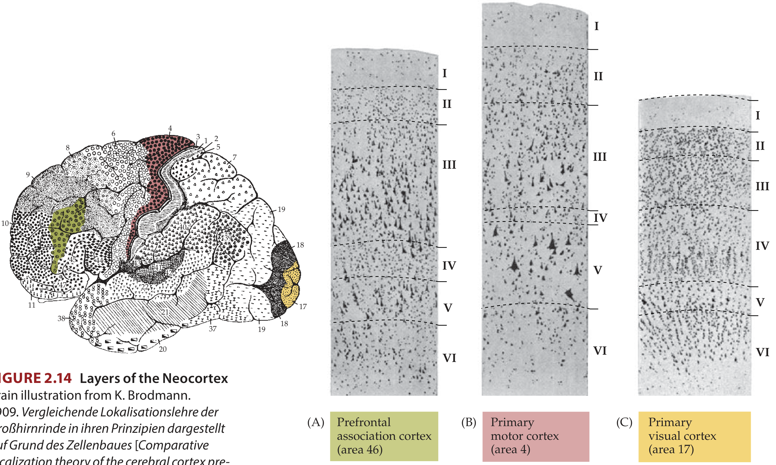
Figure 2.14: Layers of the Neocortex with Brodmann's map - Neuroanatomy through Clinical Cases, 3rd Ed.
| Layer | Name | Cell Types | Main Connections |
|---|---|---|---|
| I | Molecular layer | Cajal-Retzius cells, dendritic tufts, horizontal axons | Dendrites from deeper layers + long-range cortical inputs |
| II | External granular layer | Small pyramidal + stellate cells | Cortico-cortical (short-range) |
| III | External pyramidal layer | Medium pyramidal cells | Cortico-cortical (long-range: callosal, association) |
| IV | Internal granular layer | Stellate (spiny/aspiny) cells | Receives thalamocortical input (thickest in sensory cortex) |
| V | Internal pyramidal layer | Giant pyramidal (Betz) cells | Long descending outputs: CST, corticobulbar, corticostriate, corticopontine |
| VI | Multiform/Polymorphic layer | Spindle-shaped cells | Corticothalamic projections (feedback) |
Regional Variations
- Primary motor cortex (area 4): Agranular - layer IV absent/poorly developed; layer V enormously thick with Betz cells
- Primary visual cortex (area 17): Granular - layer IV expanded into 3 sublayers (contains line of Gennari - visible to naked eye)
- Primary somatosensory cortex (areas 3, 1, 2): Granular - thick layer IV
- Prefrontal cortex: Homotypical - all 6 layers well developed
Cell Types
-
Pyramidal neurons (~75% of neocortical neurons)
- Characteristic triangular cell body
- Single apical dendrite + basal dendrites with dendritic spines
- Excitatory projection neurons - use glutamate
- Project to other cortical areas or to subcortical structures
-
Stellate (granule/non-pyramidal) neurons (~25%)
- Local circuit neurons (interneurons)
- Most use GABA (inhibitory)
- Subtypes: chandelier cells, basket cells, double-bouquet cells
- Regulate cortical excitability; critical for columnar processing
-
Columnar organization: Cortex is organized into vertical cortical columns (~100 cells wide), sharing similar function and connections - the basic unit of cortical processing
BRODMANN'S CYTOARCHITECTONIC AREAS
In 1909, Korbinian Brodmann divided each hemisphere into 52 numbered areas based on cytoarchitecture. These correlate well with functional areas:
| Brodmann's Area(s) | Location | Function |
|---|---|---|
| 4 | Precentral gyrus | Primary motor cortex |
| 1, 2, 3 | Postcentral gyrus | Primary somatosensory cortex |
| 6 | Anterior to area 4 | Premotor + supplementary motor area |
| 8 | Middle frontal gyrus | Frontal eye fields |
| 9, 10, 11, 12 | Prefrontal cortex | Cognition, executive function |
| 17 | Calcarine sulcus | Primary visual cortex |
| 18, 19 | Occipital lobe | Secondary & tertiary visual cortex |
| 20, 21 | Inferior & middle temporal gyrus | Visual form recognition |
| 22 (posterior) | Superior temporal gyrus | Wernicke's area (language comprehension) |
| 41, 42 | Heschl's gyrus, temporal | Primary & secondary auditory cortex |
| 44, 45 | Inferior frontal gyrus | Broca's area (motor speech) |
| 39 (Angular gyrus) | Inferior parietal | Reading, writing, calculation |
| 40 (Supramarginal) | Inferior parietal | Spatial perception, praxis |
CONNECTIONS OF THE CEREBRAL CORTEX
1. ASSOCIATION FIBERS (ipsilateral cortico-cortical)
Connect areas within the same hemisphere:
- Short U-fibers (arcuate fibers): connect adjacent gyri
- Long association fasciculi:
| Fasciculus | Connects | Function |
|---|---|---|
| Superior longitudinal / Arcuate fasciculus | Broca's area ↔ Wernicke's area (frontal ↔ temporal/parietal) | Language network |
| Uncinate fasciculus | Frontal pole ↔ Temporal pole (via insula) | Memory-emotion integration |
| Cingulum | Cingulate gyrus → hippocampus, parahippocampus | Limbic system |
| Inferior longitudinal fasciculus | Occipital ↔ Temporal | Visual object recognition |
| Inferior fronto-occipital fasciculus | Frontal ↔ Occipital | Semantic processing |
2. COMMISSURAL FIBERS (inter-hemispheric)
- Corpus callosum (massive - 200-250 million fibers): connects corresponding neocortical areas
- Genu - prefrontal
- Body - motor, somatosensory, parietal
- Splenium - parietal, temporal, occipital (visual)
- Anterior commissure: connects temporal lobes and olfactory cortex
- Posterior commissure: connects pretectal nuclei (pupillary light reflex)
- Habenular commissure, hippocampal commissure
3. PROJECTION FIBERS (corticofugal/corticipetal)
Descending (efferent) from layer V and VI:
CORTEX
│
├── Corticospinal tract → ventral horn (voluntary movement)
├── Corticobulbar tract → cranial nerve motor nuclei
├── Corticopontine tract → pontine nuclei → cerebellum (areas 4, 6, parietal)
├── Corticostriate → caudate, putamen (motor learning)
├── Corticoreticular → reticular formation (muscle tone)
└── Corticothalamic (layer VI) → thalamic relay nuclei (feedback)
Ascending (afferent) to layer IV:
THALAMUS (all specific nuclei) → Layer IV of corresponding cortical area
Nonspecific thalamic projections → Layers I, II (diffuse arousal/attention)
Cholinergic (nucleus basalis of Meynert) → All cortical layers
Monoaminergic (LC, raphe) → All cortical layers (neuromodulation)
FUNCTIONAL AREAS AND THEIR FUNCTIONS
Frontal Lobe
Primary motor cortex (area 4, precentral gyrus):
- Motor homunculus (Penfield's map): face and hand occupy disproportionately large areas
- Somatotopy: leg in interhemispheric fissure, arm on lateral surface, face below
- Contains Betz cells → gives rise to CST
- Electrical stimulation → contralateral muscle contraction
Premotor area (area 6, lateral):
- Programming and planning of complex motor sequences
- Involved in preparation for voluntary movement
- Receives input from prefrontal cortex, parietal cortex, cerebellum (via VL thalamus)
Supplementary motor area (area 6, medial):
- Bilateral motor planning
- Internally generated (self-initiated) movements
- Active during mental rehearsal of movement
Frontal eye fields (area 8):
- Voluntary saccadic eye movements (contralateral)
Broca's area (areas 44, 45 - inferior frontal gyrus, left dominant hemisphere):
- Motor programming of speech
- Lesion → Broca's (expressive/non-fluent) aphasia: poor speech output, preserved comprehension
Prefrontal cortex (areas 9, 10, 11, 12, 46):
- Working memory
- Executive function: planning, decision making, impulse control
- Personality and judgment
- Orbitofrontal cortex: emotional regulation, reward
Parietal Lobe
Primary somatosensory cortex (areas 3, 1, 2 - postcentral gyrus):
- Somatosensory homunculus - mirrors motor homunculus
- Processes touch, proprioception, pain, temperature
- Areas 3a: proprioception; 3b: texture; 1: texture; 2: size/shape
Superior parietal lobule (areas 5, 7):
- Stereognosis, spatial orientation
- Visuomotor coordination
Inferior parietal lobule:
- Supramarginal gyrus (area 40): praxis (skilled movement sequences); left lesion → ideomotor apraxia
- Angular gyrus (area 39): reading, writing, calculation; lesion → Gerstmann's syndrome (dyslexia, dysgraphia, dyscalculia, finger agnosia, right-left disorientation)
Temporal Lobe
Primary auditory cortex (areas 41, 42 - Heschl's gyri):
- Tonotopic organization (high frequencies posterolateral, low frequencies anteromedial)
- Bilateral representation (each ear has bilateral cortical projection)
Wernicke's area (posterior area 22 - left hemisphere):
- Language comprehension
- Lesion → Wernicke's aphasia: fluent but paraphasic, poor comprehension
Inferior temporal cortex (areas 20, 21, 37):
- Object and face recognition (prosopagnosia from right lesion)
- "What" (ventral) visual processing stream
Hippocampus and parahippocampal gyrus:
- Declarative memory consolidation
- Spatial navigation
Occipital Lobe
Primary visual cortex (area 17 - calcarine cortex):
- Striate cortex (line of Gennari visible)
- Retinotopic mapping: macula → posterior pole; periphery → anterior
- Upper visual field → lower bank of calcarine; lower field → upper bank
Visual association cortex (areas 18, 19):
- V2, V3, V4 (color), V5/MT (motion)
- "What" pathway (V4 → inferior temporal) vs "Where/How" pathway (V5 → parietal)
CORTICAL PROCESSING HIERARCHY
PRIMARY SENSORY AREAS (areas 17, 41, 3/1/2)
Simple attributes: edges, tones, touch spots
|
▼
UNIMODAL ASSOCIATION AREAS (areas 18/19, 22, 5/7)
Single modality higher processing:
- Visual: orientation, color, motion
- Auditory: speech sounds
- Somatosensory: stereognosis
|
▼
HETEROMODAL ASSOCIATION AREAS
1. Posterior inferior parietal (angular gyrus, area 39/40)
- Cross-modal integration (visual + auditory + somatosensory)
- Language, praxis, spatial cognition
2. Prefrontal cortex (area 46)
- Working memory
- Executive function, abstract reasoning
|
▼
BEHAVIOR / CONSCIOUS EXPERIENCE
CLINICAL SYNDROMES
| Lobe/Area | Lesion | Syndrome |
|---|---|---|
| Precentral gyrus | Contralateral hemiparesis (UMN) | |
| Broca's area (left) | Non-fluent aphasia, good comprehension | Broca's aphasia |
| Wernicke's area (left) | Fluent aphasia, poor comprehension, paraphasia | Wernicke's aphasia |
| Arcuate fasciculus (left) | Fluent, good comprehension, poor repetition | Conduction aphasia |
| Angular gyrus (left) | Gerstmann's syndrome | Dyslexia, dysgraphia, dyscalculia, R-L disorientation |
| Inferior temporal (right) | Prosopagnosia | Cannot recognize faces |
| Prefrontal (bilateral) | Frontal lobe syndrome | Personality change, impulsivity, executive dysfunction |
| Primary visual cortex (bilateral) | Cortical blindness | Anton's syndrome (denial of blindness) |
| Non-dominant parietal | Hemispatial neglect | |
| Corpus callosum | Alien hand syndrome, callosal disconnection |
Q5. VISUAL PATHWAY WITH LESIONS (20 Marks)
Introduction
The visual pathway transmits visual information from the retina to the primary visual cortex in the occipital lobe. The pathway has a specific anatomical arrangement such that lesions at different levels produce characteristic, predictable visual field defects that are of great localizing value in clinical neurology.
STRUCTURE OF THE RETINA
The retina is a 10-layered neural tissue derived from the diencephalon. The photoreceptors-to-optic nerve pathway involves 3 neurons:
LIGHT
|
▼
PHOTORECEPTORS (rods and cones)
- Rods: ~120 million; scotopic (dim light), peripheral vision
- Cones: ~6 million; photopic (bright light), color, central/macular vision
|
▼
BIPOLAR CELLS
|
▼
RETINAL GANGLION CELLS
- ~1.2 million cells
- Two major types:
├── M (Magno) cells: large; motion, flicker, coarse form; → Magnocellular LGN layers
└── P (Parvo) cells: small; color, fine detail; → Parvocellular LGN layers
- Axons converge at OPTIC DISC (blind spot)
- Acquire myelin sheath after leaving retina
Visual field representation on retina:
- Nasal retina receives light from the temporal (outer) visual field
- Temporal retina receives light from the nasal (inner) visual field
- Upper retina receives light from the lower visual field (inverted + reversed)
- Lower retina receives light from the upper visual field
THE COMPLETE VISUAL PATHWAY FLOWCHART
VISUAL STIMULUS (light)
|
▼
RETINAL PHOTORECEPTORS → BIPOLAR CELLS → GANGLION CELLS
|
▼
OPTIC NERVE (CN II)
- Carries fibers from entire ipsilateral retina
- Covered by all 3 meningeal sheaths
- CNS tract (not a true nerve - myelinated by oligodendrocytes)
- ~50 mm long (intraorbital, intracanalicular, intracranial)
|
▼
OPTIC CHIASM
(at the base of the brain, above the pituitary, below the hypothalamus)
┌─────────────────────────────────────────────────────────────┐
│ NASAL (medial) retinal fibers ──────► CROSS to other side │
│ TEMPORAL (lateral) retinal fibers ──► STAY ipsilateral │
│ │
│ Result: Each optic tract contains: │
│ - IPSILATERAL temporal retina (ipsilateral eye) │
│ - CONTRALATERAL nasal retina (contralateral eye) │
│ Both representing the CONTRALATERAL visual hemifield │
└─────────────────────────────────────────────────────────────┘
|
▼
OPTIC TRACT
(passes posterolaterally around the midbrain)
┌──────────────────────────────────────────┐
│ Main pathway → LGN of thalamus │
│ Minor branches: │
│ → PRETECTAL AREA (pupillary light reflex)│
│ → SUPERIOR COLLICULUS (eye movements) │
│ → SUPRACHIASMATIC NUCLEUS (circadian) │
└──────────────────────────────────────────┘
|
▼
LATERAL GENICULATE NUCLEUS (LGN) of THALAMUS
- 6 layers (bilaminar structure):
├── Layers 1, 2 → Magnocellular (M cells) - motion, depth
└── Layers 3, 4, 5, 6 → Parvocellular (P cells) - color, fine detail
- Ipsilateral eye → layers 2, 3, 5
- Contralateral eye → layers 1, 4, 6
- NOT a simple relay: modulated by attention and feedback from visual cortex
|
▼
OPTIC RADIATIONS (Geniculocalcarine tract)
┌──────────────────────────────────────────────────────────────┐
│ UPPER fibers (dorsal radiations): │
│ Travel via PARIETAL LOBE │
│ Carry LOWER visual field │
│ → Upper bank of calcarine sulcus (cuneus) │
│ │
│ LOWER fibers (ventral radiations - MEYER'S LOOP): │
│ Loop anteriorly around temporal horn of lateral ventricle │
│ Travel via TEMPORAL LOBE (most anterior fibers at temporal │
│ tip, 4-5 cm posterior to temporal tip) │
│ Carry UPPER visual field │
│ → Lower bank of calcarine sulcus (lingual gyrus) │
└──────────────────────────────────────────────────────────────┘
|
▼
PRIMARY VISUAL CORTEX (Area 17 = V1 = Striate cortex)
Calcarine sulcus, medial occipital lobe
- Blood supply: posterior cerebral artery (PCA)
- Retinotopic organization:
├── MACULA → Posterior (occipital) pole (largest cortical representation)
├── PERIPHERAL vision → Anterior calcarine
├── Lower visual field → Upper bank (cuneus)
└── Upper visual field → Lower bank (lingual gyrus)
LABELLED DIAGRAMS
Visual pathway with lesion sites and field defects:
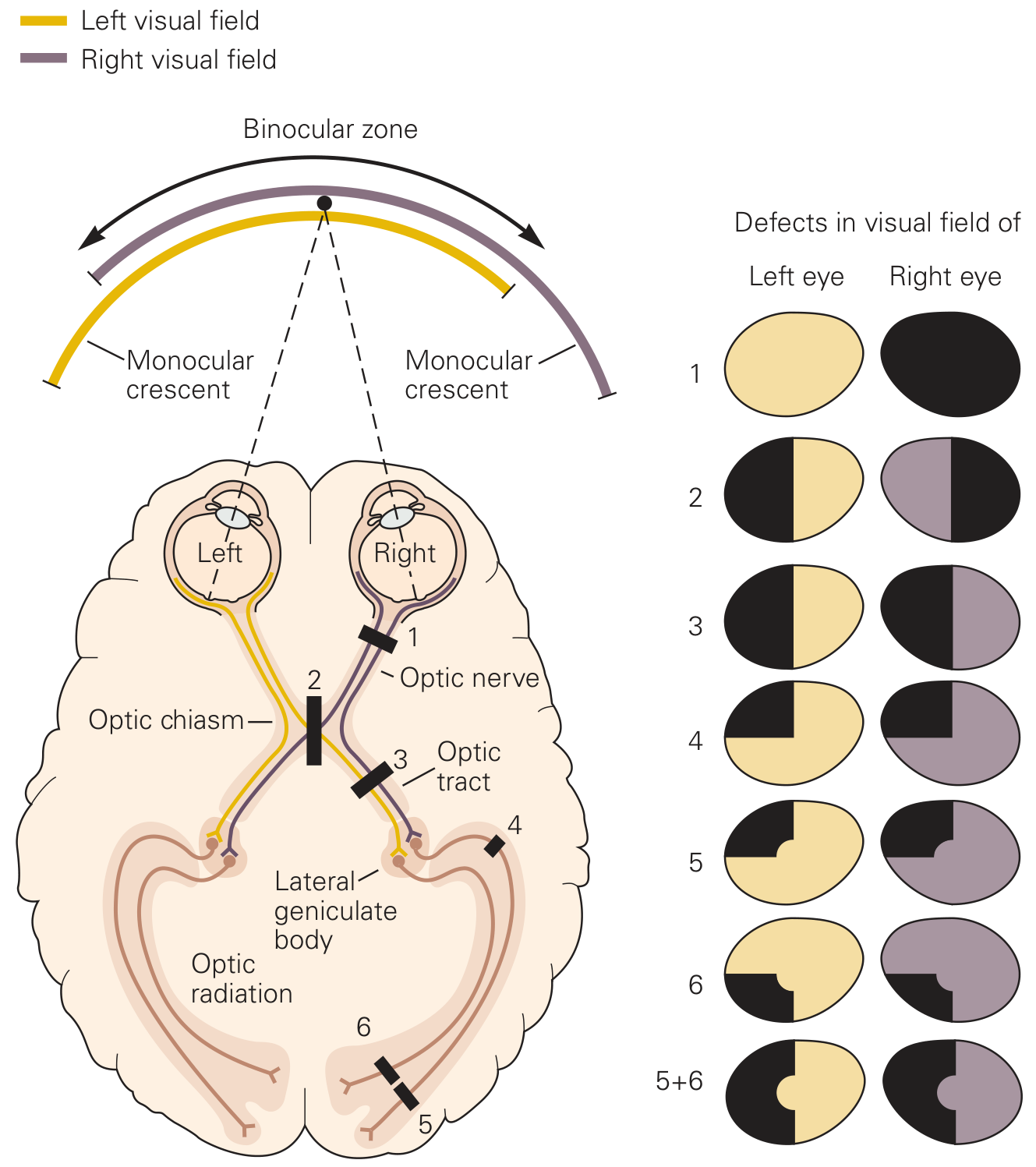
Figure 21-5: Visual pathway representation and lesion defects - Principles of Neural Science (Kandel), 6th Ed.
Visual field representation to visual cortex (right LGN processes left visual field):
Figure: Visual field representation - Gray's Anatomy for Students
Complete lesion chart:
Figure eFig 9.68: Visual Field Deficits with Lesions - Gray's Anatomy for Students
VISUAL FIELD DEFECTS WITH LESIONS (DETAILED)
Lesion 1: Optic Nerve (unilateral, partial - central scotoma)
- Cause: Optic neuritis (multiple sclerosis), ischemic optic neuropathy
- Defect: Ipsilateral central scotoma (loss of central vision in one eye)
- Mechanism: Macular fibers occupy the papillomacular bundle (central optic nerve)
Lesion 2: Complete Optic Nerve
- Cause: Optic nerve trauma, severe optic neuritis, optic nerve compression
- Defect: Complete monocular blindness - total loss of vision in ipsilateral eye
- Additional: Ipsilateral afferent pupillary defect (Marcus Gunn pupil - relative APD)
Lesion 3: Optic Chiasm (central midline)
- Cause: Pituitary adenoma (most common), craniopharyngioma, meningioma
- Mechanism: Crosses NASAL fibers from both eyes are damaged
- Defect: Bitemporal hemianopia - loss of temporal (outer) visual field in BOTH eyes
- Note: The nasal fibers from each eye carry temporal visual field input
Lesion 4: Optic Tract
- Cause: Craniopharyngioma, temporal lobe herniation, MS
- Mechanism: Both temporal retina of ipsilateral eye + nasal retina of contralateral eye are cut
- Defect: Contralateral homonymous hemianopia (incongruous - unevenly matched in the two eyes)
- A left optic tract lesion → right homonymous hemianopia
Lesion 5: Meyer's Loop (temporal lobe optic radiation)
- Cause: Temporal lobe tumor, stroke, temporal lobectomy
- Mechanism: Lower fibers (carrying upper visual field) are damaged
- Defect: Contralateral superior (upper) quadrantanopia = "Pie in the sky"
- Right temporal lesion → left upper quadrant visual field loss in both eyes
Lesion 6: Parietal Lobe Optic Radiation (upper fibers)
- Cause: Parietal lobe stroke, tumor
- Mechanism: Upper fibers (carrying lower visual field) are damaged
- Defect: Contralateral inferior (lower) quadrantanopia = "Pie on the floor"
Lesions 6+7: Complete Optic Radiation / Occipital Cortex
- Cause: PCA territory stroke (most common), occipital tumor
- Defect: Contralateral homonymous hemianopia with MACULAR SPARING
- Why macular sparing?: The macular cortical area at the occipital pole receives dual blood supply (MCA + PCA); also large macular cortical representation makes it partially spared
- Key feature: Distinguishes occipital lobe from tract/LGN lesion
Lesion 8: Bilateral Occipital Cortex
- Cause: Bilateral PCA territory strokes (vertebrobasilar disease)
- Defect: Cortical blindness (bilateral central scotoma or complete blindness)
- Anton's syndrome: Patient is cortically blind but DENIES being blind (anosognosia)
SUMMARY TABLE: VISUAL FIELD DEFECTS
| Lesion Site | Visual Field Defect | Cause |
|---|---|---|
| Optic nerve (partial) | Monocular scotoma | Optic neuritis, ischemia |
| Optic nerve (complete) | Monocular blindness + APD | Trauma, severe neuritis |
| Optic chiasm (central) | Bitemporal hemianopia | Pituitary adenoma |
| Optic chiasm (lateral) | Binasal hemianopia (rare) | Bilateral carotid aneurysms |
| Optic tract | Incongruous contralateral homonymous hemianopia | Herniation, craniopharyngioma |
| LGN | Congruous contralateral homonymous hemianopia | Rare vascular lesion |
| Meyer's loop (temporal) | Contralateral superior quadrantanopia ("pie in sky") | Temporal lobectomy, stroke |
| Parietal radiation | Contralateral inferior quadrantanopia | Parietal stroke, tumor |
| Complete radiation/occipital | Contralateral homonymous hemianopia + macular sparing | PCA stroke |
| Bilateral occipital | Cortical blindness | Bilateral PCA strokes |
PUPILLARY LIGHT REFLEX (Branching off Visual Pathway)
LIGHT → RETINAL GANGLION CELLS
|
▼ (via optic nerve and optic tract)
PRETECTAL NUCLEUS (midbrain, both sides)
|
┌─────┴──────┐
↓ ↓
Ipsilateral Contralateral
EW nucleus EW nucleus
(Edinger-Westphal - CN III)
|
▼ (via ciliary ganglion)
SPHINCTER PUPILLAE → PUPIL CONSTRICTION
Direct reflex: light in one eye → both pupils constrict
Afferent limb: CN II | Efferent limb: CN III (parasympathetic)
APD (relative): afferent limb lesion → no direct but consensual preserved in that eye
HIGHER VISUAL PROCESSING STREAMS
After V1, visual information splits into two processing streams:
V1 (Primary visual cortex, area 17)
|
┌─────┴──────┐
↓ ↓
DORSAL STREAM VENTRAL STREAM
("WHERE/HOW") ("WHAT")
V5 (area MT) → MT V4 → Inferior temporal
→ Posterior parietal → Fusiform gyrus (faces)
cortex → Parahippocampal (places)
Motion, depth Object/color/face recognition
Spatial location Object identity
Action guidance Conscious recognition
Q6. ORGAN OF CORTI (20 Marks)
Introduction
The organ of Corti (organum spirale) is the highly specialized sensory epithelium of the inner ear that converts mechanical vibrations into electrical neural signals - the process of mechanoelectrical transduction. It is housed within the scala media (cochlear duct) of the cochlea, resting on the basilar membrane and covered by the tectorial membrane.
ANATOMY OF THE COCHLEA
The cochlea is a snail-shaped bony labyrinth with 2.75 turns, ~35 mm long (range 28-40 mm) in humans. It is divided into three fluid-filled compartments:
CROSS-SECTION OF COCHLEAR DUCT
═══════════════════════════════════════════════
SCALA VESTIBULI (perilymph, Na⁺-rich, -5 mV)
← communicates with oval window
─────────── VESTIBULAR (REISSNER'S) MEMBRANE ─
SCALA MEDIA / COCHLEAR DUCT (endolymph, K⁺-rich, +80 mV)
← contains organ of Corti
← bounded below by basilar membrane
─────────────────── BASILAR MEMBRANE ──────────
SCALA TYMPANI (perilymph, Na⁺-rich, -5 mV)
← communicates with round window
← connects with scala vestibuli at HELICOTREMA (apex)
═══════════════════════════════════════════════
Basilar Membrane Properties
The basilar membrane is non-uniform - its physical properties change systematically from base to apex:
| Property | Base | Apex |
|---|---|---|
| Width | Narrow (~0.1 mm) | Wide (~0.5 mm) |
| Thickness | Thick | Thin |
| Stiffness | Stiff | Compliant/floppy |
| Best frequency | High (20,000 Hz) | Low (20 Hz) |
This creates the tonotopic map - different frequencies cause maximum vibration at different positions.
DETAILED STRUCTURE OF THE ORGAN OF CORTI
══════════════════ SCALA MEDIA (endolymph) ═══════════════════
TECTORIAL MEMBRANE (gelatinous, acellular)
│ stereocilia contact/embed
═══════════ RETICULAR LAMINA (stiff, forms barrier) ══════════
│ │ │
│ INNER HAIR │ OUTER HAIR CELLS │
│ CELLS (IHC) │ (OHC) │
│ • 1 row │ • 3-4 rows │
│ • 3,500 total │ • 12,000 total │
│ • 12 μm diameter │ • 8 μm diameter │
│ • ~100 stereocilia/cell │ • ~100 stereocilia/cell │
│ • flask-shaped │ • cylindrical │
│ │ │
│ TUNNEL OF CORTI ───┘ │
│ (inner pillar cells ←→ outer pillar cells) │
│ (filled with cortilymph = perilymph-like) │
│ SPACES OF NUEL │
│ (around OHCs) │
│ filled with perilymph│
│ INNER PHALANGEAL OUTER PHALANGEAL CELLS │
│ CELLS (Deiter cells) │
│─────────────────────────────────────────────────────────────│
BASILAR FIBERS ── BASILAR MEMBRANE
══════════════════ SCALA TYMPANI (perilymph) ═════════════════
The rods (pillars) of Corti:
- Inner and outer pillar cells form a triangular tunnel
- Composed of bundles of microtubules and filaments
- Function: structural support, create the tunnel of Corti
Phalangeal cells (Deiters cells):
- Support the outer hair cells
- Send phalangeal processes up to the reticular lamina, plugging the spaces between hair cell apices
Supporting cells:
- Cells of Hensen (lateral to OHCs)
- Cells of Claudius (even more lateral)
- Cells of Boettcher (on basilar membrane, below Claudius cells)
HAIR CELLS IN DETAIL
Stereocilia
- Each hair cell has ~100 stereocilia arranged in rows of increasing height (staircase pattern)
- Not true cilia (no 9+2 structure) - rigid bundles of actin filaments
- Interconnected by tip links (made of cadherin proteins: CDH23 and PCDH15)
- Tip links connect shorter stereocilia to the back of taller adjacent stereocilia
Mechanoelectrical Transduction (MET) at Stereocilia
The transduction mechanism diagram:
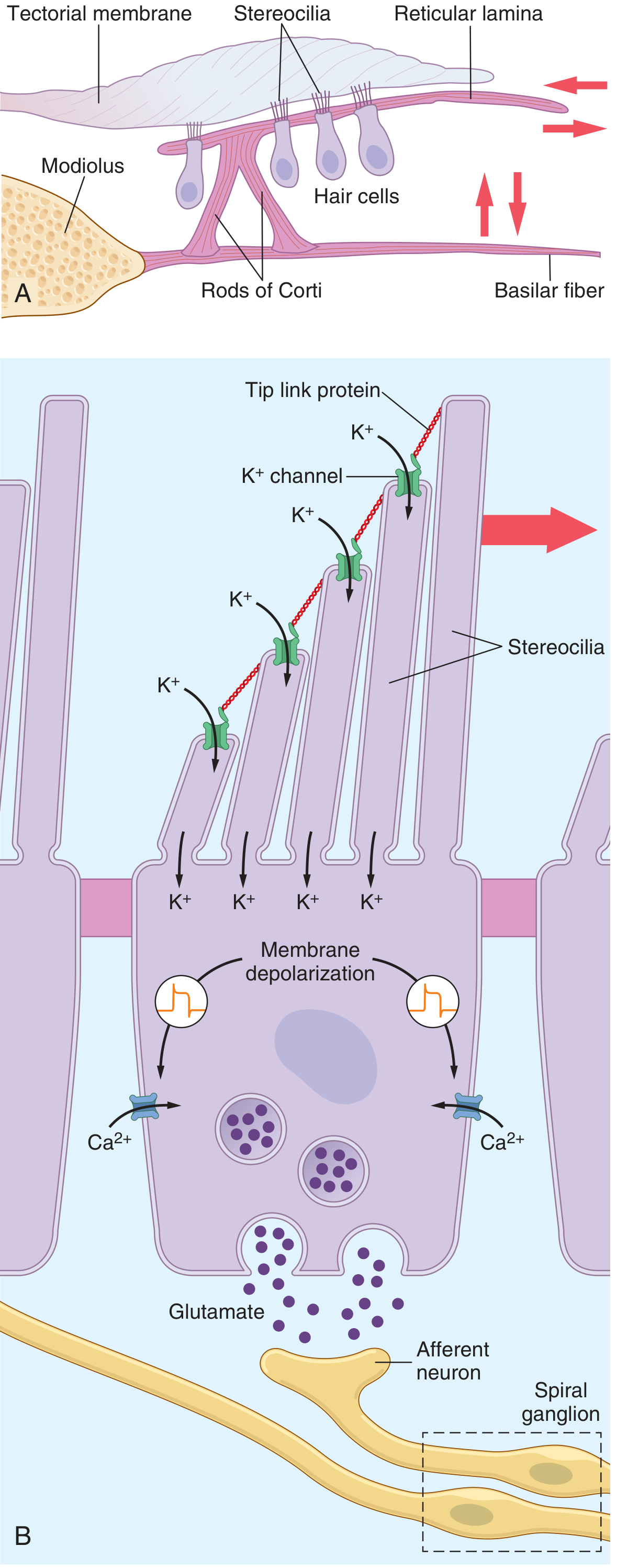
Figure 53.7A,B: Hair cell transduction - Guyton and Hall Textbook of Medical Physiology
MECHANISM OF SOUND TRANSDUCTION (Step-by-Step)
Step 1: Sound Waves to Fluid Motion
SOUND WAVES (pressure fluctuations in air)
|
▼ Directed by pinna, travels down external auditory canal
TYMPANIC MEMBRANE vibrates (pressure → mechanical vibration)
|
▼
OSSICULAR CHAIN amplifies (area ratio + lever action = ~22x amplification):
Malleus → Incus → Stapes footplate
|
▼
OVAL WINDOW → Creates pressure waves in PERILYMPH (scala vestibuli)
|
▼
TRAVELLING WAVE along BASILAR MEMBRANE
(von Bekésy's travelling wave theory - Nobel Prize 1961)
Step 2: Frequency Analysis at Basilar Membrane
Each frequency causes a TRAVELLING WAVE that peaks at a specific location:
HIGH frequency → maximum vibration at BASE
LOW frequency → maximum vibration at APEX
This is the PLACE THEORY of pitch discrimination
Step 3: Stereocilia Deflection
BASILAR MEMBRANE vibrates upward
|
▼
RETICULAR LAMINA + RODS OF CORTI move as a RIGID UNIT
|
▼
Stereocilia shear against TECTORIAL MEMBRANE
- Toward modiolus (upward BM movement) → DEPOLARIZATION
- Away from modiolus (downward BM) → HYPERPOLARIZATION
Step 4: Mechanoelectrical Transduction (MET)
Stereocilia deflect toward taller stereocilia
|
▼
TIP LINKS stretched → open MET CHANNELS (MET channels = TRPA1-related)
|
▼
K⁺ flows INTO hair cell from endolymph
(Driving force = +150 mV: endolymph is +80 mV, hair cell interior is −70 mV)
|
▼
HAIR CELL DEPOLARIZES
|
▼
Voltage-gated Ca²⁺ channels open at BASOLATERAL MEMBRANE
|
▼
Ca²⁺ influx triggers vesicle fusion → GLUTAMATE released
|
▼
AFFERENT COCHLEAR NERVE FIBER (type I - spiral ganglion) EXCITED
|
▼ REPOLARIZATION:
K⁺ exits through Ca²⁺-sensitive K⁺ channels (basolateral)
K⁺ recycled back to stria vascularis via supporting cells
ENDOCOCHLEAR POTENTIAL
The stria vascularis (highly vascularized epithelium on lateral wall of scala media) maintains:
- Endolymph composition: High K⁺ (~150 mM), Low Na⁺ (~1 mM)
- Endocochlear potential: +80 mV (scala media positive relative to perilymph)
This creates a ~150 mV driving force for K⁺ at stereocilia tips (endolymph +80 mV vs. hair cell −70 mV), making the system highly sensitive to minimal sound-induced deflections.
INNER vs OUTER HAIR CELLS - FUNCTIONAL DISTINCTION
| Feature | Inner Hair Cells (IHC) | Outer Hair Cells (OHC) |
|---|---|---|
| Number | ~3,500 (1 row) | ~12,000 (3-4 rows) |
| Shape | Flask/pear-shaped | Cylindrical |
| Stereocilia pattern | Straight line | U/W shape |
| Stereocilia height | Taller | Shorter |
| % Afferent innervation | 90-95% of cochlear nerve | 5-10% |
| Type of afferent | Type I spiral ganglion (~30,000) | Type II spiral ganglion (~3,000) |
| Efferent innervation | Sparse (indirect via afferents) | Dense (medial olivocochlear bundle) |
| Primary function | Sound detection and transduction | Cochlear amplification and frequency selectivity ("tuning") |
| Electromotility | No | Yes - OHCs change length with voltage (prestin-motor protein) |
| Damage effect | Severe hearing loss | Reduced frequency discrimination, loss of "active process" |
Cochlear Amplifier (Active Process)
OHCs possess prestin (a voltage-sensitive motor protein in their lateral wall membrane). When depolarized, OHCs shorten (contract); when hyperpolarized, they elongate. This electromotility amplifies basilar membrane vibration at the characteristic frequency by up to 40-50 dB, creating a "cochlear amplifier" that gives the ear its extraordinary sensitivity and frequency resolution. This is why OHC damage causes larger hearing loss than their 10% afferent representation would predict.
DETERMINATION OF FREQUENCY (PITCH)
1. Place Theory (Békésy):
- Each frequency activates a specific location on the basilar membrane (tonotopy)
- Brain reads out which nerve fibers are firing → frequency perceived
- Best for frequencies >200 Hz
2. Volley/Frequency Principle:
- For low frequencies (<200 Hz), nerve fibers fire in volleys synchronized to the sound waves
- Groups of fibers fire alternately to encode precise temporal information
- Used for low frequencies and pitch discrimination at low frequencies
CENTRAL AUDITORY PATHWAY
ORGAN OF CORTI → Hair cells → COCHLEAR NERVE (CN VIII, ~30,000 fibers)
|
▼
COCHLEAR NUCLEI (dorsal + ventral, pontomedullary junction)
BILATERAL representation begins here
|
├─────────────────────────────────────┐
▼ ▼
SUPERIOR OLIVARY COMPLEX Dorsal acoustic stria
(bilateral) → sound localization (crossed)
(IID - interaural intensity diff.)
(ITD - interaural time difference)
|
▼
LATERAL LEMNISCUS (bilateral fibers)
|
▼
INFERIOR COLLICULUS (midbrain tectum)
- Integration, sound localization
- Auditory reflexes (acoustic startles)
|
▼ Brachium of inferior colliculus
MEDIAL GENICULATE NUCLEUS (MGN) of THALAMUS
- Parvocellular, magnocellular, dorsal divisions
|
▼ Auditory radiation
PRIMARY AUDITORY CORTEX (Heschl's gyrus, areas 41, 42)
Tonotopy preserved
|
▼
AUDITORY ASSOCIATION CORTEX (area 22)
Left hemisphere: speech sound analysis (phonemes)
Right hemisphere: melody, prosody, music
Key feature: Bilateral representation from cochlear nucleus onward. Unilateral cortical lesion does NOT cause complete deafness in either ear; it causes deficits in sound localization and speech discrimination.
DETERMINATION OF LOUDNESS
- Firing rate of auditory nerve fibers increases with louder sounds
- Spatial summation - more hair cells activated as amplitude increases (broader area of BM vibration)
- OHC recruitment - outer hair cells only activated at higher intensities; their activation signals loud sound
- The perceived loudness follows a power law (cube root of actual intensity) - Weber-Fechner law
CLINICAL CORRELATIONS
| Condition | Mechanism | Features |
|---|---|---|
| Presbycusis | Loss of OHCs from base (age-related oxidative damage) | Progressive high-frequency sensorineural hearing loss |
| Noise-induced hearing loss | Acoustic trauma → OHC damage at 4 kHz region (resonance of ear canal) | 4 kHz notch on audiogram |
| Meniere's disease | Endolymphatic hydrops → distorted organ of Corti function | Fluctuating SNHL, tinnitus, vertigo |
| Gentamicin ototoxicity | Aminoglycosides enter endolymph, damage OHCs (esp. basal turn) | Irreversible high-frequency SNHL |
| Otosclerosis | Bony fixation of stapes footplate → reduced BM vibration | Conductive hearing loss |
| Connexin 26 (GJB2) mutation | Disrupts K⁺ recycling via gap junctions in supporting cells | Commonest genetic SNHL |
SOURCES
All content sourced from:
- Neuroanatomy through Clinical Cases, 3rd Edition (Blumenfeld) - Ascending tracts, corticospinal tract, thalamus, cerebral cortex cytoarchitecture
- Guyton and Hall Textbook of Medical Physiology, 14th Edition - Organ of Corti transduction, sound frequency/loudness, corticospinal tract fiber composition
- Principles of Neural Science, 6th Edition (Kandel) - Visual pathway, LGN structure, visual processing streams
- Gray's Anatomy for Students - Visual pathway lesions, central visual pathway
- Kaplan & Sadock's Comprehensive Textbook of Psychiatry - Cerebral cortex neuron types, Brodmann areas, cortical connections
- Scott-Brown's Otorhinolaryngology Head & Neck Surgery - Organ of Corti tonotopy and hair cell types
This is a shared conversation. Sign in to Orris to start your own chat.