
Trigger finger
Reading File
Now let me search the medical library and PubMed in parallel for trigger finger.
Finding Sources
Finding Sources
Searching PubMed
"trigger finger" AND treatment
Good - I have rich sources. Let me now read the most comprehensive sections in parallel.
Reading File
Reading File
Reading File
Now let me read the Campbell's section for treatment detail and fetch the injection technique image:
Reading File
Loading Image
Now let me read the key treatment content and grading section from Campbell's:
Reading File
I have enough content. Let me read the main treatment/surgical section of Campbell's and the Notta's node grading:
Reading File
I now have comprehensive content to write the full answer.
Trigger Finger (Stenosing Tenosynovitis)
Definition and Pathophysiology
Trigger finger (stenosing tenosynovitis) is caused by thickening of the A1 annular pulley at the level of the metacarpal head in the palm, creating a mismatch between the pulley's lumen and the flexor tendon passing through it. A nodule or fusiform swelling forms on the tendon just proximal to the narrowed pulley; this nodule catches as the tendon slides, producing the characteristic clicking, snapping, or locking. - Firestein & Kelley's Textbook of Rheumatology, p. 972
The A1 pulley sits at the level of the MCP joint. When the nodule is proximal and the pulley is stenotic, active finger flexion draws the swollen tendon into the pulley - but during extension it catches, causing the "trigger." In rheumatoid patients, the nodule may be distal to the pulley, producing the same phenomenon. - Campbell's Operative Orthopaedics 15th Ed, p. 4496
Epidemiology
- Most common digit: Thumb, followed by ring finger, then long finger.
- Primary (idiopathic): Typically in individuals over 45 years; thumb triggering is 4x more frequent in women.
- Secondary: Associated with RA, diabetes mellitus, gout, hypothyroidism, amyloidosis. In these, multiple fingers are often involved simultaneously, and co-occurrence with de Quervain's disease or carpal tunnel syndrome is common.
- Congenital/pediatric: Much less common; thumb is most affected and typically presents locked in flexion. Only ~30% resolve spontaneously. - Firestein & Kelley's, p. 972
A 2025 meta-analysis (Sallai et al., PMID 40323072) confirmed a significant co-occurrence between trigger finger and carpal tunnel syndrome, underscoring the shared predisposing conditions.
Classification (Quinnell Grading)
| Grade | Description |
|---|---|
| 0 | Normal movement |
| 1 | Uneven movement (no locking) |
| 2 | Actively correctable locking |
| 3 | Passively correctable locking |
| 4 | Fixed flexion contracture |
Clinical Presentation
- Pain at the base of the digit (proximal palmar crease level), often mistakenly attributed to the PIP joint.
- Palpable tendon nodule in the palm that moves with the tendon during finger motion.
- Snapping or clicking on active flexion/extension - typically worse in the morning upon awakening.
- Progressive limitation of motion as the condition advances.
- End-stage: locked digit that cannot be actively straightened.
Treatment
1. Conservative (Non-Surgical)
- Splinting: Night splinting of the MCP joint in extension prevents locking during sleep, when symptoms are worst.
- Heat/ice and stretching: Useful adjuncts.
- Corticosteroid injection into the tendon sheath: First-line active treatment. Success rate ~60% after a single injection; repeat injections further improve outcomes.
- Dose: 10-15 mg methylprednisolone acetate mixed with 1-2 mL lidocaine (25-27 gauge needle).
- Needle at 45-60° angle, entering just distal to the proximal palmar crease.
- Avoid intratendinous injection (risk of tendon rupture). Intrasheath placement is not strictly required for success.
- Up to 3 injections 3 weeks apart are acceptable.
- Complication: focal palmar fat atrophy. - Rheumatology (Elsevier 2022), p. 287
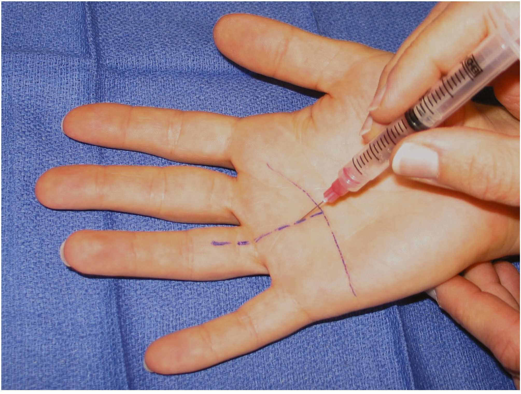
Special considerations in diabetes: Corticosteroid injections may elevate serum glucose for 5+ days. In unstable diabetes, surgery (open release) may be preferred - one cost-analysis study found it to be the most cost-effective strategy in diabetic patients. - Campbell's Operative Orthopaedics 15th Ed
2. Surgical Release
Indicated when nonoperative measures fail. Approximately 97% of patients have complete resolution after surgery. - Campbell's, p. 4497
Open release (A1 pulley release) - Technique:
- Transverse incision ~2 cm long in the palm, several mm distal to the distal palmar crease for middle/ring/small fingers; more proximal for index finger.
- Careful dissection to expose the A1 pulley.
- Longitudinal division of the A1 pulley at the MCP head level under direct vision.
- Perform under local block so triggering resolution can be confirmed intraoperatively.
- Beware: in the index finger and thumb, digital nerves overlie the A1 pulley and require careful protection.
Percutaneous release: Using a needle or push-knife under fluoroscopic or ultrasound guidance. Carries risk of incomplete release and digital nerve/tendon injury - especially in the index finger and thumb. A 2024 meta-analysis (Garcia et al., PMID 38953949) found ultrasound-guided percutaneous release had comparable outcomes to open surgery. A 2026 meta-analysis of RCTs (Huang et al., PMID 41571568) similarly showed no significant difference in efficacy between open and percutaneous release.
Persistent triggering post-release can occur due to:
- Incomplete pulley release
- Tendon catching on palmar aponeurosis transverse fibers (usually resolves spontaneously)
- Nodule from a previously partially lacerated tendon
Summary Table
| Approach | Success Rate | Notes |
|---|---|---|
| Single corticosteroid injection | ~60% | First-line; repeat injections improve rates |
| Splinting + injection | Slightly higher | Splinting may improve injection longevity |
| Open A1 pulley release | ~97% | Gold standard; safest for index/thumb |
| Percutaneous release | Comparable | Risk of nerve/tendon injury; US-guidance preferred |
Sources: Campbell's Operative Orthopaedics 15th Ed 2026, p. 4496-4498; Firestein & Kelley's Textbook of Rheumatology, p. 972; Rheumatology 2-Volume Set (Elsevier 2022), p. 287. Recent meta-analyses: Garcia et al. 2024, Huang et al. 2026, Sallai et al. 2025.
This is a shared conversation. Sign in to Orris to start your own chat.