
Spondylolisthesis etiology, causes, mechanism , clinical features, clinical signs , radiological signs, treatment options
"Spondylolisthesis"[MeSH Terms]
spondylolisthesis diagnosis imaging treatment guideline adult pediatric
lateral lumbar spine radiograph showing spondylolisthesis and pars interarticularis defect
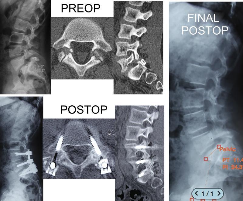
This composite diagnostic image illustrates the surgical management of spondylolisthesis in the lumbar spine across three phases: PREOP, POSTOP, and FINAL POSTOP. The PREOP section includes a lateral radiograph and sagittal/axial CT views showing anterior vertebral slippage and spondylolysis (pars interarticularis defect). The POSTOP section demonstrates surgical stabilization using percutaneous pedicle screws and rods, visible on both lateral radiography and axial/sagittal CT. The FINAL POSTOP lateral radiograph shows the spine after hardware removal, highlighting the maintenance of vertebral alignment and improved sagittal parameters. Superimposed measurements in the final stage indicate a Pelvic Tilt (PT) of 11.4° and Pelvic Incidence (PI) of 34.3°. The image effectively teaches the concept of temporary internal fixation for reduction of spondylolisthesis without permanent arthrodesis, allowing for the restoration of spinal anatomy and subsequent hardware removal once stabilization is achieved.
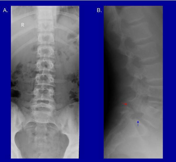
Diagnostic images consisting of an anteroposterior (AP) and lateral radiograph of the lumbar spine. Image A (AP view) displays the lumbar vertebral bodies and the sacrum in a vertical orientation, showing intact pedicles and spinous processes aligned mid-sagittally. Image B (lateral view) provides critical diagnostic detail of the lumbosacral junction. A red arrow identifies a radiolucent defect in the pars interarticularis of the L5 vertebra, diagnostic of spondylolysis. A blue arrow highlights the resulting Grade I spondylolisthesis, characterized by a mild anterior displacement of the L5 vertebral body relative to the S1 base. Key landmarks include the vertebral bodies, intervertebral disc spaces, and the pelvic brim. This comparison illustrates the typical radiographic presentation of a chronic pars defect leading to mechanical instability at the L5-S1 segment, a common cause of lower back pain in adolescents and athletes.
https://pmc.ncbi.nlm.nih.gov/articles/PMC7343250/
PMID: 40168558
Spondylolisthesis
Textbook of Family Medicine 9e, p. 857; Campbell's Operative Orthopaedics 15e, p. 2393.
Etiology and causes: Wiltse-Newman classification
| Type | Cause and mechanism | Typical setting |
|---|---|---|
| I. Dysplastic (developmental) | Congenital dysplasia of the lumbosacral junction, such as abnormal sacral facets, malformed posterior elements, spina bifida occulta, or a dysplastic pars. | Children and adolescents; can produce a high-grade L5-S1 slip. |
| II. Isthmic | Defect in the pars interarticularis, allowing the vertebral body, pedicles, and superior facets to move forward relative to the posterior arch. | Adolescents, athletes, young adults; most commonly L5-S1. |
| IIA. Lytic | Repetitive stress fracture of the pars. | Gymnastics, cricket fast bowling, football linemen, weightlifting, diving. |
| IIB. Elongated pars | Recurrent microfracture and healing causes elongation of an intact pars. | Repetitive loading during growth. |
| IIC. Acute pars fracture | Single acute traumatic fracture through the pars. | Uncommon. |
| III. Degenerative | Disc degeneration, facet osteoarthritis, capsular and ligament laxity, and weak segmental stabilizers cause forward translation without a pars defect. | Older adults, often women; classically L4-L5. |
| IV. Traumatic | Acute fracture through posterior elements other than the pars, or fracture-dislocation. | Major trauma. |
| V. Pathological | Bone weakened by tumor, infection, Paget disease, metabolic bone disease, etc. | Depends on underlying disease. |
| VI. Iatrogenic | Instability after decompression, excessive facetectomy, laminectomy, or adjacent-segment disease after fusion. | Postoperative spine. |
Mechanism
Normal restraints to anterior translation
Why the slip occurs
- The lumbar spine has normal lordosis, creating an anterior shear force at the lumbosacral junction.
- Gravity acting through the upper-body mass and pull of the erector spinae muscles increase this shear force.
- A failure of the posterior stabilizers occurs:
- pars stress fracture in isthmic disease
- disc and facet degeneration in degenerative disease
- congenital dysplasia in developmental disease
- The cephalad vertebra translates anteriorly.
- Foraminal narrowing, central canal stenosis, nerve-root compression, altered sagittal balance, and compensatory muscle spasm may follow.
Campbell's Operative Orthopaedics 15e, pp. 2393-2395.
Clinical features
A. Mechanical symptoms
- Low-back pain, often localized to the lumbosacral region
- Pain worsened by standing, walking, lumbar extension, hyperextension, or activity
- Relief with rest, sitting, or flexion, especially in stenotic degenerative disease
- Hamstring tightness and stiffness
- Fatigue with prolonged standing
B. Isthmic spondylolisthesis
- Common in adolescents and young athletes
- Extension-related low-back pain
- Often follows repetitive hyperextension and rotation activities
- Radicular pain may occur from L5 foraminal stenosis
- Symptoms commonly develop during an adolescent growth spurt
C. Degenerative spondylolisthesis
- Predominantly in older adults
- Low-back pain plus neurogenic claudication
- Leg pain, numbness, heaviness, or weakness on standing or walking
- Symptoms improve with sitting or flexing the lumbar spine
- May coexist with lumbar spinal stenosis
D. Severe or high-grade slip
- Radiculopathy
- Neurogenic claudication
- Progressive sagittal imbalance
- Rarely, cauda equina syndrome: saddle anesthesia, urinary retention or incontinence, fecal incontinence, bilateral progressive weakness
Clinical signs
Inspection and posture
- Increased lumbar lordosis
- Shortened trunk, especially in high-grade developmental slips
- Pelvic retroversion and altered gait
- “Phalen-Dickson sign”: shortened trunk with horizontal skin folds in the lumbar region in severe slips
- Possible crouched or “psoas” gait from hip and knee flexion contractures
Palpation
- Midline lumbosacral tenderness
- A palpable step-off at the slipped vertebral level, especially at L5-S1
- Paraspinal muscle spasm
Movement
- Pain on lumbar extension or single-leg hyperextension
- Restricted lumbar flexion/extension
- Tight hamstrings, often detected by reduced straight-leg raising due to hamstring contracture rather than true nerve-root tension
Neurologic examination
- Motor power: especially ankle and great-toe dorsiflexion for L5 involvement
- Sensory deficit: L5 or S1 distribution
- Reflexes: ankle jerk for S1
- Straight-leg raise and femoral stretch where relevant
- Gait, heel walking, toe walking
- Bowel/bladder function when severe compression is suspected
Radiological signs and investigations
1. Standing plain radiographs: first-line
- Forward translation of one vertebra over another
- L5-S1 is typical for isthmic disease
- L4-L5 is typical for degenerative disease
- Disc-space narrowing and facet arthropathy in degenerative disease
- Lumbosacral kyphosis and sacral verticalization in high-grade slips
- Spina bifida occulta or dysplastic posterior elements in developmental disease
2. Meyerding grading
| Grade | Slip |
|---|---|
| I | <25% |
| II | 25%-50% |
| III | 51%-75% |
| IV | 76%-100% |
| V | >100%, called spondyloptosis |
Textbook of Family Medicine 9e, p. 858.
3. Oblique lumbar radiographs
- The collar represents a pars interarticularis defect.
- It supports diagnosis of spondylolysis.
4. Flexion-extension lateral radiographs
5. CT
- Defining a pars fracture
- Assessing chronicity, sclerosis, nonunion, or bony anatomy
- Surgical planning
6. MRI
- Nerve-root compression
- Disc degeneration
- Central/lateral recess/foraminal stenosis
- Bone marrow edema in an early pars stress injury
- Alternative pathology such as tumor, infection, or fracture
Textbook of Family Medicine 9e, p. 858.
7. Bone scan or SPECT-CT
Important radiographic parameters in high-grade disease
- Slip percentage
- Slip angle
- Sacral slope
- Pelvic tilt
- Pelvic incidence
- Global sagittal balance
Campbell's Operative Orthopaedics 15e, pp. 2394-2398.
Treatment options
1. Conservative treatment: initial approach for most low-grade cases
Activity modification
- Avoid painful repetitive lumbar extension, heavy axial loading, and sport-specific triggers temporarily.
- In adolescents with pars stress injury, rest from provoking sport is followed by graded return when symptoms and function improve.
Analgesia
- Paracetamol and, when suitable, short courses of NSAIDs
- Treat neuropathic pain selectively if radicular pain is present
Physiotherapy
- Core stabilization and motor control
- Abdominal and multifidus strengthening
- Hamstring and hip-flexor flexibility
- Neutral-spine mechanics
- Graded aerobic conditioning
- Weight management where appropriate
Bracing
- May be considered short term in children/adolescents with acute symptomatic pars stress injury or low-grade isthmic slip.
- It is used less often in adults and should complement, not replace, rehabilitation.
Epidural or selective nerve-root injection
Textbook of Family Medicine 9e, p. 858.
Indications for surgical referral or surgery
- Cauda equina symptoms
- New bowel or bladder dysfunction
- Progressive motor deficit
- Severe or rapidly worsening neurologic deficit
- Suspected infection, tumor, or unstable traumatic lesion
- Persistent disabling pain despite an adequate conservative program, commonly at least 6 months for isthmic disease
- Persistent radiculopathy or neurogenic claudication with imaging-confirmed compression
- Progressive neurologic deficit
- Progressive slip in a growing child/adolescent
- High-grade slip, especially in a skeletally immature patient
- Marked sagittal imbalance or high-risk spinopelvic parameters
Surgical options
| Procedure | Main role |
|---|---|
| Direct pars repair | Selected young patients with a symptomatic pars defect, minimal slip, and preserved disc. Preserves motion segment. |
| Decompression | Relieves neural compression in stenosis or radiculopathy. Must be planned carefully because decompression alone may worsen instability. |
| Posterolateral fusion | Stabilizes the involved level, often used for isthmic slips. |
| Interbody fusion: PLIF, TLIF, ALIF, LLIF | Adds anterior-column support, restores disc height/foraminal height, and improves fusion environment. |
| Instrumented fusion | Pedicle screws with fusion for instability, high-grade slips, or when decompression could destabilize the segment. |
| In-situ fusion | Fusion without substantial reduction. Often favored in high-grade slips to avoid traction injury to nerve roots. |
| Reduction plus fusion | Selected cases with significant deformity or sagittal imbalance; needs careful neurological risk assessment. |
Practical summary
- Young athlete + extension-related pain + L5 pars defect: think isthmic spondylolisthesis.
- Older patient + L4-L5 slip + back pain with neurogenic claudication: think degenerative spondylolisthesis with spinal stenosis.
- Start with standing AP and lateral radiographs.
- Use MRI for neurologic symptoms, radicular pain, or stenosis; CT for pars-bone detail.
- Most low-grade cases improve with activity modification and rehabilitation.
- Operate for refractory disabling symptoms, progressive neurological deficit, progressive/high-grade slip, or major deformity.