
What is therapeutic ultrasound
therapeutic ultrasound physical therapy mechanisms
therapeutic ultrasound physiotherapy treatment diagram
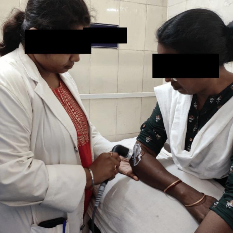
A clinical photograph depicting a physiotherapy session focused on ultrasonic therapy. A female healthcare provider, wearing a white clinical coat, is shown applying a handheld therapeutic ultrasound transducer to the lateral aspect of a female patient's proximal left forearm, near the elbow. The patient's arm is resting on a white examination surface. A visible layer of clear coupling gel is applied to the skin over the treatment site to facilitate the transmission of ultrasonic waves. The ultrasound probe is ergonomic and black, connected via a thick gray cable to a power unit (not shown). This setup is characteristic of conservative management for musculoskeletal conditions such as lateral epicondylitis or soft tissue adhesions, aiming to promote tissue healing and alleviate fibrosis through thermal or non-thermal acoustic energy. The setting is an indoor clinical environment with tiled walls.
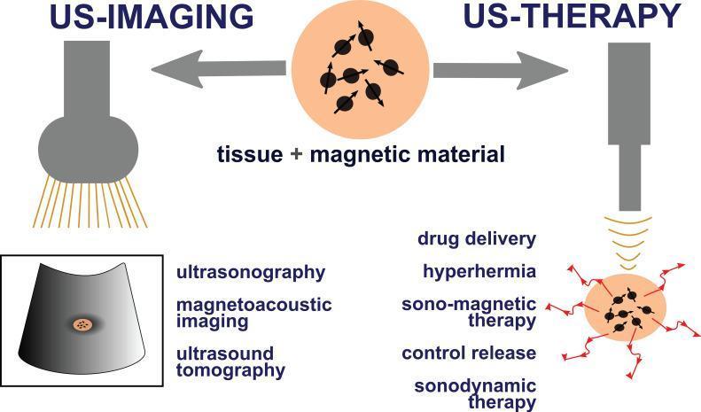
A medical infographic illustrating the concept of ultrasound-based theranostics using magnetic materials. The diagram is divided into two primary sections: 'US-IMAGING' and 'US-THERAPY', both centered around a target labeled 'tissue + magnetic material'. On the imaging side, an ultrasound transducer is shown emitting waves to visualize the tissue; associated diagnostic modalities listed include ultrasonography, magnetoacoustic imaging, and ultrasound tomography. On the therapy side, the ultrasound transducer targets the same tissue, but the illustration now includes red radiating arrows signifying therapeutic activation. Listed therapeutic applications include drug delivery, hyperthermia, sono-magnetic therapy, controlled release, and sonodynamic therapy. The magnetic materials are depicted as nanoparticles with orientation vectors, serving a dual role as contrast agents for diagnostic imaging and as mediators for localized treatment through mechanisms like heating or triggered drug release. This conceptual diagram summarizes the integration of diagnostics and therapeutics into a single medical platform.
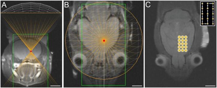
This diagnostic and planning image set illustrates MRI-guided focused ultrasound (MRgFUS) treatment planning in a rodent brain model. Panels A and B present T2-weighted MR images in coronal and axial planes, respectively, showing the focused ultrasound transducer's positioning. A white wireframe represents the transducer geometry, with orange rays converging at a specific focal point marked by a red dot within the brain's subcortical structures. A green rectangular boundary delineates the targeted treatment volume. Panel C provides an axial T2-weighted image displaying the sonication rastering protocol applied to the striatum. A grid of 15 yellow circles (3x5 arrangement) identifies the discrete sonication points. An inset diagram in the upper right corner details the serpentine sonication sequence, indicating the path and order of treatment points. The images demonstrate the spatial precision of therapeutic ultrasound targeting for neurointerventional research, specifically focusing on the striatal region.
therapeutic ultrasound physiotherapy mechanisms clinical applications 2024
Therapeutic Ultrasound
Definition
Mechanism of Action
1. Thermal Effects (Continuous Mode)
- Sound waves cause molecules in tissue to vibrate, generating heat through friction
- Raises deep tissue temperature, producing temporary vasodilation
- Increased blood flow helps wash out pro-inflammatory and pro-algogenic (pain-causing) substances
- Reduces local edema
- Increases collagen extensibility, helping loosen scar tissue and joint capsule contractures
- Best achieved with continuous (non-pulsed) ultrasound
2. Non-Thermal / Mechanical Effects (Pulsed Mode)
- Cavitation: Formation and oscillation of microscopic gas bubbles in tissue fluid. Stable cavitation alters cell membrane permeability; inertial (unstable) cavitation at high intensities can be destructive.
- Acoustic streaming: Unidirectional movement of fluid along cell membranes, increasing membrane permeability and stimulating cellular activity (e.g., fibroblast stimulation)
- Pulsed mode is used when thermal effects are not desired (e.g., acute inflammation)
Phonophoresis
- Dexamethasone
- Hydrocortisone
- Lidocaine
Dosimetry
| Parameter | Detail |
|---|---|
| Frequency | 0.8-3 MHz (higher = more superficial penetration) |
| Intensity | 1-2 W/cm² for inflamed areas |
| Duration | 5-10 minutes per session |
| Mode | Continuous (thermal) or Pulsed (non-thermal) |
Indications
- Superficial periarticular disorders: bursitis, tendinitis, ligament sprains
- Contracture of joint capsules or adhesive scars
- Neuromas
- Reflex sympathetic dystrophy
- Plantar warts
- Muscular strains, fibrosis, myositis
- Myofascial pain syndrome (trigger points) - a 2024 systematic review found large effect sizes vs sham
- Calcific tendinitis (calcium deposit reduction)
- Bone healing (low-intensity pulsed ultrasound / LIPUS)
- Knee osteoarthritis - moderate effects on pain and function with pulsed ultrasound (2024 systematic review of 21 RCTs)
Contraindications
- Tumors (risk of stimulating growth)
- Pregnancy - contraindicated over abdomen and pelvis
- Cardiac conditions - avoid over the cervical ganglia, cardiac area, or implanted pacemakers
- Thrombophlebitis
- Hemorrhagic or infected areas
- Unhealed fracture sites
- Epiphyseal plates in growing bones
- Spinal cord
- Areas of previous radiation therapy (wait 6 months)
- Eyes
Advanced / Modern Applications
| Application | Description |
|---|---|
| High-Intensity Focused Ultrasound (HIFU) | Concentrates energy to ablate tumors (liver, kidney, uterine fibroids) without incision |
| Histotripsy | Mechanical tissue ablation for cancers (liver, kidney, pancreas) - in clinical trials |
| Low-Intensity Pulsed Ultrasound (LIPUS) | Bone fracture healing acceleration; also investigated for deep-brain stimulation in Alzheimer's and depression |
| Shock wave therapy | Extracorporeal treatment for plantar fasciitis, lateral epicondylitis, calcific tendinitis (success rates 30-90%) |
| Sonophoresis / phonophoresis | Transdermal drug delivery |
Current Evidence
- Positive: Myofascial pain (large effect, 2024 SR), knee OA pain/function (moderate effect, 2024 SR), calcific tendinitis, bone healing (LIPUS)
- Negative/Uncertain: Osteoarthritis in general (NICE advises against); rotator cuff tendinopathy guidelines (2025) now recommend against it, preferring shockwave therapy
- The field is moving toward more focused and higher-intensity applications (HIFU, histotripsy, neuromodulation) where evidence is stronger
Clinical Image
The definition u have given is not good for exam give me definition in exam point of video exam oriented and easy to learn
Exam-Ready Definition of Therapeutic Ultrasound
Short Definition (1-liner for MCQs)
Therapeutic ultrasound is the use of high-frequency sound waves (0.8–3 MHz) to produce thermal and non-thermal effects in deep tissues for the purpose of pain relief, tissue healing, and reducing inflammation.
Standard Definition (for short/long answer exams)
Therapeutic ultrasound is a physiotherapy modality in which high-frequency acoustic (sound) waves are applied to body tissues through a handheld transducer using a coupling gel, producing thermal effects (via continuous mode) and non-thermal effects (via pulsed mode - cavitation and acoustic streaming), used to treat musculoskeletal conditions, soft tissue injuries, and periarticular disorders.
Memory Formula (break it down to remember easily)
| Component | What to Remember |
|---|---|
| What it uses | High-frequency sound waves (0.8–3 MHz) |
| How it's applied | Handheld transducer + coupling gel |
| Two effects | Thermal (continuous) + Non-thermal (pulsed) |
| Non-thermal = 2 things | Cavitation + Acoustic streaming |
| Purpose | Heal tissue, relieve pain, reduce inflammation |
Quick Recall Trick
"TUS = Sound + Heat + Vibration = Heal"
- Sound waves → Heat (thermal) → Vibration (cavitation/streaming) → Heal
One Line Each for Viva/Oral Exam
- Frequency: 0.8–3 MHz (higher freq = superficial; lower = deeper tissues)
- Mode: Continuous = thermal | Pulsed = non-thermal
- Coupling agent: Gel (to eliminate air between probe and skin)
- Dose: 1–2 W/cm² for 5–10 minutes
- Special use: Phonophoresis = ultrasound + drug delivery through skin