
Scleroderma detailed
scleroderma skin fibrosis clinical features
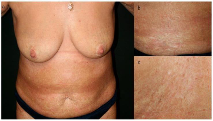
Clinical photograph series (a-c) showing the anterior torso, breasts, and abdomen of a patient after 18 months of treatment for bullous morphea. Image 'a' provides a panoramic view of the torso, demonstrating significant resolution of active bullous lesions and erosions. The skin exhibits post-inflammatory pigmentary changes, including generalized erythema, areas of hyperpigmentation, and atrophic, ivory-colored patches characteristic of sclerodermatous remission. Close-up views 'b' and 'c' highlight the residual skin texture and morphology. Image 'b' shows linear, ivory-white sclerotic markings and shiny fibrosis, while 'c' emphasizes a coarse texture with closely compacted fine lines and superficial atrophic changes. Key educational features include the absence of active bullae or acute inflammatory plaques, indicating successful therapeutic management with budesonide. The clinical presentation reflects a rare association between bullous morphea and autoimmune hepatitis, with the images illustrating the skin's progression to a stable, chronic state with persistent scleroderma-like fibrosis and skin laxity.
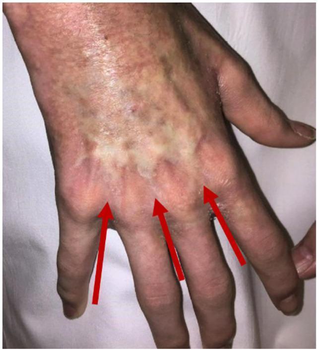
This clinical photograph displays the dorsal aspect of a human hand, illustrating the dermatological regression of sclerodermatous chronic Graft-versus-Host Disease (cGvHD). The skin exhibits significant pigmentary alterations and textural changes following treatment. Three red arrows point to residual areas of fibrotic thickening over the metacarpophalangeal joints, which appear as slightly hypopigmented, indurated patches compared to the surrounding mottled, erythematous, and hyperpigmented skin. The overall appearance demonstrates a transition from severe cutaneous fibrosis toward clinical resolution, though residual dyspigmentation and uneven skin texture persist. This image serves as an educational example of the clinical evolution of skin involvement in systemic cGvHD post-immune ablation and stem cell rescue, highlighting the characteristic scleroderma-like features and their potential for regression.
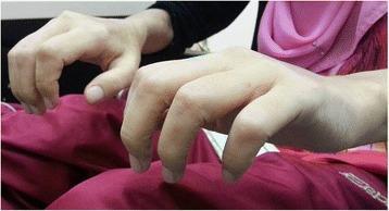
This clinical photograph displays the hands of a patient with Nephrogenic Systemic Fibrosis (NSF), a rare systemic disorder occurring in individuals with renal failure. The image prominently features scleroderma-like changes, characterized by significant tightening, thickening, and induration of the skin across the dorsum of the hands and fingers. Marked joint contractures are visible, particularly in the proximal interphalangeal (PIP) and metacarpophalangeal (MCP) joints, resulting in a 'claw-hand' deformity and severely restricted range of motion. The skin appears taut, waxy, and shiny, with a loss of normal skin creases and prominence of underlying skeletal structures. This presentation illustrates the aggressive fibrotic process of NSF following gadolinium-based contrast exposure in a patient with end-stage renal disease. The visual evidence emphasizes the debilitating physical impact of the condition on manual dexterity and joint function.
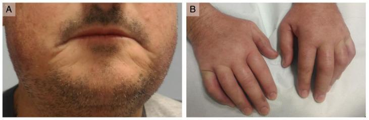
Two-panel clinical photograph demonstrating characteristic features of systemic sclerosis (SSc). Figure A shows 'scleroderma facies' in the perioral region, characterized by thin, retracted lips and skin tightening with a paucity of normal wrinkles. Visible telangiectasias are present on the malar area, and the nose appears sharpened due to skin tethering. Figure B depicts both hands with prominent 'puffy fingers,' a sign of the early edematous phase of SSc. The digits exhibit diffuse swelling and a smooth, taut skin texture. There is a lack of digital ulcers or pitting scars on the fingertips. Subtle bluish discoloration on the right hand's knuckle suggests active Raynaud’s phenomenon. These visual findings are diagnostic hallmarks of systemic sclerosis, indicating progressive dermal fibrosis and vascular dysfunction.
"systemic sclerosis" AND treatment
systemic sclerosis Raynaud phenomenon nailfold capillary pulmonary hypertension
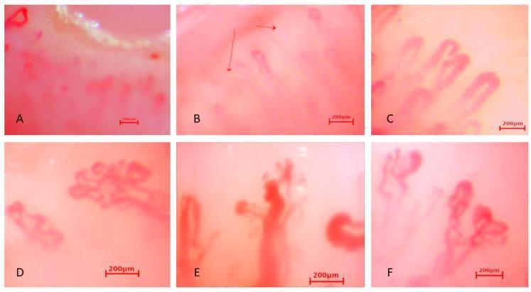
This composite image presents six nailfold capillaroscopy micrographs (labeled A-F) demonstrating various microvascular abnormalities characteristic of systemic sclerosis or secondary Raynaud's phenomenon. (A) and (B) illustrate capillary loss or rarefaction, with a significantly reduced density of vascular loops against a pale background. (C) depicts 'giant capillaries,' characterized by extreme homogeneous dilation and tortuosity of the vascular loops. (D) and (F) showcase 'bizarre capillaries,' exhibiting highly irregular, branched, and bushy morphological distortions that deviate from the standard U-shape. (E) demonstrates active neoangiogenesis, evidenced by clusters of disorganized, newly formed microvessels and exuberant branching patterns. Each frame includes a 200 µm scale bar for size reference. These visual findings are critical diagnostic markers in rheumatology for evaluating microangiopathy and connective tissue diseases.
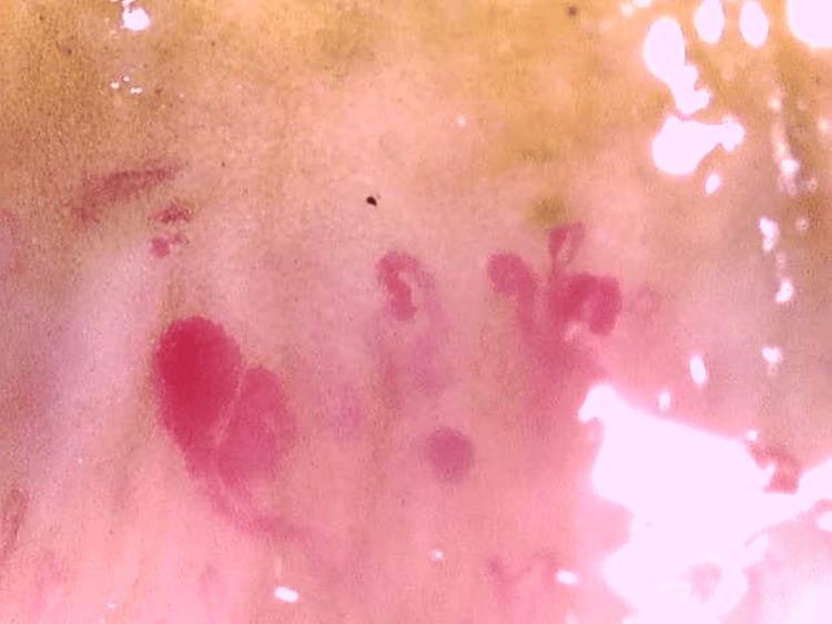
This clinical diagnostic image shows a nailfold capillaroscopy procedure, a key diagnostic tool in rheumatology for evaluating microvascular changes. The image displays classic findings associated with an 'active' scleroderma pattern, frequently seen in systemic sclerosis (SSc). Visible features include prominent giant capillaries (megacapillaries) characterized by significant, bulbous dilation and irregular vessel morphology. Several dark red focal lesions represent microhemorrhages, resulting from the rupture of these fragile, ectatic capillaries. Furthermore, the image shows a non-uniform distribution of vessels with apparent gaps, indicating capillary dropout or loss of the normal hairpin loop architecture. These microvascular abnormalities are clinically significant for the assessment of Raynaud phenomenon and the diagnosis of connective tissue diseases, reflecting the underlying vasculopathy of scleroderma-spectrum disorders.
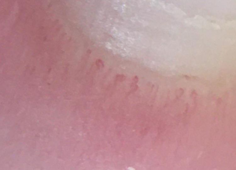
This clinical photograph displays a nailfold capillaroscopy of a patient, demonstrating classic microvascular changes associated with systemic sclerosis (SSc). The image reveals significant diagnostic abnormalities, most notably the presence of frequent 'giant capillaries' characterized by marked homogeneous dilation of the capillary loops. The overall capillary architecture shows a mild degree of disorganization compared to the typical linear 'hairpin' arrangement seen in healthy subjects. Furthermore, there is a moderate reduction in capillary density, with visible areas of capillary loss or 'dropout,' which are hallmarks of progressive microangiopathy in scleroderma-spectrum disorders. These findings serve as critical bedside diagnostic indicators for connective tissue diseases and correlate with systemic manifestations such as Raynaud's phenomenon and internal organ involvement.
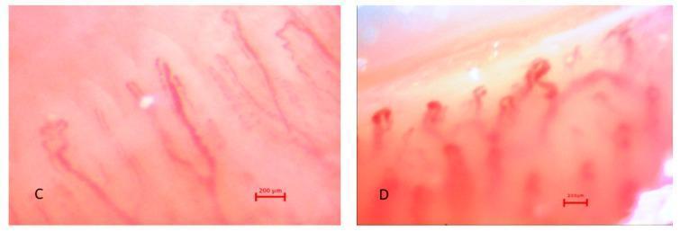
This clinical diagnostic image displays nailfold capillaroscopy frames (C and D) illustrating microvascular patterns in patients with Raynaud's Phenomenon (RP). Frame C shows spastic capillary loops characterized by elongated, slender, and relatively linear structures with mild tortuosity and organized, parallel arrangement. Frame D demonstrates advanced pathological changes, including 'giant capillaries' with significantly enlarged diameters, irregular bulbous shapes, and a disorganized, clustered spatial distribution. The imaging utilizes in vivo capillaroscopy to evaluate microangiopathy, commonly used in the assessment of systemic sclerosis and undifferentiated Raynaud's phenomenon (uRP). Key educational features include the visualization of capillary loop morphology, vessel density, and the presence of microvascular architectural distortion. Each frame includes a 200 µm scale bar for size comparison of the microvessels.
Scleroderma (Systemic Sclerosis) - Comprehensive Review
Overview
- Fibrosis of the skin and visceral organs
- Obliterative vasculopathy (microangiopathy)
- Immune dysregulation with autoantibody production
Classification & Clinical Subsets
| Feature | Limited Cutaneous SSc (lcSSc) | Diffuse Cutaneous SSc (dcSSc) |
|---|---|---|
| Skin distribution | Distal to elbows/knees; face | Proximal to elbows/knees; trunk |
| Onset | Slow, insidious | Rapid, early |
| Raynaud's onset | Years before other features | Concurrent with skin disease |
| Key autoantibodies | Anti-centromere (ACA) | Anti-topoisomerase I (Scl-70), anti-RNA Pol III |
| ILD risk | Lower (16%) | High (38-63%) |
| PAH risk | Higher | Moderate (~12-20%) |
| Renal crisis | Rare | 15-20% |
| Prognosis | Better long-term | Worse survival |
- Scleroderma sine scleroderma - internal organ involvement without skin fibrosis
- Overlap syndromes - features of other CTDs (SLE, myositis, Sjogren's)
- Very Early Diagnosis of SSc (VEDOSS) - Raynaud's + puffy fingers + positive ANA + abnormal nailfold capillaries, without skin thickening; 60-70% develop full SSc within 3-5 years
Multiorgan Involvement
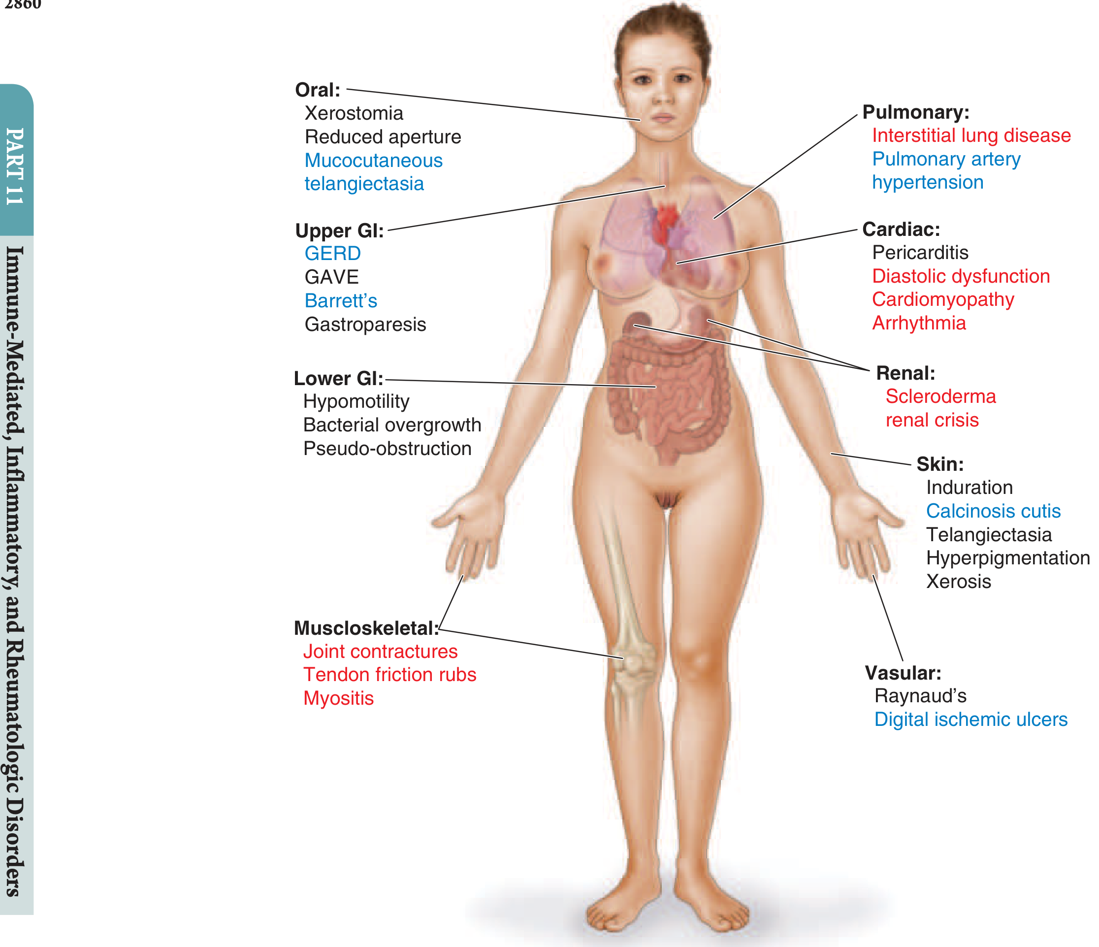
| Feature | dcSSc | lcSSc |
|---|---|---|
| Raynaud phenomenon | 95-97% | 96-99% |
| Skin hardening | 95-100% | 90% |
| GI tract | 65-90% | 60-90% |
| Lung fibrosis | 39-63% | 16-27% |
| PAH | 12-20% | 14-15% |
| Musculoskeletal | 45-49% | 35-39% |
| Kidney | 15-19% | 3-10% |
| Heart | 3-20% | 1-10% |
Pathogenesis
1. Vascular Injury
- The initiating event is likely endothelial cell injury (triggered by viruses, toxins, autoimmune attack)
- Resulting in fibroproliferative vasculopathy: intimal hyperplasia, medial hypertrophy, luminal narrowing
- Capillary loss and obliteration → ischemia, Raynaud's, digital ulcers, PAH
- EndoMT (endothelial-to-mesenchymal transition): endothelial cells in injured vessels transdifferentiate into myofibroblasts driving perivascular fibrosis
2. Immune Dysregulation
- T-cell and B-cell activation → autoantibodies, cytokine release
- IL-6, TGF-β, IL-11, IL-13, IL-23, PDGF, CXCL4, endothelin, CTGF all implicated in fibrogenesis
- Toll-like receptor (TLR4, TLR8, TLR9) activation by endogenous ligands (HMGB1, mitochondrial DNA, EDA-fibronectin) perpetuates inflammation
3. Fibrosis
- TGF-β is the central mediator - drives fibroblast proliferation, migration, and myofibroblast differentiation
- Myofibroblasts secrete excessive collagen and ECM; they persist (evade apoptosis via BCL-XL upregulation) → progressive scar formation
- Sources of myofibroblasts: tissue-resident fibroblasts, circulating mesenchymal progenitor cells, EndoMT, adipocyte-mesenchymal transition (explaining loss of subcutaneous fat in fibrotic SSc skin)
- SSc fibroblasts display an intrinsically activated "scleroderma phenotype" even in ex vivo passage - driven by autocrine TGF-β loops, deregulated microRNAs, and epigenetic modifications
ACR/EULAR 2013 Classification Criteria
| Item | Subitem | Score |
|---|---|---|
| Skin thickening proximal to MCPs (both hands) | - | 9 (sufficient) |
| Skin thickening fingers only | Puffy fingers | 2 |
| Sclerodactyly (distal to MCPs) | 4 | |
| Fingertip lesions | Digital tip ulcers | 2 |
| Fingertip pitting scars | 3 | |
| Telangiectasia | - | 2 |
| Abnormal nailfold capillaries | - | 2 |
| Lung involvement | PAH | 2 |
| ILD | 2 | |
| Raynaud's phenomenon | - | 3 |
| SSc autoantibodies | ACA, Scl-70, RNA Pol III | 3 |
Clinical Features in Detail
Skin
- Early (edematous) phase: puffy, swollen fingers and hands (non-pitting)
- Indurative phase: skin becomes thickened, taut, leathery - "hidebound" appearance
- Late (atrophic) phase: skin thins, loses appendages, may ulcerate
- Scleroderma facies: thin retracted lips, reduced oral aperture (microstomia), loss of normal skin folds, "beak-nose," prominent perioral furrows
- Sclerodactyly: skin thickening of digits causing flexion contractures
- Digital pitting scars / ulcers: ischemic necrosis at fingertips
- Calcinosis cutis: calcium deposits, especially at pressure points
- Telangiectasias: mat-like, on hands, face, lips, tongue
Raynaud's Phenomenon (RP)
- Present in 95-99% of SSc patients - often the first manifestation, preceding other features by months to years
- Triphasic color change: white (pallor/vasospasm) → blue (cyanosis) → red (reperfusion)
- In SSc, Raynaud's is secondary (structural vessel disease) unlike primary RP which is purely functional/vasospastic
- Associated with nailfold capillary abnormalities: giant capillaries, microhemorrhages, capillary dropout (pathognomonic of SSc-spectrum disease)
Pulmonary Disease
- HRCT evidence in up to 65% of patients; clinically significant ILD in 16-43%
- Risk factors: dcSSc, anti-Scl-70 antibody, male sex, African-American race, severe GERD
- Protective: anti-centromere antibody
- Pattern: typically NSIP (nonspecific interstitial pneumonia); less often UIP
- PFTs: restrictive pattern (reduced FVC, TLC, DLCO); FVC <70% predicted
- HRCT: bilateral lower lobe subpleural reticular opacities with ground-glass changes; apicobasal gradient
- Most rapid progression in first 3-5 years; FVC can decline 30%/year early on
- "Velcro" crackles on auscultation
- More common in lcSSc (15%); occurs later in disease
- Symptoms: progressive exertional dyspnea, fatigue, syncope
- Screening: annual echocardiography + PFTs; low DLCO disproportionate to FVC should raise suspicion
- Confirmed by right heart catheterization (RHC): mPAP ≥ 20 mmHg, PCWP ≤ 15 mmHg, PVR > 2-3 Wood units (2022 ESC/ERS updated thresholds)
Gastrointestinal Tract (60-90%)
- Esophageal dysmotility (most common GI manifestation): reduced peristalsis of distal 2/3 (smooth muscle); lower esophageal sphincter incompetence → GERD, Barrett's esophagus, strictures
- Gastroparesis: early satiety, nausea, bloating
- GAVE (Gastric Antral Vascular Ectasia / "watermelon stomach"): GI hemorrhage - more common in dcSSc
- Small bowel hypomotility: bacterial overgrowth, malabsorption, diarrhea
- Large bowel: wide-mouthed diverticulae, pseudo-obstruction (Ogilvie's syndrome)
Scleroderma Renal Crisis (SRC)
- Occurs in ~15-20% of dcSSc (rare in lcSSc)
- Presents with acute hypertension (often severe, >160/110), rapidly progressive renal failure, and microangiopathic hemolytic anemia
- Risk factors: rapidly progressive skin disease, anti-RNA Pol III antibody, high-dose corticosteroids (>15 mg/day prednisolone)
- Medical emergency: prompt recognition is essential
- Before ACE inhibitors: >90% mortality at 1 year; now >70% survival with prompt ACEi treatment
- Key drug: ACE inhibitors (captopril preferred for rapid titration) - continue even if dialysis needed, as up to 50% may recover renal function within 24 months
- Note: ARBs are less well supported; do NOT withhold ACEi in pregnancy if SRC occurs (life-threatening emergency)
Cardiac Involvement
- Primary cardiac disease (myocardial fibrosis, microvascular disease): diastolic dysfunction, LV/RV cardiomyopathy, arrhythmias
- Pericarditis and pericardial effusion (common but often asymptomatic)
- Conduction system fibrosis: heart block, atrial/ventricular arrhythmias
- Beta-blockers may precipitate vasospasm; use selectively
Musculoskeletal (35-49%)
- Arthralgia and arthritis: symmetric, non-erosive (usually)
- Tendon friction rubs: pathognomonic of dcSSc - leathery crepitus felt over tendons with movement; indicates inflammatory activity
- Myopathy: inflammatory (overlap with polymyositis) or fibrotic
- Acro-osteolysis: resorption of distal phalanges in long-standing lcSSc with severe RP
Autoantibodies & Their Clinical Significance
| Antibody | Prevalence | Clinical Association |
|---|---|---|
| ANA | 90-95% | General SSc marker |
| Anti-centromere (ACA) | 20-40% | lcSSc, PAH risk, lower ILD risk |
| Anti-topoisomerase I (Scl-70) | 15-30% | dcSSc, high ILD risk, worse prognosis |
| Anti-RNA Pol III | 10-25% | dcSSc, renal crisis risk, cancer association |
| Anti-U3-RNP (anti-fibrillarin) | 5-8% | dcSSc, cardiac/pulmonary/renal involvement |
| Anti-Th/To | 2-5% | lcSSc, ILD, PAH |
| Anti-Pm-Scl | 3-5% | Overlap with myositis |
| Anti-U1-RNP | ~10% | MCTD overlap |
Laboratory & Diagnostic Evaluation
- ANA: positive in ~90-95% (nucleolar, speckled, centromere patterns)
- CBC: anemia (chronic disease or microangiopathic in SRC), thrombocytopenia in SRC
- Urinalysis + creatinine: baseline and monitoring for renal involvement
- Inflammatory markers: CRP/ESR mildly elevated
- NT-proBNP/BNP: elevated in PAH or cardiac disease
- PFTs: FVC, FEV1/FVC, DLCO - baseline and serial monitoring
- HRCT chest: preferred over plain CXR for ILD detection
- Echocardiography: annual PAH screening in all SSc patients
- Nailfold capillaroscopy: giant capillaries, microhemorrhages, capillary dropout = SSc pattern
- Right heart catheterization: definitive PAH diagnosis
Treatment
Skin / General Immunosuppression
- Methotrexate: early dcSSc skin disease (shown to reduce mRSS modestly in RCTs)
- Mycophenolate mofetil (MMF): widely used for skin and ILD; well tolerated
- Cyclophosphamide (CYC): effective for ILD (SLS trial); now largely replaced by MMF + nintedanib for ILD
- Autologous HSCT: for severe, rapidly progressive early dcSSc - superior to CYC in ASTIS and SCOT trials; significant treatment-related mortality ~10%, reserved for selected patients
- Rituximab (anti-CD20): open-label data supporting skin and ILD benefit
- CAR-T (CD19): early-stage, promising for sustained remission (2024-2025 emerging data)
Pulmonary (ILD)
- Mycophenolate mofetil: first-line maintenance for SSc-ILD (SLS II trial: non-inferior to CYC with better tolerability)
- Nintedanib: tyrosine kinase inhibitor - significantly slows FVC decline in SSc-ILD (SENSCIS trial, 2019); can be combined with MMF
- Pirfenidone: less evidence in SSc specifically
- Avoid high-dose corticosteroids (renal crisis risk)
Pulmonary Arterial Hypertension
- Treat per general PAH guidelines based on WHO functional class:
- ERA (endothelin receptor antagonists): bosentan, ambrisentan, macitentan
- PDE-5 inhibitors: sildenafil, tadalafil
- Prostacyclin/analogs: epoprostenol (IV), treprostinil (SC/IV/inhaled), iloprost (inhaled)
- Riociguat: soluble guanylate cyclase stimulator
Raynaud's Phenomenon & Digital Ulcers
- First-line: long-acting dihydropyridine CCBs (amlodipine, nifedipine)
- Second-line: ARBs (losartan), PDE-5 inhibitors (sildenafil), alpha-1 blockers (prazosin)
- Digital ulcers: bosentan (reduces new ulcer formation), IV iloprost
- Severe ischemia: topical nitroglycerin, botulinum toxin intradigital injection, digital sympathectomy
- General: warm clothing, avoid cold/stress/nicotine/vasoconstrictors
Scleroderma Renal Crisis
- Captopril (or any ACE inhibitor) - start immediately and titrate aggressively
- Continue ACEi even if dialysis is required (recovery possible up to 24 months)
- Target BP: rapid normalization
- High-dose corticosteroids are a risk factor and should be minimized
GI Involvement
- GERD: PPIs (high dose), prokinetics, elevate head of bed
- Gastroparesis: metoclopramide, domperidone, erythromycin
- Small bowel bacterial overgrowth: rotating antibiotics (amoxicillin-clavulanate, rifaximin, metronidazole)
- GAVE: argon plasma coagulation, angiodysplasia treatment
Prognosis
- Overall 10-year survival: ~70-80% in lcSSc; ~55-65% in dcSSc
- Leading causes of death: ILD, PAH, cardiac disease, renal crisis (pre-ACEi era)
- Adverse prognostic factors: dcSSc, male sex, African-American race, older age at onset, anti-Scl-70 or anti-RNA Pol III antibody, low FVC/DLCO at diagnosis, PAH, renal involvement
- HSCT associated with durable remission and improved long-term survival in selected severe dcSSc
- ACE inhibitors for SRC have transformed renal mortality from >90% to <30% at 1 year (Harrison's)
Scleroderma Mimics (Differential Diagnosis)
| Condition | Distinguishing Features |
|---|---|
| Morphea | Localized, no systemic involvement, no Raynaud's |
| Eosinophilic fasciitis | Spares digits, peripheral eosinophilia, no RP, no autoantibodies |
| Scleromyxedema | Paraprotein, no RP, papular lesions |
| Nephrogenic systemic fibrosis | Renal failure + gadolinium exposure |
| GVHD (chronic) | Allogeneic HSCT history |
| Scleredema | Post-strep or diabetic, no RP, no autoantibodies |
| POEMS syndrome | Paraprotein, organomegaly, polyneuropathy |
Sources
- Harrison's Principles of Internal Medicine, 22nd Edition (2025), Chapter 372
- Firestein & Kelley's Textbook of Rheumatology, 2-Volume Set, Chapter 85
- Fitzpatrick's Dermatology, 9th Edition, Chapter 63
- Rheumatology, 2-Volume Set (Elsevier, 2022)
- Quick Compendium of Clinical Pathology, 5th Edition
Scleroderma
Scleroderma (Systemic Sclerosis)
Definition
Classification
| Feature | Limited (lcSSc) | Diffuse (dcSSc) |
|---|---|---|
| Skin distribution | Distal to elbows/knees + face | Proximal limbs + trunk |
| Onset | Slow, insidious | Rapid |
| Autoantibodies | Anti-centromere (ACA) | Anti-Scl-70, Anti-RNA Pol III |
| ILD risk | Lower (16%) | High (39-63%) |
| PAH risk | Higher | Moderate |
| Renal crisis | Rare | 15-20% |
| Prognosis | Better | Worse |
- CREST syndrome: Calcinosis, Raynaud's, Esophageal dysmotility, Sclerodactyly, Telangiectasia - the classic limited form
- Scleroderma sine scleroderma: internal organ involvement without skin fibrosis
- VEDOSS (Very Early Diagnosis): Raynaud's + puffy fingers + ANA + abnormal nailfold capillaries - 60-70% develop full SSc within 3-5 years
Pathogenesis (3 Arms)
- Vascular injury - endothelial cell damage → fibroproliferative vasculopathy → capillary loss, obliteration, Raynaud's
- Immune dysregulation - T/B cell activation, cytokine release (TGF-β, IL-6, IL-13, PDGF, endothelin), TLR-driven innate immune signaling
- Fibrosis - TGF-β drives myofibroblast differentiation; myofibroblasts persist (BCL-XL anti-apoptosis), secreting excess collagen; sources include tissue fibroblasts, EndoMT, circulating progenitors, adipocyte-mesenchymal transition
ACR/EULAR 2013 Classification Criteria (Score ≥ 9 = definite SSc)
| Item | Score |
|---|---|
| Skin thickening proximal to MCPs (both hands) | 9 (sufficient alone) |
| Sclerodactyly (distal to MCPs) | 4 |
| Fingertip pitting scars | 3 |
| Raynaud's phenomenon | 3 |
| SSc autoantibodies (ACA / Scl-70 / RNA Pol III) | 3 |
| Puffy fingers | 2 |
| Digital tip ulcers | 2 |
| Telangiectasia | 2 |
| Abnormal nailfold capillaries | 2 |
| PAH or ILD | 2 each |
Multiorgan Involvement
Skin
- Edematous phase: puffy, non-pitting swollen digits (early)
- Indurative phase: taut, leathery, "hidebound" skin
- Atrophic phase: thin skin, ulceration, loss of appendages
- Scleroderma facies: microstomia, beak-nose, perioral furrows, loss of skin folds
- Sclerodactyly, calcinosis cutis, telangiectasias, digital pitting scars/ulcers
Raynaud's Phenomenon (95-99%)
- Almost always the first manifestation, preceding systemic disease by years
- Triphasic: white → blue → red
- Secondary RP in SSc = structural vessel disease (vs. purely functional primary RP)
- Nailfold capillaroscopy: giant capillaries, microhemorrhages, capillary dropout = pathognomonic SSc pattern
Pulmonary (leading cause of SSc mortality)
- HRCT evidence in up to 65%; clinically significant in 16-43%
- Risk factors: dcSSc, Scl-70 antibody, male sex, African-American, severe GERD
- Pattern: NSIP most common; restrictive PFT pattern (↓FVC, ↓TLC, ↓DLCO)
- HRCT: bilateral lower lobe subpleural reticular opacities + ground-glass, apicobasal gradient
- Most rapid progression in first 3-5 years (FVC can decline 30%/year)
- More common in lcSSc; later in disease course
- Screen annually: echocardiography + PFTs; disproportionate ↓DLCO is a red flag
- Confirm with RHC: mPAP ≥ 20 mmHg, PCWP ≤ 15 mmHg, PVR > 2 Wood units
GI Tract (60-90%)
- Esophagus (most common): reduced peristalsis, LES incompetence → GERD, Barrett's, strictures
- Gastroparesis: early satiety, nausea
- GAVE ("watermelon stomach"): gastric antral vascular ectasia → GI bleeding
- Small bowel: bacterial overgrowth, malabsorption, diarrhea
- Large bowel: wide-mouthed diverticulae, pseudo-obstruction
Scleroderma Renal Crisis (SRC)
- Occurs in 15-20% of dcSSc; rare in lcSSc
- Triad: acute severe hypertension + rapidly progressive renal failure + microangiopathic hemolytic anemia
- Risk factors: rapid skin progression, anti-RNA Pol III, high-dose steroids (>15 mg/day prednisolone - a precipitant)
- Pre-ACEi era: >90% mortality at 1 year → now >70% survival with prompt treatment
- Treatment: ACE inhibitors immediately (captopril preferred); continue even on dialysis - up to 50% recover renal function within 24 months
Cardiac
- Myocardial fibrosis → diastolic dysfunction, cardiomyopathy
- Conduction system disease: arrhythmias, heart block
- Pericarditis/pericardial effusion (often asymptomatic)
Musculoskeletal (35-49%)
- Arthralgia, non-erosive arthritis
- Tendon friction rubs - pathognomonic of active dcSSc
- Inflammatory myopathy (overlap)
- Acro-osteolysis: distal phalangeal resorption in long-standing lcSSc
Autoantibodies
| Antibody | Association |
|---|---|
| ANA (90-95%) | General SSc marker |
| Anti-centromere (ACA) | lcSSc, PAH risk, lower ILD risk |
| Anti-Scl-70 (anti-topoisomerase I) | dcSSc, high ILD risk, poor prognosis |
| Anti-RNA Pol III | dcSSc, renal crisis, cancer association |
| Anti-U3-RNP (anti-fibrillarin) | dcSSc, cardiac/pulmonary/renal |
| Anti-Th/To | lcSSc, ILD + PAH |
| Anti-Pm-Scl | Myositis overlap |
Management (Organ-Specific)
Skin / Immunosuppression
- Methotrexate: early dcSSc skin disease
- Mycophenolate mofetil (MMF): skin + ILD; well tolerated, widely used
- Cyclophosphamide: severe ILD (largely replaced by MMF + nintedanib)
- Autologous HSCT: selected severe early dcSSc (ASTIS, SCOT trials - superior to CYC; ~10% treatment mortality)
- Rituximab: skin and ILD (open-label data)
- CAR-T (CD19 targeting): emerging, promising for sustained remission
Pulmonary ILD
- MMF: first-line maintenance (non-inferior to CYC, better tolerated - SLS II trial)
- Nintedanib: tyrosine kinase inhibitor, slows FVC decline (SENSCIS trial); combine with MMF
PAH
- ERA: bosentan, ambrisentan, macitentan
- PDE-5 inhibitors: sildenafil, tadalafil
- Prostacyclins: epoprostenol (IV), treprostinil, iloprost
- Riociguat: soluble guanylate cyclase stimulator
Raynaud's & Digital Ulcers
| Severity | Drug |
|---|---|
| First-line | Long-acting CCBs (amlodipine, nifedipine) |
| Second-line | ARBs (losartan), PDE-5i (sildenafil), alpha-blockers (prazosin) |
| Digital ulcers | Bosentan (prevents new ulcers), IV iloprost |
| Severe ischemia | Topical nitrates, botulinum toxin, digital sympathectomy |
Scleroderma Renal Crisis
- Captopril (ACE inhibitor) - immediate, aggressive titration
- Continue ACEi even on dialysis
- Avoid/minimize corticosteroids >10-15 mg/day
GI
- GERD: high-dose PPIs, prokinetics
- Gastroparesis: metoclopramide, domperidone, erythromycin
- Bacterial overgrowth: rotating antibiotics (rifaximin, amoxicillin-clavulanate)
- GAVE: argon plasma coagulation
Prognosis
- 10-year survival: lcSSc ~80%; dcSSc ~55-65%
- Leading causes of death: ILD, PAH, cardiac disease
- Poor prognosis markers: dcSSc, anti-Scl-70/RNA Pol III, male, African-American, low FVC/DLCO, renal or cardiac involvement
- SRC: ACEi transformed 1-year mortality from >90% to <30%
There is a case presentation on scleroderma limited type what questions to be asked in seminar
Seminar Questions: Limited Cutaneous Scleroderma (lcSSc) Case Presentation
🔵 SECTION 1: History & Clinical Presentation
Basic / Recall Level
- What is the first and most common presenting symptom of lcSSc, and how long before other features does it typically appear?
- Describe the triphasic color change in Raynaud's phenomenon and the underlying vascular mechanism for each phase.
- What does CREST stand for, and which features are present in this patient?
- How does the skin distribution in lcSSc differ from dcSSc?
- What does "sclerodactyly" mean clinically, and how would you detect it on examination?
Intermediate
- This patient presents with Raynaud's for 5 years before developing other features. How does this timeline fit with lcSSc vs. dcSSc?
- The patient reports difficulty opening her mouth wide and swallowing. What are the structural bases for these complaints?
- What is "scleroderma sine scleroderma" and could this patient have had that phase earlier?
- The patient has puffy fingers but no definitive skin thickening proximal to the MCPs. Can she still be diagnosed with SSc? How?
🟡 SECTION 2: Pathogenesis
Basic
- What are the three major pathological processes underlying SSc?
- Which cytokine is the central mediator of fibrosis in SSc, and what does it do to fibroblasts?
- What is a myofibroblast and why is its persistence a problem in SSc?
Intermediate / Higher-Order
- Why do SSc patients lose subcutaneous fat in fibrotic skin? What cellular process explains this?
- What is EndoMT (endothelial-to-mesenchymal transition) and what role does it play in SSc vasculopathy?
- SSc fibroblasts maintain an activated phenotype even when cultured ex vivo away from the patient. What does this tell us about the disease mechanism?
- How do Toll-like receptors (TLR4, TLR8, TLR9) contribute to non-resolving fibrosis in SSc?
🟠 SECTION 3: Autoantibodies & Classification
Basic
- Which autoantibody is characteristically associated with lcSSc?
- Are anti-centromere and anti-Scl-70 antibodies ever found together in the same patient? Why or why not?
- What ANA pattern corresponds to anti-centromere antibodies on immunofluorescence?
Intermediate
- This patient is anti-centromere positive. What specific organ complications does this antibody predict she is at higher risk for?
- What is the significance of anti-RNA Pol III antibody, and why is it important this patient does NOT have it?
- A patient with lcSSc is anti-Th/To positive. How does this change your monitoring strategy compared to an ACA-positive patient?
- The patient's ANA is positive in a speckled pattern with low-titer anti-Scl-70. How do you interpret this?
🔵 SECTION 4: ACR/EULAR Classification Criteria
- Walk us through the 2013 ACR/EULAR classification criteria for SSc. What is the minimum score for classification?
- This patient scores: puffy fingers (2) + Raynaud's (3) + ACA (3) + telangiectasias (2) + abnormal nailfold capillaries (2) = 12. What does this mean clinically?
- What is the single sufficient criterion that alone classifies a patient as having definite SSc, regardless of other features?
- What is VEDOSS and at what score/stage is this patient considered "very early SSc"?
🟡 SECTION 5: Investigations
What to Order and Why
- What is the first-line imaging investigation for detecting ILD in this patient, and why is plain CXR insufficient?
- What specific PFT pattern would you expect if this patient has ILD, and which individual parameter is most sensitive for early vascular disease?
- The patient's DLCO is 52% predicted but her FVC is 78% predicted. How do you interpret this discrepancy?
- What is the role of nailfold capillaroscopy in lcSSc, and what specific patterns would you expect to find?
- When is a right heart catheterization indicated in this patient, and what hemodynamic values define PAH?
- What annual screening protocol would you institute for this patient at the time of diagnosis?
Investigations Table Question
- Fill in the blanks: In lcSSc, annual echocardiography screens for ___; annual PFTs screen for ___; HRCT chest at diagnosis screens for ___; urinalysis + BP monitoring screens for ___.
🔴 SECTION 6: Organ-Specific Complications (lcSSc Focus)
Pulmonary Arterial Hypertension
- Why is PAH more common in lcSSc than dcSSc despite lcSSc being "milder"?
- This patient develops exertional dyspnea with a reduced 6-minute walk distance and elevated NT-proBNP. What is your next step?
- What is the DETECT algorithm, and when is it used in scleroderma?
- Name three classes of drugs used to treat SSc-associated PAH and give one example of each.
- Why is anticoagulation with warfarin controversial in SSc-PAH compared to idiopathic PAH?
GI Tract
- What is the most common GI manifestation of lcSSc? What is the structural basis for it?
- The patient complains of bloating, diarrhea, and weight loss despite a good appetite. What complication has developed, and how do you manage it?
- What is GAVE, how does it present, and what is the treatment of choice?
- How does chronic GERD in SSc contribute to worsening of ILD?
Digital Ulcers
- What is the stepwise approach to managing digital ulcers in lcSSc?
- Bosentan is used for digital ulcers in SSc. What is its mechanism, and does it heal existing ulcers or prevent new ones?
- When would you consider digital sympathectomy in this patient?
Calcinosis
- Where does calcinosis cutis typically occur in lcSSc? What are the complications?
- What limited treatment options exist for calcinosis in SSc?
🔵 SECTION 7: Management
Raynaud's Phenomenon
- What lifestyle measures do you advise this patient regarding Raynaud's?
- What is the first-line pharmacological therapy for Raynaud's in SSc, and what are the limiting side effects?
- Why are ACE inhibitors NOT preferred for Raynaud's in SSc despite being used for SRC?
- The patient's Raynaud's is uncontrolled on amlodipine 10 mg. What do you add next?
Immunosuppression
- Does this patient with lcSSc need immunosuppressive therapy? Under what circumstances would you start it?
- What is the evidence for mycophenolate mofetil in SSc-ILD (name the trial)?
- What is the role of nintedanib in SSc-ILD, and what trial established this?
- When would you consider autologous HSCT in a scleroderma patient, and is this patient a candidate?
Renal Crisis
- This patient is on 20 mg prednisolone for inflammatory myopathy overlap. What complication are you now worried about, and what monitoring would you institute?
- She develops a BP of 168/110 with rising creatinine. What is your immediate management?
- Why must ACE inhibitors be continued even if this patient requires dialysis?
🟠 SECTION 8: Prognosis & Counseling
- What is the approximate 10-year survival for lcSSc compared to dcSSc?
- What are the leading causes of death in lcSSc specifically?
- This patient is a 35-year-old woman asking about pregnancy. What counseling do you give her regarding SSc and pregnancy?
- Is SSc associated with an increased risk of malignancy? If so, which cancers and with which autoantibodies?
- The patient asks: "Will my skin ever improve?" How do you counsel her about the natural history of skin disease in lcSSc vs. dcSSc?
🔴 SECTION 9: High-Order / Viva-Style Questions
- Compare and contrast lcSSc and dcSSc across 5 domains: skin distribution, autoantibody profile, organ complications, time course, and prognosis.
- A patient with long-standing CREST syndrome develops sudden acute shortness of breath with a BP of 190/110, creatinine of 4.2, and microangiopathic hemolytic anemia. What has happened, is this expected in lcSSc, and how do you manage it?
- Why does anti-centromere positivity confer lower ILD risk but higher PAH risk? What does this tell us about the pathogenic mechanism of these two complications?
- A 42-year-old woman has Raynaud's for 3 years, puffy fingers, positive ANA (centromere pattern), and abnormal nailfold capillaries but no skin thickening. Does she have SSc? What do you do?
- The patient's HRCT shows honeycomb pattern with traction bronchiectasis. Does this change your management approach compared to ground-glass-predominant disease? Why?
- Explain why high-dose corticosteroids are relatively contraindicated in SSc despite this being an autoimmune disease routinely treated with steroids in other CTDs.
🟡 SECTION 10: Examiner's Favourite Traps
- "The patient has Raynaud's, telangiectasia, and a positive ANA. That means she has SSc." - Agree or disagree, and why?
- What is the difference between morphea and systemic sclerosis (scleroderma)?
- Why should you NOT call CREST a "benign" variant of scleroderma?
- A patient with lcSSc has normal renal function and blood pressure - does this mean her kidneys are safe from scleroderma renal crisis forever?
- The nailfold capillaroscopy report says "early pattern." What does this mean, and how does it differ from active and late patterns?
Quick Reference: Key Numbers to Know
| Parameter | Value |
|---|---|
| ACR/EULAR score for definite SSc | ≥ 9 |
| Most common first symptom | Raynaud's (~95-99%) |
| ILD prevalence in lcSSc | 16-27% |
| PAH prevalence in lcSSc | 14-15% |
| Renal crisis risk in lcSSc | <5% |
| PAH definition (RHC) | mPAP ≥ 20 mmHg, PCWP ≤ 15, PVR > 2 WU |
| SRC survival pre-ACEi | <10% at 1 year |
| SRC survival post-ACEi | >70% at 1 year |
| 10-year survival lcSSc | ~80% |
| 10-year survival dcSSc | ~55-65% |