
Septoplasty steps
septoplasty surgical steps deviated nasal septum correction
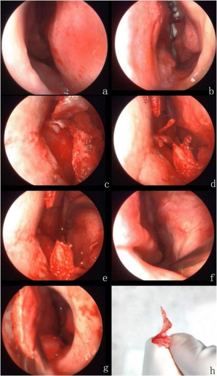
A multi-panel series of intraoperative endoscopic photographs and a clinical specimen photograph illustrating the steps of an endoscopic septoplasty for a deviated nasal septum. Panels (a) and (b) show the preoperative right and left nasal cavities, respectively, with (a) demonstrating a prominent septal deviation to the right accompanied by an obstructive horizontal spur. Panels (c) through (g) document the surgical progression: (c) demonstrates the Killian incision and elevation of the mucoperichondrium; (d) and (e) show the progressive separation and resection of the deviating septal bone and offending spur; (f) confirms the integrity of the contralateral right nasal mucosa after resection; and (g) displays the repositioned (reset) septal mucosa following correction. Panel (h) provides a gross photograph of the excised septal bone and spur specimen. The series serves as an educational guide for Otorhinolaryngology residents on the surgical management of nasal airway obstruction, highlighting anatomical landmarks such as the nasal septum, mucosal flaps, and the junction between septal cartilage and bone.
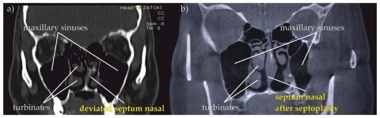
This Comparison Chart features two coronal Computed Tomography (CT) scans of the paranasal sinuses, providing a side-by-side visualization of a deviated nasal septum before and after surgical correction (septoplasty). Scan (a) demonstrates a prominent 'deviated septum nasal' with a distinct lateral curvature, causing asymmetry in the nasal airway and potentially obstructing the middle and inferior turbinates. Scan (b) illustrates the 'septum nasal after septoplasty,' showing a surgically straightened and midline-aligned septal wall, which restores bilateral patency to the nasal cavity. Both images highlight key anatomical landmarks including the maxillary sinuses (air-filled cavities lateral to the nasal walls) and the turbinates. These diagnostic images are essential for otorhinolaryngology education, specifically for demonstrating the clinical manifestation of upper respiratory tract (URT) obstruction and the efficacy of corrective surgical intervention in treating conditions like sleep apnea or chronic nasal congestion.
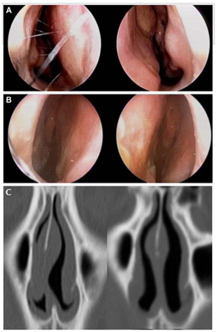
This composite medical image provides a preoperative and postoperative comparison of a nasal septoplasty procedure for correcting a deviated nasal septum. Row A shows preoperative endoscopic views, demonstrating a significant septal deviation and pronounced asymmetry between the bilateral nasal passages, with one side appearing severely narrowed. Visible suture-like structures in the left image likely represent intraoperative markers or temporary stabilizing materials. Row B displays the postoperative endoscopic results, showing a straighter septum and improved symmetry in the nasal airway. Row C presents coronal CT images of the nasal cavity. The preoperative CT scan (left) reveals a marked deviation of the perpendicular plate of the ethmoid (PPE) and upper cartilaginous septum, which encroaches on the internal nasal valve (INV) space. The postoperative CT scan (right) demonstrates the surgical correction, with the nasal septum now centrally aligned, leading to a visible increase in the cross-sectional area and volume of the bilateral nasal passages. This sequence illustrates the anatomical restoration of nasal airflow and airway symmetry.
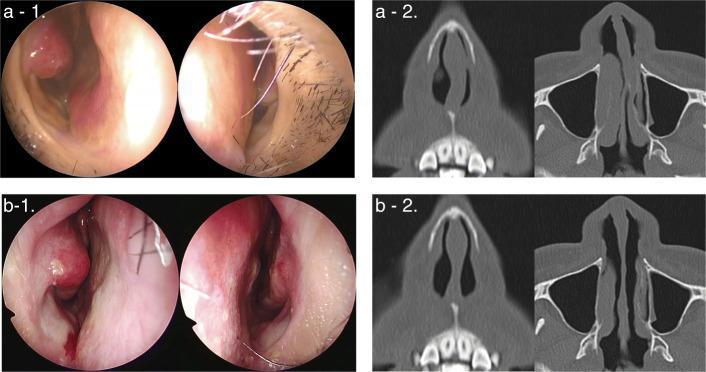
This composite medical image provides a comparative study of a patient before and three months after undergoing a modified cutting and suture technique (MCAST) to correct a deviated nasal septum. Top Row (a-1, a-2): Preoperative findings. Panel a-1 shows clinical endoscopic photographs where a significant caudal septal deviation to the left side severely constricts the left nasal airway. Panel a-2 presents axial computed tomography (CT) scans confirming a sharp C-shaped septal curvature and substantial narrowing of the left nasal cavity compared to the patent right side. Bottom Row (b-1, b-2): Postoperative findings. Panel b-1 displays endoscopic images following surgical correction, showing a straightened septum and significantly improved bilateral patency; visible sutures and mild mucosal erythema are noted. Panel b-2 contains postoperative axial CT scans demonstrating a midline septal position, symmetric enlargement of the nasal passages, and successful correction of the previous caudal deviation. This material serves as an educational resource for otolaryngology and plastic surgery, illustrating the efficacy of septoplasty techniques in restoring nasal symmetry and airflow.
Septoplasty - Surgical Steps
Pre-operative Setup
- Anesthesia: General or local with sedation. Topical vasoconstrictor is applied first - 4% cocaine solution or oxymetazoline on cotton pledgets. This is followed by infiltration with lidocaine + epinephrine along the septum, columella, and anterior nasal floor for vasoconstriction and hydrodissection.
- Pre-treatment with IV fentanyl and midazolam by anaesthesia team counters the cardiovascular effects of cocaine/vasoconstrictors.
- Patient positioned supine with head-ring support, slight head elevation (~15-20°) to reduce venous congestion.
Step 1 - Incision
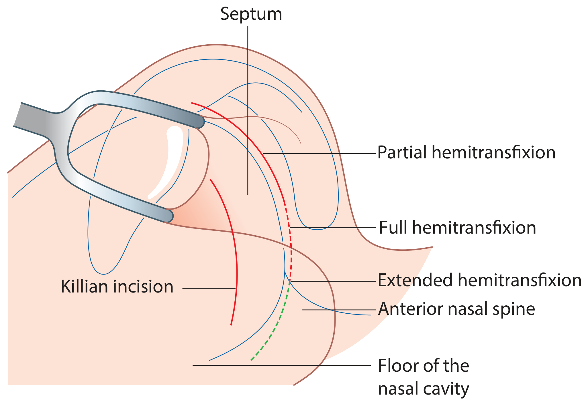
| Incision | Use |
|---|---|
| Killian incision | 1-1.5 cm from caudal septum; provides access to body of septum, preserves columellar support; used when caudal L-strut is not involved |
| Partial hemitransfixion | At the caudal edge; used when posterior parts are not needed |
| Full hemitransfixion | Placed at caudal edge of septum; gives complete access to the caudal arm of the L-strut |
| Extended hemitransfixion | Extends to floor of nasal cavity when floor access is needed |
| Bilateral transfixion | For open access (external approach) or complex S-shaped deformities |
Step 2 - Mucoperichondrial Flap Elevation
- A Cottle elevator (or similar) is inserted into the subperichondrial plane - this is the critical avascular plane that, when correctly entered, gives minimal bleeding and protects the mucosa.
- Caudally, the perichondrium adheres tightly to cartilage - scoring the area helps identify the correct dissection plane.
- Usually only the concave side flap is raised - this preserves the opposite mucosa and reduces perforation risk.
- Both flaps are raised when: surgeon wants to place through-and-through sutures, for S-shaped deformities (scoring needed on both sides), or for bilateral access.
- Extent of elevation depends on the surgical plan - can be raised as far cephalically as needed to access the perpendicular plate of ethmoid for graft harvesting.
- Back-to-front (cephalad to caudal): Flap raised anteriorly at caudal septum then directed cephalically, then sweeping back caudally posteriorly. Maintains correct subperiosteal plane.
- Front-to-back (tunnel technique): Anterior tunnel raised over quadrilateral cartilage, then a separate posterior tunnel raised behind the spur. The mucosa over the spur tip is released last by sharp dissection, avoiding tearing under tension.
Step 3 - Addressing the Deformity
A. Cutting Techniques
- Scoring: Parallel incisions/grooves made on the concave side of deviated cartilage - releases intrinsic tension and allows the septum to spring to the midline. The resulting scar tissue locks it in position. Unreliable alone; best combined with a splinting graft.
- Swinging door technique (caudal dislocation): A strip of excess cartilage is excised from the caudal L-strut, allowing the septum to swing back to midline like a door. The septum is then sutured back to the anterior nasal spine (ANS) with 4-0 PDS through a drill hole in the ANS.
- Osseocartilaginous junction disarticulation: When the dorsal L-strut deviation is due to deviation at its bony attachment to the perpendicular plate of ethmoid, the junction is fully disarticulated (sometimes including 1-1.5 mm of the last attachment to upper lateral cartilages).
- Bony septum: The perpendicular plate of ethmoid and vomer are addressed with a Jansen-Middleton forceps, Kerrison rongeur, or powered microdebrider - they are removed or reshaped. Spurs at the vomerine groove are similarly removed.
B. Grafting Techniques
- A deviated segment can be kept straight by splinting it against a batten graft.
- Graft materials: harvested septal cartilage, perpendicular plate of ethmoid (stronger and thinner than cartilage - thinned with a 5 mm diamond burr, then perforated with a Fissure burr before placement), or vomer.
- The graft is sutured on the concave side a few mm from the septal edge (to avoid palpability and avoid blocking future spreader graft placement).
C. Suturing Techniques
- Through-and-through mattress sutures placed across the septum to reshape and hold cartilage in the midline.
- Used for caudal fixation to ANS (4-0 PDS), to fix relocated segments, and to quilt the mucosal flaps at closure.
- Transcartilaginous fixation sutures can be used in the extracorporeal technique after reimplantation.
D. Relocating Techniques
- Extracorporeal septoplasty: The entire deviated cartilaginous septum is removed from the nose, reshaped/carved on the back table, and reimplanted. Used for the most complex deformities. Requires meticulous reimplantation with fixation sutures.
- Allows three-dimensional correction not possible with in situ techniques.
Step 4 - Closure
- The mucoperichondrial flap(s) are repositioned over the corrected septum.
- Bilateral quilting sutures (continuous locking or mattress, 3-0 or 4-0 absorbable - e.g. chromic gut or Vicryl) are placed through both sides of the septum to re-appose the mucosal layers, obliterate dead space, and prevent haematoma.
- Alternatively, nasal splints/packs can be used to hold flaps in position.
- The original mucosal incision is closed with interrupted or continuous absorbable sutures (4-0 Vicryl or chromic).
Step 5 - Packing / Splinting
- Internal nasal splints (silicone Doyle splints) and/or nasal packing (BIPP, merocel, or dissolvable haemostatic agents) are placed to:
- Maintain septal position
- Control bleeding
- Prevent synechia formation
- Splints are usually removed at 1-2 weeks post-op.
Endoscopic Septoplasty (Variation)
Intraoperative Endoscopic Photographs
Summary Mnemonic - "I FRAME C"
| Step | Action |
|---|---|
| Incision | Killian / hemitransfixion |
| Flap elevation | Subperichondrial / subperiosteal |
| Remove / reshape | Bony septum (PPE, vomer, spur) with rongeur |
| Address cartilage | Score / cut / relocate quadrilateral cartilage |
| Midline | Fix to ANS if needed (swinging door, 4-0 PDS) |
| Extracorporeal | If complex - remove, reshape, reimplant |
| Close | Quilting sutures + pack/splint |
- Scott-Brown's Otorhinolaryngology Head & Neck Surgery, Vol 1 - Chapter 103 (Septoplasty), pp. 1186-1188
- Cummings Otolaryngology Head and Neck Surgery - Chapter 29 (Septoplasty Technique and Evolution), pp. 571-572