
Explain the motor action of trigeminal nerve with diagram
trigeminal nerve motor nucleus muscles of mastication diagram
Motor Action of the Trigeminal Nerve (CN V)
1. Motor Nucleus and Origin
- The trigeminal motor nucleus lies in the upper-to-mid pons, medial to the main sensory nucleus, near the floor of the fourth ventricle.
- It receives bilateral (predominantly crossed and uncrossed) corticobulbar input from the lower third of the precentral gyrus (motor cortex). These fibers travel through the corona radiata, internal capsule, and cerebral peduncle, decussating in the pons before reaching the nucleus.
- Because the cortical control is bilateral, a unilateral upper motor neuron lesion (e.g., one-sided stroke) usually causes no significant jaw weakness. Bilateral corticobulbar lesions, however, can produce a hyperactive jaw jerk reflex.
2. Course of the Motor Root (Portio Minor)
- Motor fibers exit the pons as a small motor root (portio minor), separate from and medial to the much larger sensory root (portio major).
- It runs forward, passes through the posterior and middle cranial fossa beneath the trigeminal (Gasserian) ganglion in Meckel's cave, without synapsing in the ganglion (motor fibers bypass the sensory ganglion).
- It exits the skull through the foramen ovale, joining the mandibular division (V3) almost immediately outside the skull.
- From there it travels with V3 to reach its target muscles.
3. Muscles Supplied (Branchial Motor / Special Visceral Efferent)
- Muscles of mastication: masseter, temporalis, medial pterygoid, lateral pterygoid (these elevate, retrude/protrude, and move the jaw side to side)
- Tensor tympani (dampens ossicular movement in the middle ear)
- Tensor veli palatini (tenses the soft palate)
- Mylohyoid
- Anterior belly of the digastric
4. Clinical Testing
- Ask the patient to clench the jaw while you palpate the masseter and temporalis for bulk and strength.
- Ask the patient to open the jaw and move it side to side against resistance (pterygoids); with a unilateral lower motor neuron lesion, the jaw deviates toward the weak side because the intact contralateral pterygoid muscles push the jaw over.
- The jaw jerk reflex (tap the chin with mouth slightly open) tests the motor nucleus and its afferent limb (via the mesencephalic nucleus, which carries proprioception from the muscles of mastication) - it is normally absent or minimal but becomes brisk with bilateral UMN lesions.
Diagram
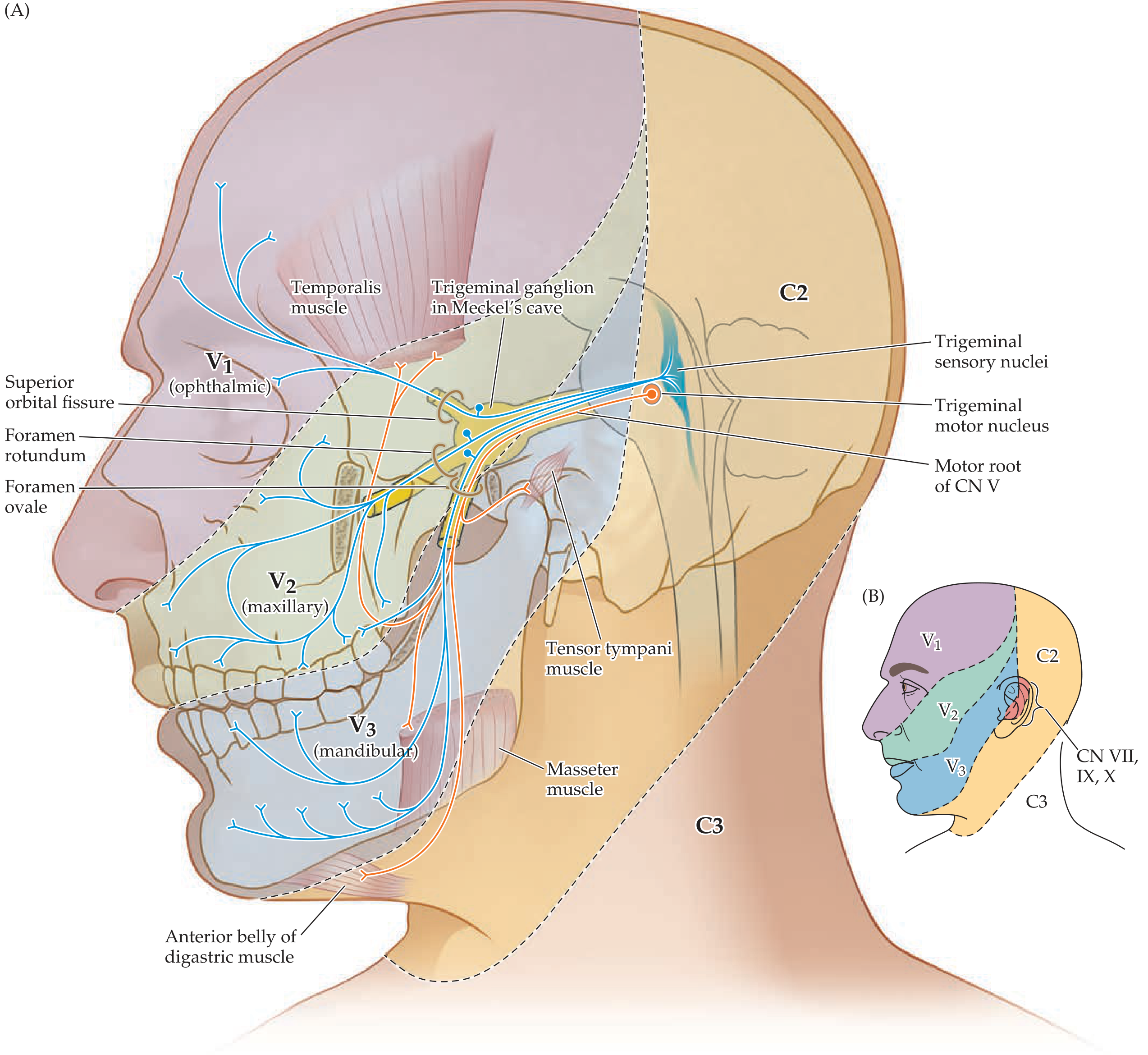
- Neuroanatomy through Clinical Cases, 3rd ed., Fig 12.7, p. 535-536
- Localization in Clinical Neurology, 8th ed., p. 739-740
- Gray's Anatomy for Students, p. 1037
Search for clinical images of trigeminal nerve lesions
trigeminal nerve lesion clinical presentation
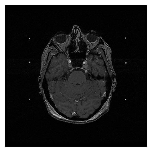
This diagnostic image is an axial T1-weighted post-gadolinium enhanced MRI of the head at the level of the skull base and brainstem. The image displays a well-circumscribed, hyperintense (brightly enhancing) mass located along the course of the left trigeminal nerve. The lesion involves the preganglionic segment and extends toward the cavernous sinus region. This enhancement pattern suggests high vascularity or a breach in the blood-nerve barrier, characteristic of a trigeminal schwannoma or similar neoplastic process. Surrounding anatomical landmarks are visible, including the pons, cerebellum, bilateral temporal lobes, orbits, and paranasal sinuses. The brain parenchyma shows normal signal intensity relative to the enhancing lesion. This image serves as a clinical example for neuro-oncology and radiology, illustrating the typical presentation of a cranial nerve sheath tumor and its anatomical relationship to the brainstem and middle cranial fossa.
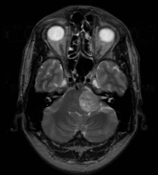
**Imaging Modality:** Magnetic Resonance Imaging (MRI), T2-weighted axial sequence.
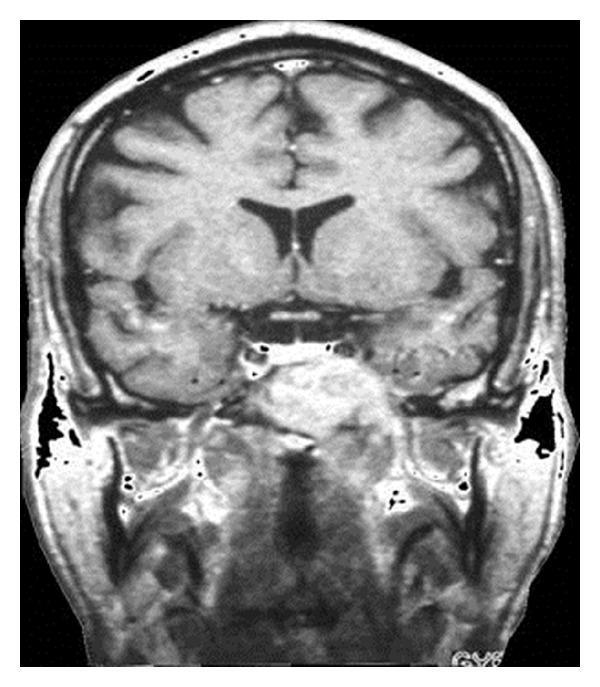
This diagnostic image is a coronal T1-weighted cranial MRI scan showing the brain and skull base. The image demonstrates a large, hyperintense, well-demarcated lesion localized in the left cranial base, measuring approximately 4 cm. The mass involves the region of the sphenoid sinus and extending toward the foramina lacerum and ovale, causing visible anatomical distortion and bone destruction compared to the contralateral side. Superiorly, the cerebral hemispheres, lateral ventricles, and interhemispheric fissure appear intact but the lesion lies in close proximity to the temporal lobe and pituitary region. The clinical significance of this finding represents Langerhans cell histiocytosis (eosinophilic granuloma) of the cranial base, which in this case presented with fifth cranial nerve (trigeminal nerve) hypesthesia. The image serves as a baseline for radiotherapy planning and illustrates the typical aggressive local presentation of histiocytic lesions in adult patients within the neurosurgical and oncological specialty.
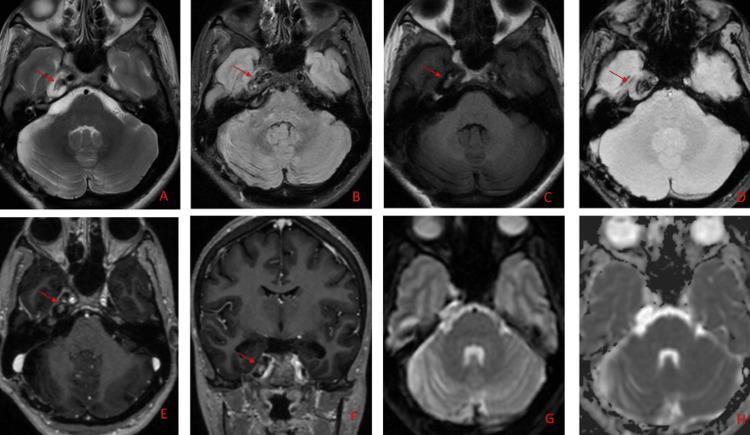
A multi-paneled figure (A-H) displaying post-operative MRI sequences of the brain in axial and coronal planes, demonstrating a residual extra-axial lesion in the right Meckel’s cave. Panel A (T2), B (FLAIR), and D (Gradient/GRE) show the lesion as hypointense relative to gray matter. Panel C (T1-weighted) reveals intrinsic hyperintensity, suggesting high protein, melanin, or paramagnetic content. Axial (E) and coronal (F) post-gadolinium sequences demonstrate minimal contrast enhancement. Diffusion-weighted imaging (G) and corresponding ADC map (H) show no restricted diffusion. Anatomically, the lesion (red arrows) is localized to the right trigeminal cistern, abutting the cavernous segment of the internal carotid artery and the vestibulocochlear-facial (VII/VIII) nerve complex. The presentation is consistent with heavily pigmented neurovascular pathologies, such as melanotic neoplasms. This imaging serves as a clinical example for neuro-oncology and neuroradiology, emphasizing the multi-sequence signal analysis of posterior fossa and skull base lesions.
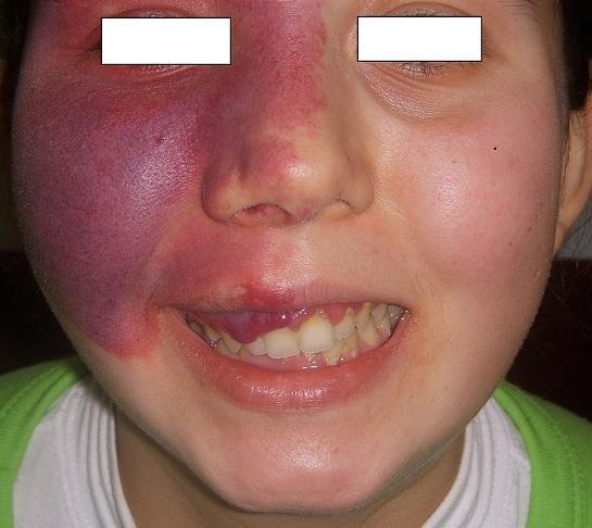
This clinical photograph displays a large, unilateral capillary malformation, commonly known as a port-wine stain (nevus flammeus), on the right side of a patient's face. The lesion is characterized by a deep purple to violaceous hue, with areas of lighter red towards the midline. Its distribution follows the V1 (ophthalmic) and V2 (maxillary) branches of the trigeminal nerve, covering the forehead, periorbital region, right nasal ala, and cheek. The lesion appears congestive and lacks sharply defined borders, blending into the surrounding skin. Additionally, the image shows intraoral manifestations: significant gingival hyperplasia is visible on the right side of the maxillary arch, appearing as bright red, bulbous overgrowths of the gingival tissue. The combination of a facial port-wine stain in the V1/V2 distribution and ipsilateral gingival vascular involvement is a classic clinical presentation of Sturge-Weber syndrome (encephalotrigeminal angiomatosis). This visual is essential for identifying vascular phakomatoses and understanding their dermatological and oral surgical implications.
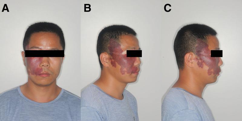
This clinical photograph series displays three views (frontal, 45° lateral, and 90° lateral) of a male patient presenting with a prominent facial capillary malformation, commonly referred to as a port-wine stain. The lesion is strictly unilateral, affecting the right side of the face. It involves the distribution of the first (ophthalmic) and second (maxillary) divisions of the trigeminal nerve, extending from the forehead and periorbital region across the cheek and nasal sidewall to the upper cutaneous lip and preauricular area. The lesion exhibits a characteristic dark red to 'grape-colored' erythema with irregular but well-defined borders. Visually, the affected skin shows signs of hypertrophy and textural changes, including areas of thickening and a slightly cobble-stoned or flaky surface. This presentation is a hallmark cutaneous sign often associated with neurocutaneous syndromes such as Sturge-Weber syndrome. The images demonstrate the significant facial asymmetry caused by the vascular malformation and are used for clinical diagnosis and documentation of disease extent.
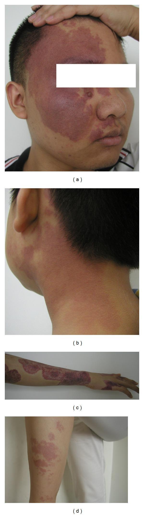
This set of clinical photographs illustrates the varied presentation of Port-Wine Stains (PWS), a congenital vascular malformation of the dermal capillaries. Image (a) depicts a large, confluent, reddish-purple macule on the face, following a distribution that involves the ophthalmic (V1) and maxillary (V2) branches of the trigeminal nerve. Small nodular elevations within the patch suggest early soft tissue or vascular hypertrophy. Image (b) shows smaller, lighter-colored erythematous patches on the neck with less defined borders. Images (c) and (d) demonstrate PWS on the extremities; the arm and hand show dark, irregularly shaped, and partially interconnected plaques, while the leg displays more discrete, scattered patches. The lesions are non-blanching and represent classic dermatological manifestations of capillary malformations, which may be isolated or associated with systemic syndromes like Sturge-Weber. These images serve as educational references for identifying lesion morphology, color intensity, and anatomical distribution across different body regions.
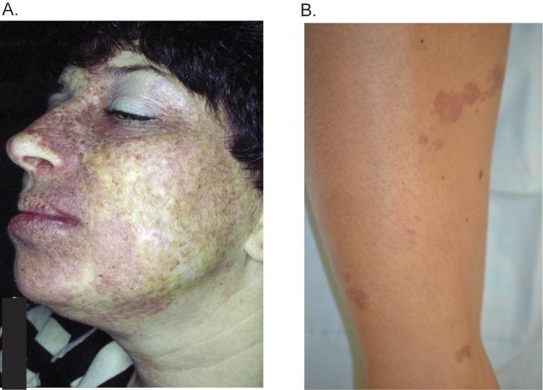
This clinical photograph comparison displays two presentations of port-wine stains (PWS), or nevus flammeus, which are congenital capillary malformations. Image A shows a diffuse facial PWS on a female patient, primarily involving the left maxillary (V2) and mandibular (V3) dermatomes of the trigeminal nerve. The lesion presents as a confluent, mottled area of pink, red, and dusky purple discoloration extending across the cheek, chin, upper lip, and nose. Image B shows a more localized PWS on the shin of a different individual. This presentation consists of discrete, irregularly shaped, light-red to purple-tan patches arranged in a linear distribution against unaffected skin. These images illustrate the phenotypic variability of PWS in different anatomical locations and their potential association with somatic mutations, such as GNAQ variants. The educational focus is on clinical morphology, dermatomal distribution, and the distinction between confluent facial lesions and localized extremity lesions in pediatric and adult dermatology.
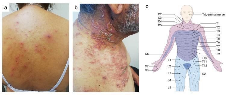
A three-panel figure demonstrating the clinical presentation and anatomical context of varicella-zoster virus (VZV) infections. Panel (a) is a clinical photograph of a patient's back showing a generalized rash characterized by polymorphous lesions, including erythematous papules, vesicles on an erythematous base (the 'dewdrop on a rose petal' appearance), and crusts in various stages of evolution, typical of primary varicella (chickenpox). Panel (b) shows a clinical photograph of a localized, painful rash on the neck and upper chest of another patient, featuring clustered vesicles and confluent crusting on an erythematous background, following a dermatomal distribution. Panel (c) is an anatomical diagram of the human torso and head mapping the cervical (C2-C8), thoracic (T1-T12), lumbar (L1-L5), and sacral (S2) dermatomes, as well as the trigeminal nerve distribution. This figure illustrates the clinical distinction between primary varicella and herpes zoster (shingles) based on lesion morphology and dermatomal versus generalized distribution.
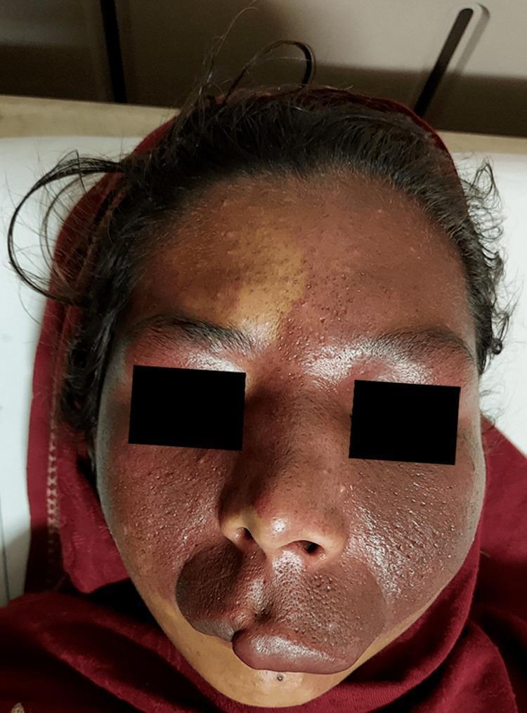
Clinical photograph of a patient's face displaying a large port-wine stain (PWS), a congenital capillary malformation. The lesion exhibits characteristic deep red to purple discoloration with a predominant distribution on the left side of the face. Anatomically, the stain involves the dermatomes associated with the ophthalmic (V1) and maxillary (V2) divisions of the trigeminal nerve on the left, while also crossing the midline to involve the maxillary division on the right side. The affected areas include the forehead, periorbital regions, bilateral cheeks, nose, upper lip, and chin. Visually, the PWS demonstrates varying color intensity and textural irregularities, including subtle surface nodularity and enlarged pores, which are typical of aging vascular lesions. Notably, there is a distinct midline demarcation on the forehead. This clinical presentation is highly suggestive of Sturge-Weber syndrome, especially when associated with neurological or ocular findings. This image is an educational example of segmental vascular malformations and their dermatomal correlations.
Trigeminal Neuralgia (most common CN V disorder)
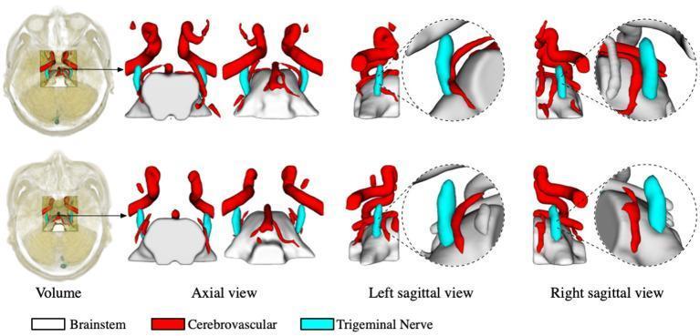
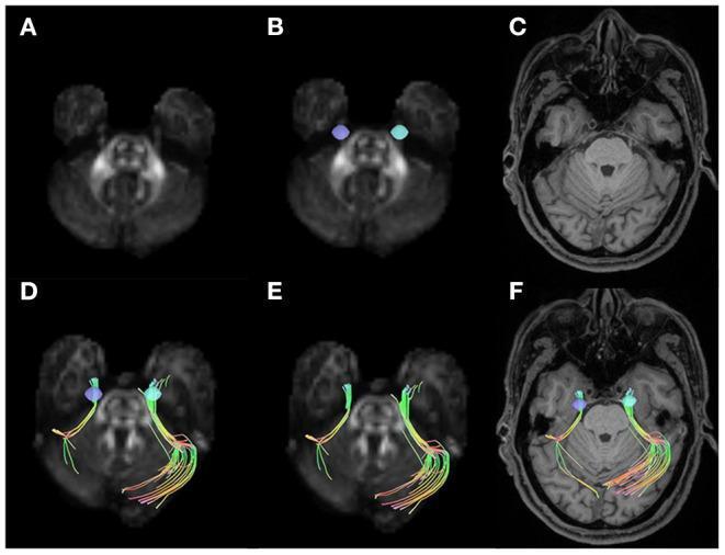
Mass Lesions Compressing the Nerve
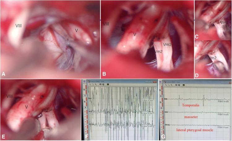
Motor Root Disorders (Masticatory Muscles)
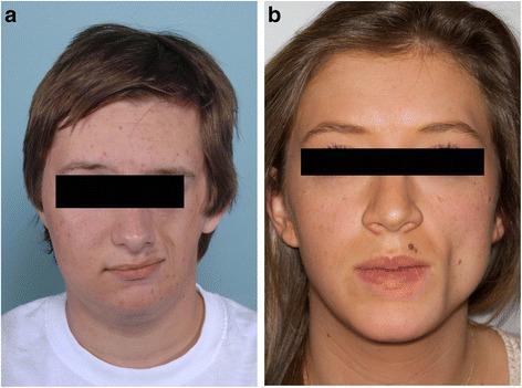
Sensory Dermatome-Related Skin Lesions (V1/V2/V3 distribution)
Notes on interpretation
- Sensory lesions (numbness, neuralgia) dominate the clinical picture because the sensory root is much larger than the motor root - this is reflected in most available clinical imagery being MRI/tractography of the sensory/trigeminal ganglion region rather than photographs of motor deficits.
- Pure motor deficits (masseter/temporalis wasting, jaw deviation) are uncommon in isolation since most lesions affecting CN V (schwannoma, neuralgia, zoster) preferentially involve the sensory root or ganglion; isolated motor involvement points more toward a pontine lesion at the motor nucleus or a mandibular nerve (V3) lesion distal to where the motor root joins it.
- Images sourced from PMC Clinical VQA / MedPix collections in the medical image library.
Conus medullar8s and cauda equina explain with lesions extends and jerks to dfferentiate and lesions
conus medullaris cauda equina syndrome anatomy diagram
Conus Medullaris Syndrome vs Cauda Equina Syndrome
Anatomy
- The spinal cord narrows and ends as the conus medullaris, typically at the L1 vertebral level (range: T12 disc space to mid-L2), varying between individuals.
- Below this, the neural canal contains only the lumbosacral nerve roots (L2-S5) descending to their exit foramina, bathed in CSF and surrounded by the filum terminale - this bundle of roots is the cauda equina ("horse's tail").
- Because the conus is the transition point between CNS (spinal cord = upper motor neuron territory) and PNS (nerve roots = lower motor neuron territory), lesions here often produce a mixed UMN + LMN picture, whereas lesions purely below it (cauda equina) give a pure LMN picture.
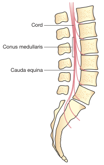
Key Differentiating Features
| Feature | Conus Medullaris Syndrome | Cauda Equina Syndrome |
|---|---|---|
| Level of injury | Vertebral injury T12-L2 (conus itself) | Vertebral injury distal to L2 (roots below conus) |
| Motor neuron involvement | Mixed UMN + LMN | Pure LMN |
| Motor weakness pattern | Symmetrical, often less severe | Asymmetrical, often more severe |
| Onset | Usually sudden/rapid | Can be sudden or gradual (e.g. slow disc herniation) |
| Reflexes | Knee jerk may be preserved (L2-L4 arc intact), ankle jerk absent; Babinski/UMN signs may be present because corticospinal tract fibers are still in the cord | All deep tendon reflexes (knee and ankle) absent/reduced; no Babinski sign (pure LMN, no UMN component) |
| Bulbocavernosus / anal reflex | Absent - and importantly permanently absent (unlike transient loss in spinal shock, which returns within ~48 hours) | Absent |
| Sensory loss | Symmetrical saddle anesthesia (perianal, perineal), often sharply demarcated | Asymmetrical, patchy, dermatomal (L2-S5 distribution), saddle anesthesia if bilateral roots involved |
| Bladder/bowel/sexual dysfunction | Present - flaccid, areflexic (low-pressure, high-capacity) bladder; loss of reflexogenic erection | Present, similarly areflexic, but may be less symmetric |
| Pain | Less prominent | More prominent - radicular, often severe (root compression) |
| Prognosis | Guarded; roots have limited regenerative capacity here | Better - peripheral nerve roots regenerate more readily than cord tissue, though sacral roots are delicate and can be permanently damaged |
The "Jerks" (Reflex) Differentiator - the Clinical Shortcut
- Knee jerk (L3-L4):
- Conus lesion: may be preserved (reflex arc is above/at the conus, still spinal-cord mediated) or even brisk if UMN fibers are affected.
- Cauda equina lesion: absent if L3-L4 roots are involved (pure LMN disruption of the arc).
- Ankle jerk (S1-S2): Absent in both syndromes, since S1-S2 fibers pass through/near the conus and cauda equina.
- Babinski sign (extensor plantar response):
- Present in conus medullaris syndrome if corticospinal (UMN) fibers within the cord are damaged.
- Never present in pure cauda equina syndrome (no UMN component) - a flexor or absent plantar response is expected.
- Bulbocavernosus reflex and anal wink: Absent in both, and in true conus/cauda injury this loss is permanent - this is what separates it from spinal shock, where these reflexes typically return within 48 hours.
Common Causes (both)
- Burst fractures / fracture-dislocations of the thoracolumbar spine (most common traumatic cause)
- Large central disc herniation (classically L4-L5 or L5-S1 causing cauda equina syndrome)
- Spinal tumors (e.g., myxopapillary ependymoma - characteristically arises right at the conus/filum terminale)
- Spinal stenosis, epidural abscess/hematoma, spondylolisthesis
- Bradley and Daroff's Neurology in Clinical Practice, Table 63.2, p. 1307
- Bradley and Daroff's Neurology in Clinical Practice, "Conus Medullaris and Cauda Equina," p. 484-485
- Neuroanatomy through Clinical Cases, 3rd ed. (bulbocavernosus/cremasteric reflex discussion)
Extent of them
Extent of the Spinal Cord, Conus Medullaris, and Cauda Equina
- Extends from the foramen magnum down to the conus medullaris, ending at approximately the T12-L1 disc space to mid-L2 vertebral body (most commonly around the L1 level in adults).
- This is shorter than the vertebral column because the vertebral column grows faster than the cord during development, causing the cord's relative "ascent."
- The tapering, cone-shaped terminal end of the spinal cord itself.
- Located at the T12/L1 to L2 vertebral level (varies by individual; most texts cite the inferior border of L1 as the average adult level).
- Contains the sacral and coccygeal spinal cord segments compressed into this short segment - hence why a small lesion here affects many segmental functions (bladder, bowel, sexual, saddle sensation) at once.
- A thin fibrous (pial) extension continuing down from the tip of the conus medullaris.
- Runs from the conus (~L1-L2) down through the sacral canal and attaches to the posterior surface of the coccyx.
- Below S2, it is covered by the dura and often called the coccygeal ligament.
- Formed by the bundle of lumbar (L1-L5), sacral (S1-S5), and coccygeal nerve roots that must travel down inside the dural sac to reach their respective intervertebral/sacral foramina, since the cord itself has already ended above them.
- Extends from the conus medullaris (~L1-L2) down to approximately S2, where the dural sac (thecal sac) narrows and ends - though individual nerve roots continue further to exit at their own foramina (as low as the sacral hiatus/coccyx for the coccygeal nerve).
- So the cauda equina occupies roughly L2 through the sacrum, i.e., the lower third to half of the vertebral canal.
Quick Reference Table
| Structure | Starts | Ends |
|---|---|---|
| Spinal cord | Foramen magnum | ~T12/L1-L2 (conus medullaris) |
| Conus medullaris | ~T12-L1 disc space | ~L2 vertebral body |
| Filum terminale | Tip of conus (~L1-L2) | Coccyx |
| Dural (thecal) sac | Foramen magnum | ~S2 |
| Cauda equina (nerve roots) | ~L1-L2 (below conus) | Sacrum/coccyx (each root exits at its own foramen) |
Clinical relevance of this extent
-
Because the cord ends around L1-L2, lumbar puncture is safely performed at L3-L4 or L4-L5 - below the conus but within the cauda equina, where a needle simply displaces the mobile nerve roots rather than injuring cord tissue.
-
The variability in conus termination level (as low as L2-L3 in some normal individuals, or lower in tethered cord syndrome) is why imaging (MRI) is used to confirm the level before certain spinal procedures.
-
A vertebral injury/fracture at T12-L2 is likely to damage the conus (mixed UMN/LMN picture), while an injury distal to L2 damages only nerve roots of the cauda equina (pure LMN picture) - this is the anatomical basis for the differentiating features discussed previously.
-
Neuroanatomy through Clinical Cases, 3rd ed., p. (Segmental Organization of the Nervous System)
-
Miller's Review of Orthopaedics, 9th ed., Ch. 7 (Nerves)
-
Sabiston Textbook of Surgery, "Spinal Anatomy"
-
Campbell-Walsh-Wein Urology, 3-Vol Set (sacral spinal cord/conus levels)
Why cauda is always LMN
Why Cauda Equina Lesions Are Always Pure LMN
1. The corticospinal tract terminates inside the spinal cord
2. Below the conus, there is no CNS tissue left
- Axons of anterior horn cells (motor roots) that already left the cord higher up and are simply traveling down inside the dural sac to reach their exit foramen
- Axons of dorsal root ganglion cells (sensory roots)
3. Contrast with the conus medullaris
- Flaccid weakness (not spastic)
- Hyporeflexia/areflexia (not hyperreflexia)
- No Babinski sign
- Muscle atrophy over time (denervation)
- Asymmetric, root-pattern distribution (since individual roots are affected, not the whole cord cross-section)
Simple analogy
Myelopathy
Myelopathy: Overview
- Clinical presentation & localization - UMN signs, sensory level pitfalls, dissociated sensory loss, autonomic/bladder involvement
- Compressive causes - degenerative disease (spondylosis/spinal stenosis), disc herniation, spondylolisthesis, tumor, epidural abscess, epidural hematoma
- Non-compressive / vascular causes - spinal cord infarction, dural AV fistula, decompression sickness, radiation myelopathy
- Inflammatory / demyelinating causes - transverse myelitis, multiple sclerosis, neuromyelitis optica, sarcoidosis, lupus myelopathy, Behcet's
- Infectious causes - HIV-associated myelopathy, HTLV-1 (tropical spastic paraparesis), schistosomiasis, viral myelitis
- Metabolic / toxic causes - subacute combined degeneration (B12 deficiency), copper deficiency (hypocupric myelopathy), zinc toxicity, hepatic myelopathy
- Traumatic causes - MVAs, falls, gunshot wounds, sports injuries, electrical injury
- Cervical spondylotic myelopathy (CSM) - specific deep dive, since it's the most common cause of spastic paraparesis in patients over 55
- Diagnostic workup - MRI first-line, then LP/CSF studies, B12/copper levels, further testing if imaging is negative
- Hereditary myelopathies - hereditary spastic paraparesis and related genetic disorders
Which aspect of myelopathy do you want to go deeper into?