
Upper limb mbbs detailed
Reading File
The upper limb is a major anatomy topic in MBBS, covering bones, joints, muscles, nerves, vessels, and clinical correlations. Let me pull comprehensive content from the medical library.
Searching sources...
Finding Sources
Finding Sources
Finding Sources
Finding Sources
Finding Sources
Reading File
Reading File
Reading File
Reading File
Reading File
Reading File
Reading File
Reading File
Reading File
Finding Sources
Finding Sources
Loading Image
Loading Image
Loading Image
Loading Image
Now I have comprehensive material. Let me compile the full detailed notes.
Upper Limb - Detailed MBBS Notes
Source: Gray's Anatomy for Students (Gray's AFS), Chapter 7
1. OVERVIEW
The upper limb is designed for two main functions:
- Positioning the hand in space (shoulder, arm, and forearm)
- Manipulating objects - both as a mechanical tool (grip/grasp) and a sensory tool (fine touch, proprioception)
Component Parts
- Bones and joints: Pectoral girdle (clavicle + scapula) → humerus → radius + ulna → carpals → metacarpals → phalanges
- Regions: Shoulder, axilla, arm, cubital fossa, forearm, wrist, hand
- Innervation: Via cervical (C5-C8) and upper thoracic (T1) nerves through the brachial plexus
2. BONES OF THE UPPER LIMB
Pectoral Girdle
| Bone | Key Features |
|---|---|
| Clavicle | S-shaped; medial 2/3 convex forward, lateral 1/3 concave. Connects sternum to scapula. Most commonly fractured bone |
| Scapula | Flat triangular bone; has spine, acromion, coracoid process, glenoid cavity. Supraspinous + infraspinous fossae for rotator cuff muscles |
Arm
- Humerus: Head articulates with glenoid (glenohumeral joint). Greater + lesser tubercles for rotator cuff insertions. Spiral groove (radial nerve). Medial + lateral epicondyles. Capitulum (for radius), trochlea (for ulna).
Forearm
- Radius: Head at proximal end; styloid process distally. Biceps inserts on radial tuberosity.
- Ulna: Olecranon proximally (triceps insertion). Coronoid process. Styloid process distally.
Wrist & Hand
- Carpals (8): Two rows - proximal (Scaphoid, Lunate, Triquetrum, Pisiform), distal (Trapezium, Trapezoid, Capitate, Hamate). Mnemonic: "Some Lovers Try Positions That They Can't Handle"
- Metacarpals (5) + Phalanges (14: 2 in thumb, 3 in fingers)
3. JOINTS
Glenohumeral (Shoulder) Joint
- Type: Ball-and-socket (multiaxial synovial)
- Articular surfaces: Head of humerus + glenoid cavity of scapula (deepened by fibrocartilaginous glenoid labrum)
- Stabilizers:
- Static: Joint capsule, glenohumeral ligaments (superior, middle, inferior), coracohumeral ligament, glenoid labrum
- Dynamic: Rotator cuff muscles (SITS - Supraspinatus, Infraspinatus, Teres minor, Subscapularis)
- Movements: Flexion/Extension, Abduction/Adduction, Medial/Lateral rotation, Circumduction
- Dislocation: Anteroinferior most common (arm forced into abduction + lateral rotation). Bankart lesion (avulsion of anteroinferior glenoid labrum) + Hill-Sachs lesion (impression fracture of humeral head) in recurrent cases
Elbow Joint
- Type: Hinge + pivot synovial joint (one capsule, 3 articulations)
- Humeroulnar: hinge (flexion/extension)
- Humeroradial: hinge + pivot
- Proximal radioulnar: pivot (pronation/supination)
- Capsule: Reinforced by ulnar (medial) collateral ligament and radial (lateral) collateral ligament; the anular ligament of radius secures the radial head
- Synovial membrane: Lines coronoid fossa, olecranon fossa, radial fossa with fat pads
- Movements: Flexion 0-145°; Supination/Pronation at radioulnar joints
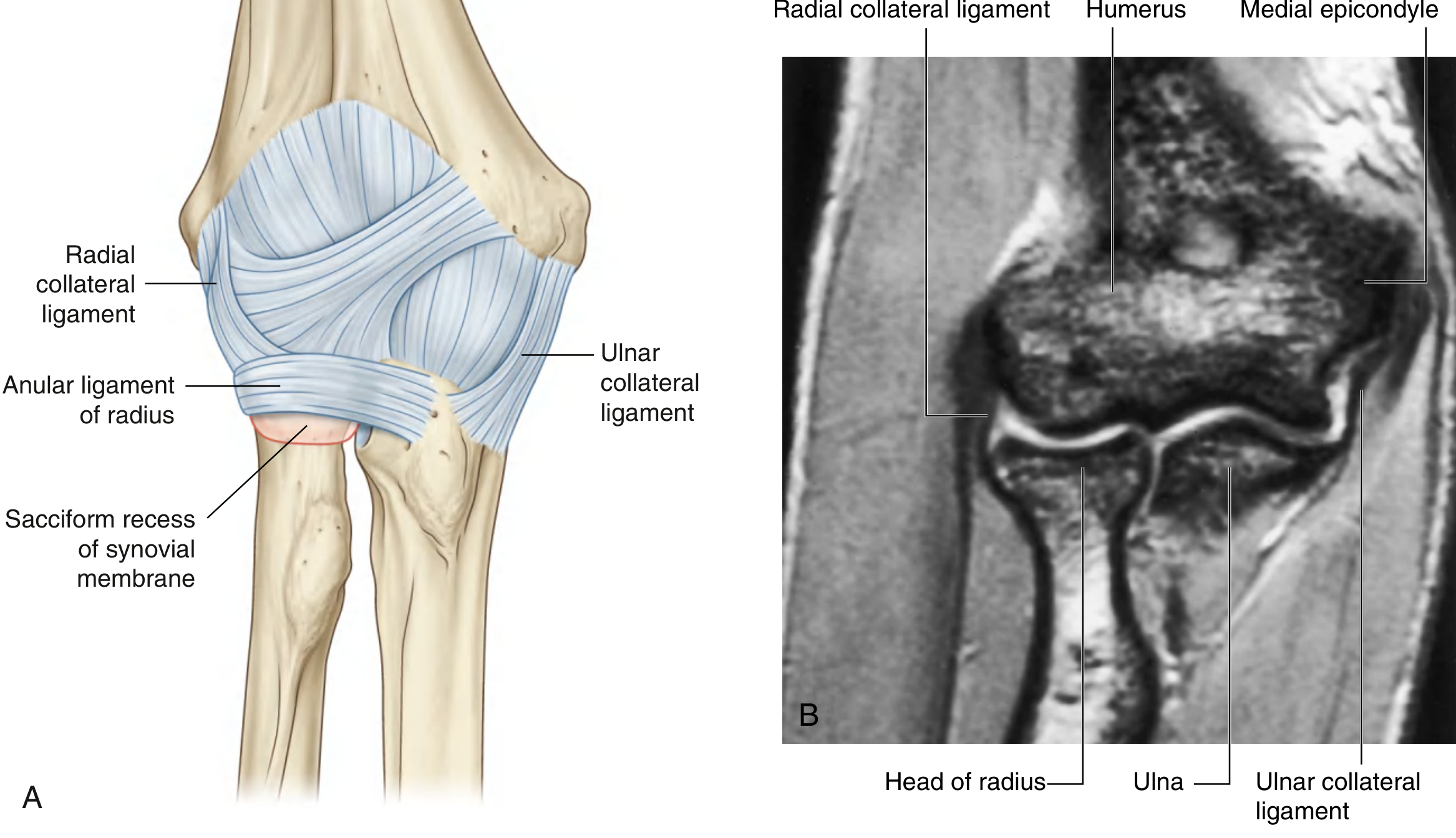
Elbow joint: (A) Capsule and ligaments; (B) MRI coronal plane - Gray's AFS
Wrist (Radiocarpal) Joint
- Type: Ellipsoid (condyloid) synovial
- Between: Distal end of radius + articular disc of DRUJ (above) and proximal carpal row - scaphoid, lunate, triquetrum (below)
- Movements: Flexion, extension, abduction (radial deviation), adduction (ulnar deviation), circumduction
4. THE AXILLA
The axilla is the gateway to the upper limb - an irregularly shaped pyramidal space with 4 walls, an inlet, and a floor.
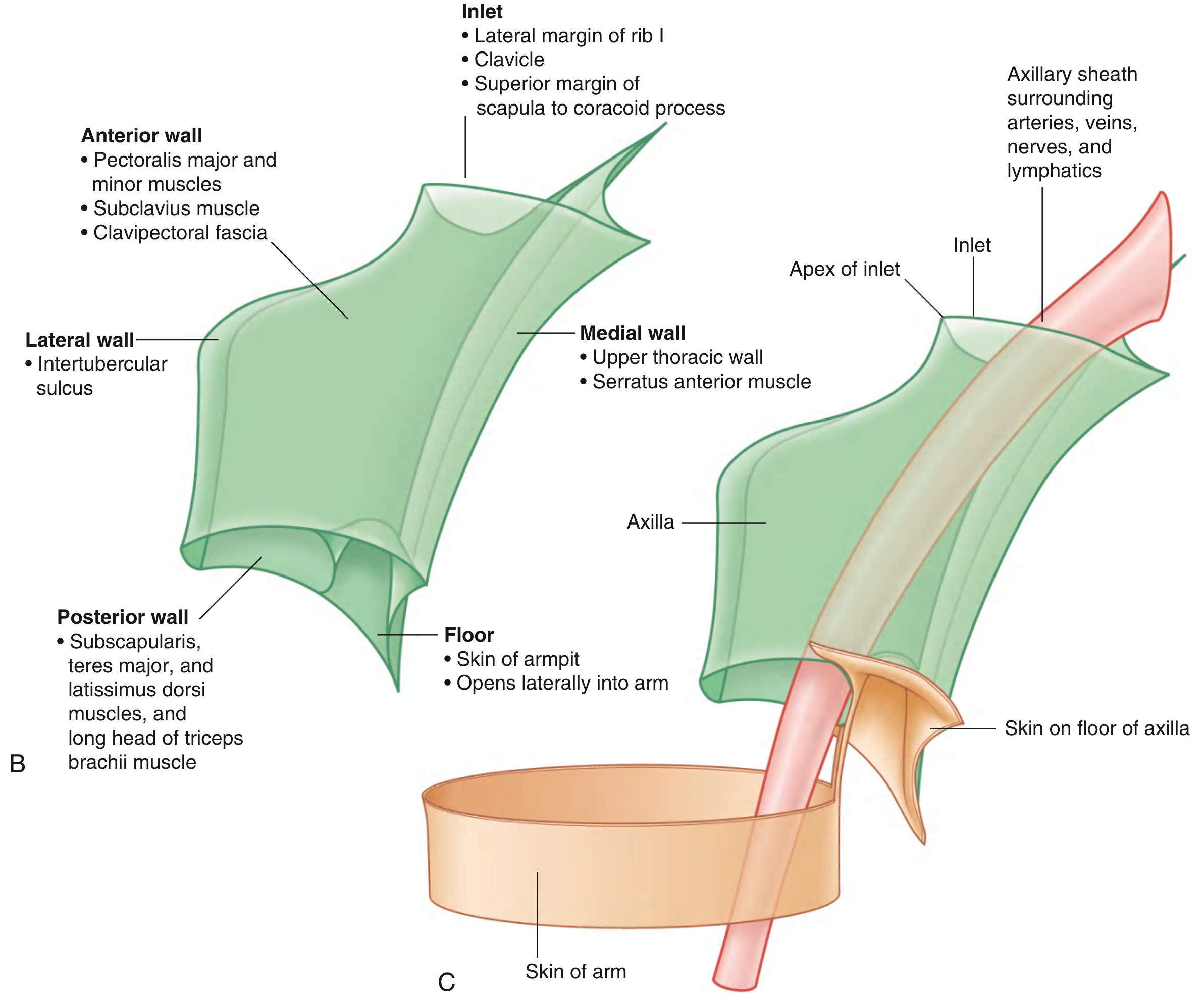
Axilla walls and continuity with the arm - Gray's AFS
| Wall | Structures |
|---|---|
| Anterior | Pectoralis major + minor, subclavius, clavipectoral fascia |
| Posterior | Subscapularis, teres major, latissimus dorsi, long head of triceps |
| Medial | Upper thoracic wall + serratus anterior |
| Lateral | Intertubercular (bicipital) groove of humerus |
| Inlet (Apex) | Clavicle + rib I + superior border of scapula to coracoid |
| Floor | Skin of armpit |
Openings in the Posterior Wall
| Space | Boundaries | Contents |
|---|---|---|
| Triangular space | Teres major (below), teres minor (above), long head of triceps (lateral) | Circumflex scapular artery |
| Quadrangular space | Teres major (below), teres minor (above), long head of triceps (medial), surgical neck of humerus (lateral) | Axillary nerve + posterior circumflex humeral artery |
| Triangular interval | Below teres major, lateral head of triceps (lateral), long head of triceps (medial) | Radial nerve + profunda brachii artery |
5. BRACHIAL PLEXUS
The single most important topic in upper limb anatomy for MBBS.

Brachial plexus: roots to cords in the neck and axilla - Gray's AFS

Complete brachial plexus schematic showing all terminal branches - Gray's AFS
Formation: Roots → Trunks → Divisions → Cords → Branches
Mnemonic: "Real Teenagers Drink Cold Beer"
| Level | Details |
|---|---|
| Roots | Anterior rami of C5, C6, C7, C8, T1 (between anterior + middle scalene muscles) |
| Superior trunk | C5 + C6 |
| Middle trunk | C7 alone |
| Inferior trunk | C8 + T1 |
| Anterior divisions → lateral + medial cords (supply anterior/flexor compartments) | |
| Posterior divisions → posterior cord (supply posterior/extensor compartments) | |
| Lateral cord | Anterior divisions of superior + middle trunk (C5-C7) |
| Medial cord | Anterior division of inferior trunk (C8, T1) |
| Posterior cord | All 3 posterior divisions (C5-T1) |
Branches
From Roots:
- Dorsal scapular nerve (C5) - rhomboids + levator scapulae
- Long thoracic nerve (C5, C6, C7) - serratus anterior (winged scapula if injured)
- Contribution to phrenic nerve (C5)
From Superior Trunk:
- Suprascapular nerve (C5, C6) - supraspinatus + infraspinatus
- Nerve to subclavius (C5, C6)
From Lateral Cord:
- Lateral pectoral nerve (C5-C7) - pectoralis major
- Musculocutaneous nerve (C5-C7)
- Lateral root of median nerve
From Medial Cord:
- Medial pectoral nerve (C8, T1)
- Medial cutaneous nerve of arm (T1)
- Medial cutaneous nerve of forearm (C8, T1)
- Ulnar nerve (C7, C8, T1)
- Medial root of median nerve
From Posterior Cord:
- Upper subscapular nerve (C5, C6) - subscapularis
- Thoracodorsal nerve (C6-C8) - latissimus dorsi
- Lower subscapular nerve (C5, C6) - subscapularis + teres major
- Axillary nerve (C5, C6)
- Radial nerve (C5-C8, T1)
6. MAJOR TERMINAL NERVES
Musculocutaneous Nerve (C5, C6, C7)
- From lateral cord; pierces coracobrachialis
- Supplies: Coracobrachialis, biceps brachii, brachialis (arm flexors)
- Continues as lateral cutaneous nerve of forearm
- Injury: Weak elbow flexion, sensory loss over lateral forearm
Axillary Nerve (C5, C6)
- Exits through quadrangular space with posterior circumflex humeral artery
- Supplies: Deltoid, teres minor; skin over deltoid (upper lateral arm)
- Injury: With surgical neck of humerus fracture or shoulder dislocation → inability to abduct arm 15-90° (supraspinatus initiates 0-15°), loss of roundness of shoulder
Radial Nerve (C5-C8, T1)
- Largest branch; from posterior cord
- Path: Exits triangular interval → spiral groove of humerus → lateral epicondyle → divides into superficial (sensory) and deep (posterior interosseous nerve) branches
- Supplies: All posterior compartment muscles of arm + forearm; skin on posterior arm, forearm, and dorsal hand (lateral 3.5 digits)
- Injury sites:
- At spiral groove (mid-shaft humerus fracture) → wrist drop (loss of wrist + finger extension), loss of sensation over anatomical snuffbox (first dorsal web space)
- At lateral epicondyle → weakness of finger extension (posterior interosseous nerve)
- "Saturday night palsy" (compression in axilla/spiral groove)
Median Nerve (C6-T1)
- From medial + lateral cords (C6, C7 from lateral; C8, T1 from medial)
- Path: Medial to brachial artery in arm → enters forearm between heads of pronator teres → passes through carpal tunnel under flexor retinaculum
- Supplies:
- Arm: nothing
- Forearm: most flexors (except FCU and medial 1/2 FDP), pronators
- Hand (via recurrent branch): LOAF muscles - Lateral 2 lumbricals, Opponens pollicis, Abductor pollicis brevis, Flexor pollicis brevis
- Sensory: Palmar surface of lateral 3.5 fingers
- Injuries:
- High injury (above elbow): "Hand of benediction" (pointing index finger on attempted fist) + loss of pronation, wrist flexion, thumb opposition, sensory loss over lateral palm
- Low injury (at wrist/carpal tunnel): "Ape hand" deformity (thenar wasting, cannot oppose thumb), sensory loss lateral 3.5 digits; no wrist flexion loss
Ulnar Nerve (C7, C8, T1)
- From medial cord
- Path: Posterior to medial epicondyle (groove) → between heads of FCU → Guyon's canal at wrist → superficial + deep branches
- Supplies:
- Forearm: FCU, medial half of FDP (ring + little fingers)
- Hand: Hypothenar muscles, medial 2 lumbricals, all dorsal + palmar interossei, adductor pollicis, deep head of FPB
- Sensory: Medial 1.5 fingers (palm + dorsum)
- Injury:
- At medial epicondyle: "Claw hand" (ring + little fingers flexed at IPJ, extended at MCPJ); loss of grip, interossei weakness (cannot separate/adduct fingers), wasting of hypothenar + interossei; Froment's sign (flexes IPJ of thumb when gripping paper due to adductor pollicis weakness)
- Note: Paradox - high lesion produces less claw (FDP also paralysed), low lesion more pronounced claw
7. MUSCLES (Key Tables)
Rotator Cuff - SITS
| Muscle | Origin | Insertion | Action | Nerve |
|---|---|---|---|---|
| Supraspinatus | Supraspinous fossa | Greater tubercle (superior facet) | Initiates abduction (0-15°) | Suprascapular (C5,C6) |
| Infraspinatus | Infraspinous fossa | Greater tubercle (middle facet) | Lateral rotation | Suprascapular (C5,C6) |
| Teres minor | Lateral border scapula | Greater tubercle (inferior facet) | Lateral rotation | Axillary (C5,C6) |
| Subscapularis | Subscapular fossa | Lesser tubercle | Medial rotation | Upper + lower subscapular (C5-C7) |
Arm Muscles
| Muscle | Action | Nerve |
|---|---|---|
| Biceps brachii | Flexion + supination (powerful) | Musculocutaneous (C5,C6) |
| Brachialis | Flexion (main flexor) | Musculocutaneous (C5,C6) |
| Coracobrachialis | Flexion + adduction | Musculocutaneous (C6,C7) |
| Triceps brachii | Extension | Radial (C6-C8) |
| Anconeus | Extension | Radial (C7,C8) |
Forearm - Anterior Compartment (Flexors)
Superficial (medial epicondyle origin): Pronator teres, Flexor carpi radialis, Palmaris longus, Flexor carpi ulnaris, Flexor digitorum superficialis
Deep: Flexor digitorum profundus (FDP), Flexor pollicis longus, Pronator quadratus
Forearm - Posterior Compartment (Extensors - all via radial nerve)
Superficial: Brachioradialis, Extensor carpi radialis longus (ECRL), Extensor carpi radialis brevis (ECRB), Extensor digitorum, Extensor digiti minimi, Extensor carpi ulnaris
Deep: Supinator, Abductor pollicis longus (APL), Extensor pollicis brevis (EPB), Extensor pollicis longus (EPL), Extensor indicis
Intrinsic Muscles of the Hand
| Group | Muscles | Nerve |
|---|---|---|
| Thenar | Abductor pollicis brevis, Opponens pollicis, Flexor pollicis brevis (superficial head) | Median (recurrent branch) |
| Hypothenar | Abductor digiti minimi, Opponens digiti minimi, Flexor digiti minimi | Ulnar (deep branch) |
| Lumbricals (4) | Lateral 2 = median; Medial 2 = ulnar | Mixed |
| Interossei | 4 dorsal (abduct), 3 palmar (adduct) | Ulnar |
| Adductor pollicis | - | Ulnar |
Mnemonic for palmar interossei (PAD) vs dorsal (DAB): Palmar ADduct, Dorsal ABduct
8. BLOOD SUPPLY
Axillary Artery (3 parts relative to pectoralis minor)
| Part | Branches |
|---|---|
| 1st (medial to pec minor) | Superior thoracic artery |
| 2nd (behind pec minor) | Thoracoacromial artery, Lateral thoracic artery |
| 3rd (lateral to pec minor) | Subscapular artery (→ circumflex scapular + thoracodorsal), Anterior circumflex humeral, Posterior circumflex humeral (with axillary nerve through quadrangular space) |
Brachial Artery
- Continuation of axillary below lower border of teres major
- Lies medial in arm, courses with median nerve (medial in upper arm → anterior at elbow)
- Divides at neck of radius into radial + ulnar arteries
- Branch: Profunda brachii (with radial nerve through triangular interval → spiral groove)
Palmar Arches
- Superficial palmar arch: Mainly ulnar artery; gives common palmar digital arteries
- Deep palmar arch: Mainly radial artery (from dorsum through 1st interosseous space); gives palmar metacarpal arteries and perforating branches
- Allen's test confirms radial-ulnar anastomosis before radial artery cannulation
Veins
- Cephalic vein: Lateral dorsal hand → lateral forearm → deltopectoral groove → pierces clavipectoral fascia → axillary vein
- Basilic vein: Medial dorsal hand → medial forearm → perforates deep fascia in mid-arm → axillary vein
- Median cubital vein: Connects cephalic to basilic in cubital fossa (used for venepuncture)
9. IMPORTANT CLINICAL SPACES
Cubital Fossa
- Boundaries: Base = line between medial + lateral epicondyles; Lateral = brachioradialis; Medial = pronator teres; Roof = bicipital aponeurosis + deep fascia
- Contents (lateral to medial): Radial nerve (superficial branch), Biceps tendon, Brachial artery, Median nerve - Mnemonic: "Really Need Beer To Be At My Best" (R.N.B.T.B.A.M.N. → Radial N., Biceps Tendon, Brachial Artery, Median N.)
Carpal Tunnel
- Boundaries: Flexor retinaculum (roof), carpal bones (floor + walls)
- Contents: Median nerve + 9 flexor tendons (FDS ×4, FDP ×4, FPL ×1)
- Note: Ulnar nerve and radial artery are NOT in the carpal tunnel (they pass through Guyon's canal and superficially, respectively)
- Carpal Tunnel Syndrome: Median nerve compression → pain/paraesthesia in lateral 3.5 digits (worse at night), thenar wasting, positive Tinel's and Phalen's signs
Anatomical Snuffbox
- Borders: Lateral - APL + EPB tendons; Medial - EPL tendon; Floor - scaphoid + trapezium
- Contents: Radial artery, superficial branch of radial nerve, cephalic vein origin
- Clinical: Scaphoid fracture (tender snuffbox after fall on outstretched hand; avascular necrosis risk); radial pulse can be felt here
10. KEY NERVE INJURY SUMMARY
| Nerve | Site of Injury | Deformity | Tests |
|---|---|---|---|
| Axillary | Surgical neck humerus fracture, shoulder dislocation | Flattened deltoid; lost abduction 15-90° | Cannot abduct arm against resistance |
| Radial | Spiral groove (mid-shaft humerus Fx) | Wrist drop | Cannot extend wrist/fingers; lost sensation first dorsal web space |
| Median (high) | Supracondylar fracture | Hand of benediction (index doesn't flex on attempting fist) | Loss of thumb opposition, pronation |
| Median (low) | Carpal tunnel | Ape hand (thenar wasting) | Phalen's + Tinel's signs; LOAF weakness |
| Ulnar (low) | Medial epicondyle fracture, Guyon's canal | Claw hand (ring + little fingers) | Froment's sign; cannot separate fingers |
| Long thoracic | Neck dissection, axilla | Winged scapula | Scapula protrudes on pushing against wall |
| Suprascapular | Shoulder compression | Supraspinatus/infraspinatus wasting | Weak initiation of abduction + lateral rotation |
11. CLINICAL CORRELATIONS (MBBS High-Yield)
Fractures
| Fracture | Nerve at Risk | Artery at Risk | Classic Presentation |
|---|---|---|---|
| Clavicle (middle 1/3, most common) | Brachial plexus (rarely) | Subclavian | Arm droops, held adducted |
| Surgical neck humerus | Axillary nerve | Anterior circumflex humeral | Flattened deltoid, "policeman's tip" |
| Mid-shaft humerus | Radial nerve (spiral groove) | Profunda brachii | Wrist drop |
| Supracondylar humerus (children) | Median nerve, anterior interosseous | Brachial artery (Volkmann's ischaemia) | "Dinner fork" deformity (posterior), Hand of benediction |
| Medial epicondyle | Ulnar nerve | - | Claw hand |
| Scaphoid | - | Radial artery (AVN) | Snuffbox tenderness, often missed on X-ray |
| Colles' fracture (distal radius, FOOSH in elderly) | - | - | Dinner fork deformity |
| Smith's fracture (distal radius, flexion injury) | - | - | Garden spade deformity |
De Quervain's Tenosynovitis
- Inflammation of APL + EPB in first extensor compartment (lateral wrist)
- Positive Finkelstein's test (ulnar deviation with thumb in fist causes pain)
Rotator Cuff Tears
- Supraspinatus most commonly torn (degenerative or acute trauma)
- Presents with pain on abduction, especially 60-120° (painful arc)
- Supraspinatus wasting on examination
Shoulder Dislocation
- Anterior (95%+): arm in abduction + external rotation, neurovascular injury risk (axillary nerve, anterior circumflex humeral artery)
- Bankart lesion (torn anteroinferior labrum) + Hill-Sachs lesion in recurrent cases
12. SURFACE ANATOMY LANDMARKS
- Cephalic vein in deltopectoral groove - useful for IV access
- Biceps tendon palpable in cubital fossa - Biceps reflex (C5, C6)
- Brachial artery - medial bicipital groove; blood pressure measurement; divides at neck of radius
- Radial pulse - lateral to flexor carpi radialis tendon at wrist, or in anatomical snuffbox
- Ulnar pulse - lateral to flexor carpi ulnaris at wrist
- Medial epicondyle - subcutaneous, ulnar nerve palpable behind it; "funny bone"
- Olecranon - tip of elbow (triceps insertion)
Source: Gray's Anatomy for Students, Chapter 7 - Upper Limb. All diagrams from Gray's AFS via textbook library.
This is a shared conversation. Sign in to Orris to start your own chat.