
Acute expectoration of copd
"COPD exacerbation" AND management
COPD exacerbation management algorithm
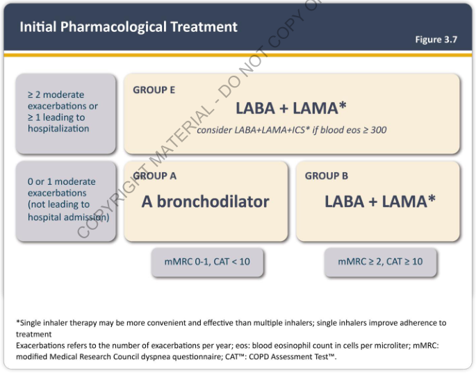
Summary : This figure presents an initial pharmacological treatment algorithm for patients with chronic obstructive pulmonary disease (COPD), stratified by exacerbation history and symptom severity, and grouped into A, B, and E for treatment recommendations. flowchart: # Nodes : • "≥ 2 moderate exacerbations or ≥ 1 leading to hospitalization" (rectangle, top left) • "0 or 1 moderate exacerbations (not leading to hospital admission)" (rectangle, bottom left) • "GROUP E: LABA + LAMA* (consider LABA+LAMA+ICS* if blood eos ≥ 300)" (rectangle, top right) • "GROUP A: A bronchodilator" (rectangle, bottom center) • "GROUP B: LABA + LAMA*" (rectangle, bottom right) • "mMRC 0-1, CAT < 10" (rectangle, below GROUP A) • "mMRC ≥ 2, CAT ≥ 10" (rectangle, below GROUP B) # Connectors : • Arrow from "≥ 2 moderate exacerbations or ≥ 1 leading to hospitalization" to "GROUP E: LABA + LAMA*" • Arrow from "0 or 1 moderate exacerbations (not leading to hospital admission)" splits to "GROUP A: A bronchodilator" and "GROUP B: LABA + LAMA*" • "GROUP A: A bronchodilator" is associated with "mMRC 0-1, CAT < 10" • "GROUP B: LABA + LAMA*" is associated with "mMRC ≥ 2, CAT ≥ 10" # Layout : • Two main branches based on exacerbation history (top left and bottom left). • Severe exacerbation branch leads directly to GROUP E (top right). • Mild exacerbation branch splits into GROUP A (center) and GROUP B (right), further differentiated by symptom scores (mMRC and CAT). • Symptom score boxes are placed below their respective treatment groups. # Treatment Recommendations : • GROUP E: LABA + LAMA*; consider LABA+LAMA+ICS* if blood eosinophils ≥ 300. • GROUP A: A bronchodilator. • GROUP B: LABA + LAMA*. • *Note: Single inhaler therapy may be more convenient and effective than multiple inhalers; single inhalers improve adherence to treatment. # Definitions & Criteria : • Exacerbations refer to the number per year. • Blood eosinophil count in cells per microliter. • mMRC: modified Medical Research Council dyspnea questionnaire. • CAT™: COPD Assessment Test™. # Analysis : • The flowchart stratifies COPD patients by exacerbation history and symptom severity (mMRC, CAT). • Patients with frequent or severe exacerbations (≥2 moderate or ≥1 hospitalization) are recommended dual bronchodilator therapy (LABA + LAMA), with possible addition of ICS if eosinophils are high. • Patients with low exacerbation risk are further divided by symptom scores: those with low symptoms (mMRC 0-1, CAT < 10) receive a single bronchodilator, while those with higher symptoms (mMRC ≥ 2, CAT ≥ 10) receive dual bronchodilator therapy. • The algorithm emphasizes tailored therapy based on risk and symptom burden, with consideration for adherence and convenience.
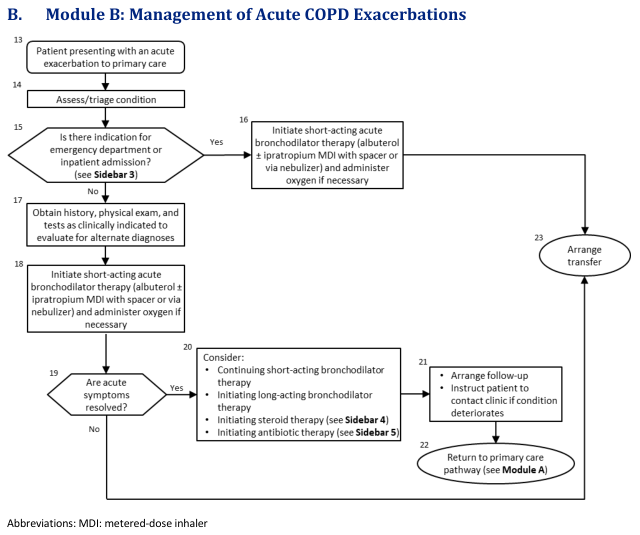
Summary : This flowchart outlines the stepwise management of acute COPD exacerbations in a primary care setting, including assessment, triage, therapy initiation, follow-up, and criteria for transfer or escalation. flowchart: # Nodes : • Patient presenting with an acute exacerbation to primary care (rectangle) • Assess/triage condition (rectangle) • Is there indication for emergency department or inpatient admission? (diamond) • Initiate short-acting acute bronchodilator therapy (rectangle) • Obtain history, physical exam, and tests as clinically indicated to evaluate for alternate diagnoses (rectangle) • Initiate short-acting acute bronchodilator therapy (rectangle) • Are acute symptoms resolved? (diamond) • Consider: Continuing short-acting bronchodilator therapy, Initiating long-acting bronchodilator therapy, Initiating steroid therapy, Initiating antibiotic therapy (rectangle) • Arrange follow-up; Instruct patient to contact clinic if condition deteriorates (rectangle) • Return to primary care pathway (rectangle) • Arrange transfer (ellipse) # Connectors : • Patient presenting → Assess/triage condition (down arrow) • Assess/triage condition → Is there indication for emergency department or inpatient admission? (down arrow) • Is there indication? → Yes → Initiate short-acting acute bronchodilator therapy (right arrow) • Initiate short-acting acute bronchodilator therapy → Arrange transfer (right arrow) • Is there indication? → No → Obtain history, physical exam, and tests (down arrow) • Obtain history, physical exam, and tests → Initiate short-acting acute bronchodilator therapy (down arrow) • Initiate short-acting acute bronchodilator therapy → Are acute symptoms resolved? (down arrow) • Are acute symptoms resolved? → Yes → Consider: Continuing therapy, etc. (right arrow) • Consider: Continuing therapy, etc. → Arrange follow-up; Instruct patient to contact clinic if condition deteriorates (down arrow) • Arrange follow-up; Instruct patient to contact clinic if condition deteriorates → Return to primary care pathway (down arrow) • Are acute symptoms resolved? → No → Initiate short-acting acute bronchodilator therapy (loop back arrow) • Arrange transfer (ellipse) is a terminal node for the emergency pathway. # Layout : • The flowchart is arranged vertically, with decision diamonds branching right for emergency pathways and continuing downward for non-emergency management. • Loops are present for unresolved acute symptoms, returning to therapy initiation. • Terminal nodes are "Arrange transfer" (for emergency) and "Return to primary care pathway" (for resolved cases). # Analysis : • The flowchart provides a clear, stepwise approach for managing acute COPD exacerbations in primary care, emphasizing initial assessment, triage for emergency needs, and therapy initiation. • Decision points ensure that patients with severe symptoms are promptly transferred, while those with resolved symptoms receive follow-up and return to routine care. • The loop for unresolved symptoms highlights the need for ongoing therapy and reassessment. • The chart references additional sidebars for detailed therapy guidance, indicating integration with broader clinical protocols.
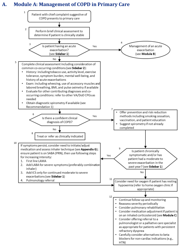
Summary : This flowchart outlines the stepwise management of Chronic Obstructive Pulmonary Disease (COPD) in primary care, including initial assessment, diagnosis, risk reduction, medication adjustment, and referral decisions. flowchart: # Nodes : • Start (rectangle): "Patient with chief complaint suggestive of COPD presents to primary care" • Rectangle: "Perform brief clinical assessment to determine if patient is clinically stable" • Diamond: "Is patient having an acute exacerbation? (see Sidebar 1)" • Ellipse: "Management of an acute exacerbation (see Module B)" • Rectangle: "Complete clinical assessment including consideration of common co-occurring conditions (see Sidebar 2): History, Exam, Evaluate for other contributing diagnoses, Obtain diagnostic spirometry if available" • Diamond: "Is there a confident clinical diagnosis of COPD?" • Rectangle: "Offer prevention and risk reduction methods including smoking cessation, vaccination, and patient education; Suggest spirometry if not already completed" • Rectangle: "Treat or refer as clinically indicated" • Diamond: "Is patient chronically symptomatic and/or has patient had a moderate to severe exacerbation in the past year? (see Sidebar 1)" • Rectangle: "If symptoms persist, consider need to initiate/adjust medication and assess inhaler technique (see Appendix G); ensure patient is on SABA (PRN), then use following steps for increasing intensity: 1. First line LAMA, 2. Add LABA for severe symptoms (preferably combination inhaler), 3. Add ICS only for continued moderate to severe exacerbations (see Sidebar 1), 4. Pulmonology referral" • Rectangle: "Consider need for oxygen if patient has resting hypoxemia (refer to home oxygen clinic if appropriate)" • Rectangle: "Continue follow-up and monitoring; Reassess severity periodically; Consider pulmonary rehabilitation; Consider medication adjustment if patient is on an inhaled corticosteroid (see Module C); Consider offering referral to a pulmonologist or a palliative care specialist as appropriate for patients with persistent refractory dyspnea; Carefully consider alternatives to beta blockers for non-cardiac indications (e.g., HTN)" # Connectors : • Arrows connect nodes in a top-down sequence, with diamonds representing decision points. • From "Is patient having an acute exacerbation?" (diamond): Yes → "Management of an acute exacerbation (ellipse)"; No → "Complete clinical assessment" (rectangle). • From "Is there a confident clinical diagnosis of COPD?" (diamond): Yes → "Offer prevention and risk reduction methods" (rectangle); No → "Treat or refer as clinically indicated" (rectangle). • From "Offer prevention and risk reduction methods" (rectangle) → "Is patient chronically symptomatic and/or has patient had a moderate to severe exacerbation in the past year?" (diamond). • From "Is patient chronically symptomatic..." (diamond): Yes → "If symptoms persist, consider need to initiate/adjust medication..." (rectangle); No → "Consider need for oxygen if patient has resting hypoxemia" (rectangle). • Both "If symptoms persist..." and "Consider need for oxygen..." nodes lead to "Continue follow-up and monitoring..." (rectangle). # Layout : • The flowchart is arranged vertically, with decision diamonds branching to different management paths. • Merges occur after medication adjustment and oxygen consideration, leading to a unified follow-up and monitoring node. • Sidebars and appendices are referenced for further detail at several steps. # Analysis : • The workflow prioritizes acute exacerbation management first, then proceeds to comprehensive assessment and diagnosis. • Preventive strategies and risk reduction are emphasized early for confirmed COPD cases. • Medication adjustment and specialist referral are considered for persistent symptoms or recent exacerbations. • Oxygen therapy is considered for patients with resting hypoxemia. • Ongoing follow-up, severity reassessment, and consideration of rehabilitation or specialist referral are recommended for all patients. • The flowchart supports a systematic, evidence-based approach to COPD management in primary care, with clear decision points and escalation pathways.
Acute Exacerbation of COPD (AECOPD)
Definition
Causes / Triggers
- Respiratory infections - viral (rhinovirus, influenza) and bacterial - account for >75% of cases; bacteria alone account for ~50%
- Air pollution / environmental irritants
- Cold weather
- Medications: beta-blockers, opioids, sedative-hypnotics
- Gastro-esophageal reflux disease (risk factor, though treating it has not been shown to reduce exacerbations)
- Common bacterial pathogens: Haemophilus influenzae, Streptococcus pneumoniae, Moraxella catarrhalis (±Pseudomonas aeruginosa in severe/high-risk cases)
Pathophysiology
- Bronchoconstriction
- Pulmonary vasoconstriction
- Mucus hypersecretion
Clinical Features
| Feature | Details |
|---|---|
| Dyspnea | Predominant symptom; orthopnea may occur |
| Cough | Often productive |
| Sputum | Increased volume or purulence |
| Hypoxemia | SaO2 <90%; presents with tachypnea, tachycardia, cyanosis, altered mentation |
| Hypercapnia | CO2 retention, respiratory acidosis |
| Use of accessory muscles | Up-and-forward posture, pursed-lip breathing |
| Pulsus paradoxus | Systolic BP drop >10 mmHg on inspiration |
Diagnosis & Workup
- Pulse oximetry - screen for hypoxemia
- ABG - best tool: assesses oxygenation (PaO2 <60 mmHg = failure), ventilation, acid-base (pH <7.35 = uncompensated acidosis)
- ECG - rule out cardiac ischemia/arrhythmia
- CXR - rule out pneumonia, pneumothorax, pleural effusion, CHF
- BNP / NTproBNP - if left heart failure suspected
- CBC, BMP - baseline labs
- Point-of-care CRP - can guide antibiotic use, reducing prescriptions without harm
- Contrast CT chest if pulmonary embolism is a concern
Differential Diagnosis
Severity Assessment: Criteria for Hospitalization
| Criteria for Hospital Admission | Criteria for ICU Admission |
|---|---|
| Significant increase in symptom severity | Need for invasive mechanical ventilation |
| Severe underlying COPD (low FEV1) | Hemodynamic instability |
| Significant comorbidities | Severe dyspnea not responding to therapy |
| Failure to respond to initial management | Mental status changes |
| Diagnostic uncertainty | Persistent or worsening hypoxemia/hypercapnia/respiratory acidosis despite O2 + NIV |
| Insufficient home support |
Management
1. Bronchodilators (First-Line)
- SABAs (short-acting beta-2 agonists) are the cornerstone:
- Albuterol: MDI 2-4 puffs q1-4h OR Nebulizer 2.5 mg q1-4h
- Short-acting anticholinergics added if inadequate SABA response:
- Ipratropium: MDI 2 puffs q4h OR Nebulizer 0.5 mg q4h
- Nebulization preferred in acute settings since optimal MDI technique may be impaired
- Methylxanthines (theophylline) - avoid in acute exacerbations due to serious side effects; if patient uses chronically, do NOT discontinue (risk of decompensation)
- Long-acting bronchodilators should be initiated once the patient is stable
2. Systemic Corticosteroids
- Recommended for all inpatients and most outpatients with exacerbations
- Prednisone 40 mg/day x 5 days - equally effective as longer regimens
- Benefits: shorter hospital stay, improved lung function, reduced relapse rate
- Systemic corticosteroids decrease treatment failures by ~46% - Textbook of Family Medicine
3. Antibiotics
- All three of: increased dyspnea + increased sputum volume + sputum purulence are present
- Two of the above including sputum purulence
- Need for assisted ventilation
| Patient Profile | Pathogens | Antibiotic (choose one) |
|---|---|---|
| No risk factors | H. influenzae, S. pneumoniae, M. catarrhalis | Macrolide (azithromycin 500 mg/day), 2nd/3rd-gen cephalosporin, doxycycline, TMP-SMX, amoxicillin-clavulanate 875/125 mg BID |
| Risk factors present* | Above + gram-negatives including Pseudomonas | Antipseudomonal fluoroquinolone (levofloxacin 750 mg/day) or antipseudomonal beta-lactam |
- Duration: 5-7 days (up to 7-10 days for pseudomonal coverage)
- Post-infection azithromycin 250 mg every 2 days x 3 months significantly reduces risk of treatment failure
4. Supplemental Oxygen
- Target SpO2 88%-92% (avoid hyperoxia, which can worsen hypercapnia by releasing hypoxic vasoconstriction and the Haldane effect)
5. Noninvasive Ventilation (NIV / BiPAP) - First Ventilatory Mode
- Moderate-severe dyspnea with increased work of breathing
- Acute respiratory acidosis: pH ≤7.35 and/or PaCO2 >45 mmHg
- Respiratory rate >25/min
- Respiratory arrest
- Hemodynamic instability
- Altered mental status / inability to cooperate
- High aspiration risk
- Viscous/copious secretions
- Recent facial or gastroesophageal surgery
- Craniofacial trauma, extreme obesity, burns
6. Invasive Mechanical Ventilation
7. Additional Measures
- DVT prophylaxis - increased VTE risk in hospitalized COPD patients
- Continuous monitoring of O2 saturation, respiratory rate, ABG
Management Algorithm (Primary Care Setting)
Initial Pharmacological Treatment (Stable COPD - GOLD Groups)
Quick Reference Summary
| Intervention | Agent / Dose | Notes |
|---|---|---|
| SABA | Albuterol 2.5 mg neb q1-4h | First-line |
| SAMA | Ipratropium 0.5 mg neb q4h | Add if SABA inadequate |
| Steroid | Prednisone 40 mg PO x 5 days | All inpatients |
| Antibiotic | Azithromycin / Amoxiclav / Levofloxacin | If purulent sputum or ventilated |
| O2 | Titrate to SpO2 88-92% | Avoid hyperoxia |
| NIV | BiPAP - first ventilatory mode | If pH ≤7.35 or RR >25 |
| DVT prophylaxis | LMWH | All hospitalized patients |
Recent evidence note (2025): A systematic review (PMID 40220861) found SGLT-2 inhibitors may reduce COPD exacerbations in patients with comorbid type 2 diabetes - an emerging therapeutic consideration. Additionally, meta-analyses on monoclonal antibodies (anti-IL-5, anti-TSLP) in eosinophilic COPD show promise for exacerbation reduction (PMID 39589286), though these are not yet standard acute management tools.