
Boundaries of canine space infection
Reading File
The canine space (also called the infraorbital or maxillary canine fossa space) is a fascial space in the face related to the canine tooth. Its boundaries are defined by surrounding anatomical structures.
Searching sources...
Finding Sources
Finding Sources
Reading File
Reading File
Reading File
Finding Sources
Finding Sources
Reading File
I now have the core textbook content. Let me also fetch the clinical image of canine space infection for visual clarity.
Loading Image
Here is a complete, textbook-sourced answer on the boundaries of canine space infection:
Canine Space Infection - Boundaries
The canine space (also called the infraorbital or canine fossa space) is a potential fascial space of the face overlying the anterior surface of the maxilla, specifically in the region of the canine tooth.
Anatomical Boundaries
| Wall | Boundary |
|---|---|
| Anterior (superficial) | Levator labii superioris (quadratus labii superioris) muscle |
| Posterior (deep) | Anterior surface of the maxilla (canine fossa) |
| Medial | Levator labii alaeque nasi |
| Lateral | Zygomaticus major and minor muscles |
| Inferior | Levator anguli oris (caninus) muscle - the key landmark |
The canine space is located specifically between the anterior surface of the maxilla and the levator labii superioris muscle.
Source of Infection
The canine space is infected almost exclusively by apical infection of the root of the maxillary canine tooth. A critical anatomical requirement: the root must be long enough so that its apex lies superior to the insertion of the levator anguli oris muscle. If the apex perforates inferiorly to this muscle insertion, infection spreads into the vestibular sulcus rather than the canine space.
Clinical Presentation
- Swelling lateral to the nose and in the infraorbital region
- Loss (flattening) of the ipsilateral nasolabial fold - the hallmark sign
- Obliteration of the infraorbital sulcus
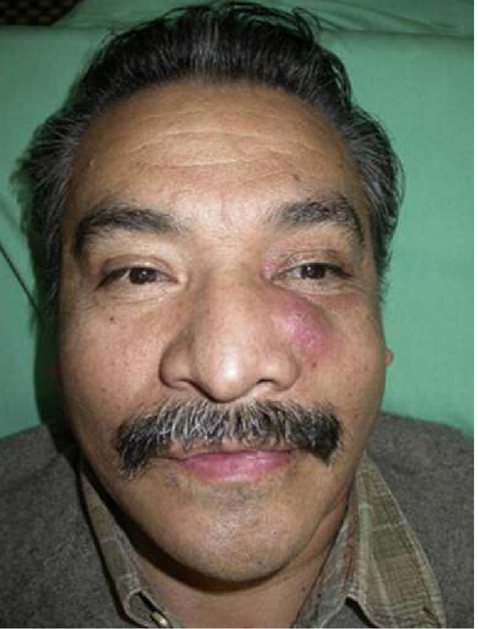
Major Complication
The most feared complication of canine space infection is cavernous sinus thrombosis, due to the angular vein's connection to the cavernous sinus via the inferior ophthalmic vein. This route is facilitated by the lack of valves in the facial venous system.
Management
Drainage is most often accomplished with an intraoral stab incision into the canine fossa region.
Sources:
- Cummings Otolaryngology Head and Neck Surgery - Maxillary Spaces section, pp. 201-203
- Rosen's Emergency Medicine: Concepts and Clinical Practice - Chapter 56, Deep Neck Infections, pp. 863-865
This is a shared conversation. Sign in to Orris to start your own chat.