

aa77fa1b-d945-497b-afee-08f0dd74cb50.jpg
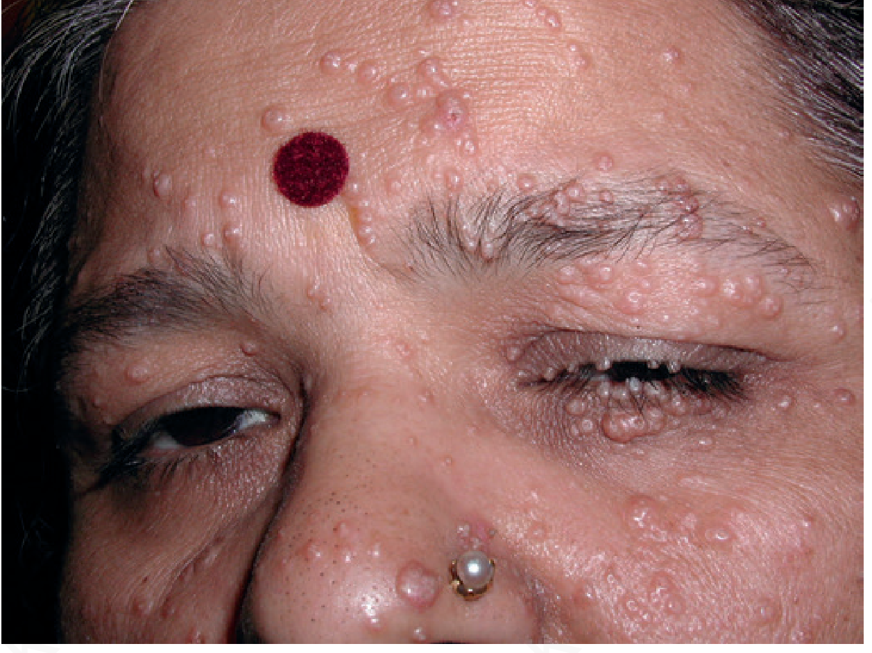
18 year female with such papular rash with itching over neck and chest since a month Provisional diagnosis?
analyse_media
Reading File
Now let me search the medical library for molluscum contagiosum details in parallel with a web search.
Finding Sources
Searching the Web
molluscum contagiosum adults neck chest treatment 2024
Reading File
Reading File
Loading Image
Provisional Diagnosis: Molluscum Contagiosum
Clinical Analysis
The image shows the following on AI analysis:
| Feature | Finding in This Patient |
|---|---|
| Lesion type | Multiple discrete, dome-shaped papules |
| Surface | Smooth, shiny, waxy/pearlescent |
| Central umbilication | Present (characteristic dimple) |
| Size | 1-4 mm diameter |
| Distribution | Dense cluster on upper chest/clavicular region, neck |
| Duration | 1 month |
| Symptom | Pruritus |
This is a textbook presentation of Molluscum Contagiosum (MC).
About Molluscum Contagiosum
Etiology: Caused by a poxvirus (Molluscum Contagiosum Virus, MCV types 1-4). MCV-1 is the most common worldwide; MCV-2 is more commonly sexually transmitted in adolescents and young adults.
Epidemiology: Three groups are primarily affected - young children (1-4 yrs), sexually active young adults (20-29 yrs), and immunosuppressed persons. This 18-year-old fits the young adult group.
Hallmark lesion: Small, smooth, firm, dome-shaped, pearly papule averaging 3-5 mm with a central umbilication or white core - exactly as seen here. (Andrews' Diseases of the Skin, p. 453)
Distribution in adults (non-STI): Upper body, trunk, neck, and axillae are common sites via direct skin-to-skin contact or autoinoculation from scratching.
Why itching? About 40% of cases develop "molluscum dermatitis" - a mild eczematous reaction around individual lesions, especially in atopic individuals. Scratching also promotes autoinoculation and spread.
Here is a reference image from the textbook showing typical MC:
Differential Diagnoses to Consider
| Condition | Points Against |
|---|---|
| Verruca plana (flat warts) | Warts are flat-topped, not dome-shaped/umbilicated; no pearlescent quality |
| Milia | Milia are tiny 1-2 mm white cysts, not umbilicated; not clustered this way |
| Folliculitis | Follicle-based pustules, erythematous base, not dome-shaped pearly papules |
| Comedonal acne | No umbilication; no pearlescent sheen |
| Varicella (early) | Widespread distribution; systemic symptoms; evolves rapidly |
Investigations
- Usually a clinical diagnosis - the umbilicated pearly papules are pathognomonic.
- If in doubt: dermoscopy shows a central pore with polylobular white/yellow amorphous structures ("cauliflower" pattern).
- Tzanck smear from the caseous core: Henderson-Paterson (molluscum) bodies (large eosinophilic cytoplasmic inclusions).
- In a young sexually active adult: screen for other STIs if lesions are in/near genital area.
- If extensive/atypical: consider HIV testing.
Management
First line:
- Cryotherapy (liquid nitrogen) - effective, well-tolerated in adults; usually 2-3 sessions
- Curettage - physical removal of each lesion; fast and effective
- Cantharidin (topical, applied in-office) - FDA-approved (July 2023) for ages 2+ years; causes blistering under the lesion
Second line / home-based:
- Imiquimod 5% cream (off-label) - immune response modulator
- Tretinoin cream - irritant mechanism
- Potassium hydroxide (KOH) 10% solution - dissolves lesion keratin; commonly used in South Asia/India
Supportive:
- Avoid scratching to prevent autoinoculation
- Avoid sharing towels/clothing
- Note that lesions are self-limiting - resolve in months to ~1 year in immunocompetent individuals
Key point for this patient: In an 18-year-old with chest and neck involvement and no mention of genital lesions, transmission is likely through direct contact or fomites (shared clothing, towels). Genital area should be examined. If extensive or atypical in distribution, check immune status.
This is a shared conversation. Sign in to Orris to start your own chat.