
Elderly male patient known case of heart block with pacemaker implantation presented with acute onset SOB to rule out ADHF and with AKI ?Acute on CKD with ALI ... evaluation and management
Reading File
Now let me run parallel searches across the key clinical domains in this complex case.
Finding Sources
Finding Sources
Finding Sources
Searching PubMed
"acute decompensated heart failure" AND "acute kidney injury" AND management
Good results. Now let me read the key textbook sections in parallel.
Reading File
Reading File
Reading File
Finding Sources
Excellent content. Let me now read the cardiorenal syndrome sections and ADHF management algorithm in parallel.
Reading File
Reading File
Reading File
Finding Sources
Now let me search for ALI (Acute Limb Ischemia) and pacemaker-related considerations.
Finding Sources
Finding Sources
Finding Sources
Reading File
Now let me read the ALI section and also the Harrison's ADHF table of drugs, plus fetch the key management flowchart image.
Reading File
Reading File
Loading Image
Loading Image
I now have comprehensive content from the textbooks. Let me also read the Goldman-Cecil ALI management section and the Sabiston ALI section for complete vascular management.
Reading File
Reading File
I now have sufficient information to construct a comprehensive clinical response. Let me compile everything.
Elderly Male with Pacemaker: ADHF + AKI/?Acute-on-CKD + ALI - Evaluation & Management
This is a high-acuity, multi-organ clinical scenario requiring simultaneous but prioritized workup. The three problems are interrelated through the cardiorenal syndrome axis and through the underlying cardiovascular risk burden that drives both heart failure and peripheral vascular disease.
I. INITIAL STABILIZATION & TRIAGE
Before detailed evaluation, immediate priorities:
| Priority | Action |
|---|---|
| Airway/Breathing | SpO2, high-flow O2 or NIV (CPAP/BiPAP) for SOB/hypoxia |
| Circulation | IV access x2, BP (both arms), heart rate, ECG |
| Monitoring | Continuous cardiac monitoring (with pacemaker - check capture/sensing) |
| Position | Sit upright, legs dependent if not ALI-threatened |
Limb salvage assessment first - if the limb with suspected ALI shows motor deficit or complete pulselessness, urgent vascular surgery consultation must happen simultaneously with cardiac stabilization.
II. ACUTE DECOMPENSATED HEART FAILURE (ADHF) - Evaluation
A. Identify the Precipitant (MANDATORY FIRST STEP)
Per Harrison's 22E, the first principle of ADHF management is to identify and address precipitating factors:
- Non-adherence to medications or dietary salt restriction
- Medications worsening HF: NSAIDs, thiazolidinediones, TNF inhibitors, cold/flu preparations
- Coronary ischemia - must be systematically investigated in all atherosclerotic patients
- Arrhythmias - atrial/ventricular arrhythmias are common contributors
- Pacemaker issues - pacemaker-mediated cardiomyopathy, lead dislodgement, loss of CRT synchrony, pacemaker syndrome (RV pacing inducing dyssynchrony), or pacemaker dysfunction triggering the decompensation
- Valvular disease - echocardiography is key
- Infection or pulmonary embolism
B. Diagnostic Workup
ECG:
- Assess pacemaker function: is there appropriate pacing spike + capture?
- Check for loss of pacing, failure to capture, undersensing/oversensing
- Identify underlying rhythm (especially AF - relevant to ALI etiology)
- Signs of new ischemia (may be masked by paced rhythm - use Sgarbossa criteria)
Biomarkers:
- NT-proBNP/BNP - elevated; confirms HF but also tracks response
- Troponin - elevated in demand ischemia or type 2 MI
- Poor prognostic markers: BUN >43 mg/dL, creatinine >2.75 mg/dL, SBP <115 mmHg (Harrison's 22E)
Imaging:
- CXR: pulmonary congestion (perihilar haziness, Kerley B lines, cardiomegaly, pleural effusion), pacemaker lead positions
- Bedside echo (urgent): EF assessment (HFrEF vs HFpEF), valvular disease, pericardial effusion, wall motion abnormalities, IVC plethora (volume status), lead position
- Echo also assesses for thrombus in left atrium/ventricle (source for limb embolism)
Labs:
- FBC, electrolytes, creatinine, BUN, eGFR, LFTs, LDH, uric acid
- ABG (if hypoxic), lactate (if suspect low-output/cardiogenic shock or ischemic limb)
- Urine output monitoring, urine Na, urine creatinine, FENa
- D-dimer (if PE suspected)
C. ADHF Phenotyping (determines treatment)
The management algorithm below from Harrison's 22E is key:
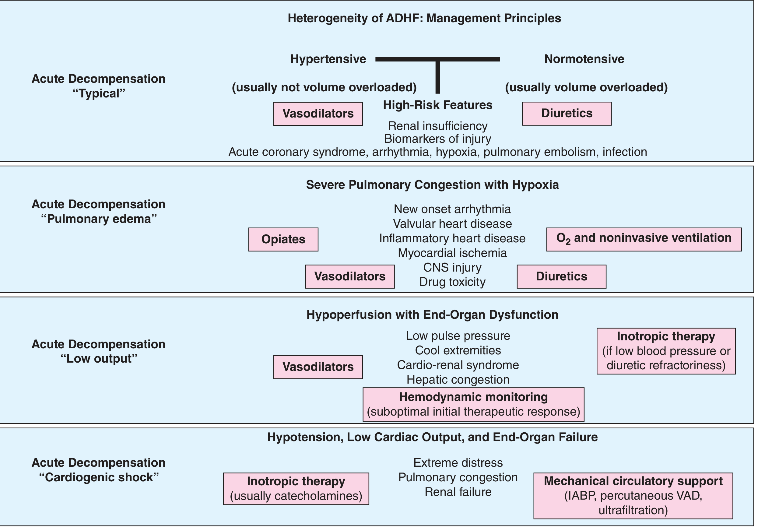
In this patient, the most likely phenotype is "Low output" or "Pulmonary edema" (normotensive/volume overloaded) given pacemaker-associated cardiac dysfunction. However, always consider:
- Hypertensive ADHF - vasodilators first
- Low-output ADHF - cardiorenal syndrome, cool extremities (overlap with ALI!)
- Cardiogenic shock - if BP <90 mmHg, poor perfusion, rising lactate
III. CARDIORENAL SYNDROME (CRS) - The Linking Pathophysiology
This patient has the classic CRS Type 1 picture (acute cardiac event causing acute kidney injury).
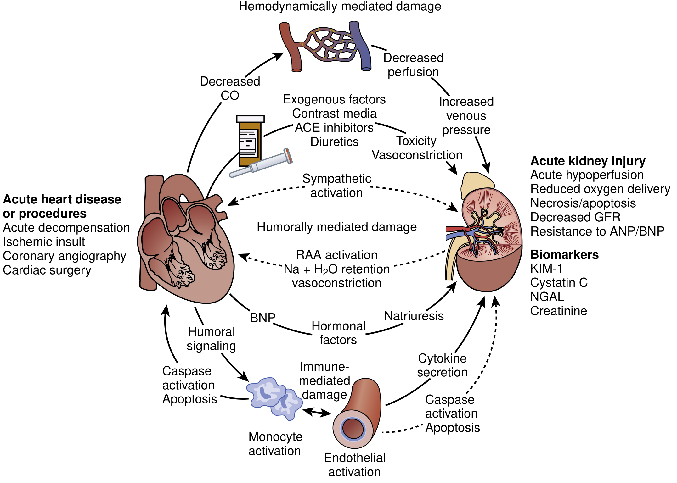
Pathophysiology (NKF Primer on Kidney Diseases, 8e)
The dominant modern understanding is that venous congestion, not reduced cardiac output, is the primary driver of kidney dysfunction in ADHF:
- Elevated right atrial pressure → renal venous hypertension → impaired renal venous outflow
- Increased intraabdominal pressure → renal interstitial pressure → back-pressure on Bowman's capsule → reduced GFR
- This was confirmed in the ESCAPE trial: only right atrial pressure (not cardiac index) correlated with renal dysfunction
- Superimposed neurohormonal activation: RAAS, sympathetic, AVP → sodium and water retention, vasoconstriction
- Pre-existing CKD significantly amplifies vulnerability: rightward and downward shift of the diuretic dose-response curve (Brenner & Rector)
AKI/Acute-on-CKD Evaluation
Determine AKI category:
| Type | Likely Cause in this Patient |
|---|---|
| Pre-renal | Diuretic over-treatment, reduced cardiac output |
| Intrinsic renal | Cardiorenal syndrome (venous congestion), contrast nephropathy (if recent imaging), NSAID use |
| Post-renal | Less likely; rule out with bladder scan |
Investigations for AKI:
- Urine microscopy: granular casts → ATN; bland sediment → prerenal
- FENa: <1% prerenal; >2% ATN (caveat: unreliable with diuretics - use FEUrea instead)
- Renal ultrasound: echogenicity (CKD background), obstruction, resistive indices
- Urine protein:creatinine ratio - baseline CKD proteinuria
- Trend creatinine at 6-12 hourly intervals
- Novel biomarkers if available: NGAL, KIM-1, cystatin C
Supportive AKI Management (Brenner & Rector Table 29.7):
| Problem | Treatment |
|---|---|
| Volume overload | Salt restriction <1-2g/day; water restriction <1L/day; diuretics if non-oliguric; ultrafiltration if refractory |
| Hyperkalemia | Calcium gluconate IV if ECG changes; Insulin + glucose; salbutamol nebulizer; kayexalate; loop diuretics; RRT if severe |
| Metabolic acidosis | NaHCO3 if HCO3 <15 mmol/L; dietary protein restriction; RRT if severe |
| Hyperphosphatemia | Phosphate binders (calcium carbonate, sevelamer) |
| Nutrition | 20-30 kcal/kg/day; protein 0.8-1.0 g/kg/day (non-dialysis); enteral preferred |
| Drug dosing | Adjust all doses for eGFR - especially low-molecular-weight heparin, milrinone, antibiotics |
IV. ADHF MANAGEMENT - Drug Therapy
1. Diuresis - Primary Therapy
The cornerstone of therapy is IV loop diuretics to relieve venous congestion.
Dose equivalency (NKF Primer):
- Furosemide 80 mg oral = Furosemide 40 mg IV = Torsemide 20 mg oral = Bumetanide 1 mg oral/IV
Practical approach:
- Administer IV route (oral absorption unreliable in ADHF)
- Double the home oral dose as IV furosemide as a starting point
- In patients with GFR <30 mL/min requiring high doses: bumetanide preferred (minimally impacted by kidney dysfunction, less ototoxicity)
- The DOSE trial: no clear advantage of continuous infusion vs. bolus; no significant difference between low-dose and high-dose strategies in general
- Assess response: urine sodium in first 2 hours >50-70 mEq/L suggests adequate response
Diuretic resistance (common in CRS):
- Add thiazide (metolazone) - "sequential nephron blockade" - monitor K+/Mg2+ closely
- Mineralocorticoid receptor antagonist (spironolactone) - caution with hyperkalemia, especially with RAAS blockers
- SGLT2 inhibitor (e.g., empagliflozin) - emerging evidence in acute CRS for proximal nephron blockade - 2025 Systematic Review PMID 40435978
- Ultrafiltration - controlled fluid removal, electrolyte-neutral, reduces neurohormonal activation
Torsemide vs. furosemide: Torsemide has >90% oral bioavailability vs. furosemide's erratic 10-90% (mean 50%). The TRANSFORM-HF trial showed no mortality/morbidity advantage, but torsemide may reduce hospital readmissions in some subgroups.
2. Vasodilators
- IV nitroglycerin: first-line in hypertensive ADHF with pulmonary edema; reduces preload and afterload; counteracts sympathetic/RAAS/AVP effects
- IV nitroprusside: more potent; beware cyanide toxicity with prolonged use
- Nesiritide (recombinant BNP): reduces filling pressures; modest diuresis; not shown to improve outcomes vs. standard therapy
Caution: Vasodilators should be avoided or used with extreme care if SBP <100 mmHg.
3. Inotropes (if low-output state or refractory to diuretics)
- Dobutamine: first-line positive inotrope; reduces SVR; increases CO; beware arrhythmias
- Milrinone: PDE-3 inhibitor; greater reduction in SVR than dobutamine; better if on beta-blockers (acts downstream from beta receptor); requires dose reduction in renal failure (renally excreted, long half-life)
- Dopamine: dose-dependent effects; "renal-dose" dopamine is NOT recommended (ROSE-AHF: no benefit over standard diuretics)
- Levosimendan: calcium sensitizer; not approved in the US; mixed trial results
⚠️ Long-term inotrope use increases arrhythmia risk and mortality - use only as bridge therapy.
4. GDMT (Guideline-Directed Medical Therapy) - In-Hospital Continuation
- Beta-blockers: Do NOT abruptly stop in admitted ADHF patients - worsens outcomes; can reduce dose if signs of low output
- ACE inhibitor/ARB/ARNI: Hold or reduce if AKI worsening or hypotension; restart at lowest dose when stable
- MRA (spironolactone/eplerenone): Hold if K+ >5.0 or creatinine rising rapidly
- SGLT2 inhibitors: Growing evidence supports continuation or initiation in stable ADHF with renal dysfunction
V. PACEMAKER-SPECIFIC CONSIDERATIONS
- Check device function immediately: Device interrogation (via programmer) - assess pacing thresholds, sensing, lead impedances, battery status, pacing mode
- Pacemaker-induced cardiomyopathy: Chronic right ventricular pacing (especially >40% burden) causes dyssynchrony and ventricular dysfunction - may require upgrade to CRT (Cardiac Resynchronization Therapy)
- Pacemaker syndrome: When ventricle paces before atrium contracts properly → loss of AV synchrony → reduced cardiac output → can precipitate ADHF
- Rate response settings: Ensure pacemaker rate is appropriate for patient's current metabolic demands
- Device lead thrombosis: In patients with cardiac implantable electronic devices, consider subclavian/axillary vein thrombosis contributing to ipsilateral limb symptoms
- Electromagnetic interference: Avoid MRI without pacemaker-safe protocol; defibrillation pads placement considerations
VI. ACUTE LIMB ISCHEMIA (ALI) - Evaluation & Management
Likely etiology in this patient: Cardiac embolism (from AF, LV thrombus, or valvular disease in setting of heart failure) OR in-situ thrombosis on background atherosclerosis.
Clinical Assessment - "The 6 Ps"
| Sign | Significance |
|---|---|
| Pain | Earliest symptom - distal to obstruction |
| Pallor | Early finding |
| Pulselessness | Localizes level of occlusion |
| Paresthesias | Indicates sensory nerve ischemia |
| Paralysis | Indicates motor nerve ischemia - urgent revascularization |
| Poikilothermia | Cool limb vs. contralateral warm limb |
Rutherford Classification (for management decisions):
| Grade | Category | Finding | Action |
|---|---|---|---|
| I | Viable | No sensory/motor loss, audible Doppler | Not immediately threatened |
| IIa | Marginally threatened | Minimal sensory loss, no motor deficit | Urgent |
| IIb | Immediately threatened | Sensory + motor deficit present | Emergent |
| III | Irreversible | Profound anesthesia, paralysis, skin mottling | Amputation |
Investigations
- Ankle-brachial index (ABI): <0.9 confirms limb ischemia; not always feasible acutely
- Urgent arterial duplex Doppler: Map level of occlusion
- CT angiography (preferred if available): Define level and extent of occlusion, plan intervention - NOTE: contrast load will worsen AKI - weigh risk vs. benefit carefully, consider withholding nephrotoxic drugs, IV hydration
- ECG and echo: Identify cardiac source (AF, LV thrombus)
Management
Step 1 - Immediate anticoagulation:
- UFH IV bolus (5000 units) → continuous infusion targeting aPTT 60-100 sec
- Prevents clot propagation; do not wait for imaging if clinical diagnosis is clear
- UFH preferred over LMWH in AKI (renally cleared LMWH has unpredictable levels)
Step 2 - Revascularization strategy (Goldman-Cecil):
| Condition | Preferred Strategy |
|---|---|
| Viable or marginally threatened (Rutherford I-IIa), onset <14 days | Endovascular: catheter-directed thrombolysis (rtPA 0.05-0.1 mg/kg/hr intra-arterially) ± thrombectomy ± stenting |
| Immediately threatened (Rutherford IIb), onset <14 days | Surgical embolectomy (Fogarty catheter) - faster than thrombolysis when limb time-critical |
| Onset >14 days | Surgical revascularization/bypass preferred |
| Irreversible (Rutherford III) | Amputation - no benefit from revascularization; reperfusion injury dangerous |
Step 3 - Post-revascularization monitoring:
- Watch for reperfusion injury: compartment syndrome (compartment pressure >30 mmHg → fasciotomy), hyperkalemia, myoglobinuria, metabolic acidosis, renal failure worsening
- Myoglobinuria: aggressive IV fluids, urinary alkalinization; this will directly worsen AKI
- Repeat neurological/vascular exam hourly post-intervention
Step 4 - Anticoagulation for the cardiac source:
- If AF confirmed: long-term anticoagulation (DOACs preferred, but dose-adjust for CKD)
- If LV thrombus: warfarin (target INR 2-3) for 3-6 months
VII. INTEGRATED MANAGEMENT CHALLENGES
| Dilemma | Solution |
|---|---|
| Diuresis needed for ADHF but worsens AKI | Continue diuresis as long as venous congestion persists - "aggressive decongestion" associated with improved survival even if creatinine rises transiently (Brenner & Rector) |
| Contrast CT needed for ALI but worsens AKI | Use minimum contrast volume; pre-hydrate with isotonic crystalloid; hold nephrotoxins 24-48h; monitor creatinine closely; consider alternative (duplex/MRA) if available |
| Anticoagulation for ALI/AF risks bleeding | UFH preferred - titratable, reversible; avoid LMWH in AKI; monitor anti-Xa levels |
| Inotropes may worsen arrhythmias with pacemaker | Careful; dobutamine preferred; milrinone requires dose reduction in renal failure |
| Milrinone dosing in CKD | Reduce dose, lengthen interval; monitor carefully |
| ALI compartment syndrome vs. AKI from myoglobin | Fasciotomy if needed; aggressive myoglobinuria management with fluids + bicarbonate |
| Fluid balance tension | In low-output state, UF may be preferred over diuretics to achieve precise fluid removal without electrolyte disturbance |
VIII. INDICATIONS FOR RENAL REPLACEMENT THERAPY (RRT)
Consider RRT (CRRT preferred in hemodynamically unstable patients) if:
- Refractory volume overload despite diuretics/UF
- Severe hyperkalemia (K+ >6.5 mEq/L or ECG changes) unresponsive to medical therapy
- Severe metabolic acidosis (pH <7.15)
- Uremic symptoms (encephalopathy, pericarditis)
- Inability to clear fluid to achieve adequate decongestion
CRRT is preferred over intermittent HD in hemodynamically compromised patients (avoid BP swings that would worsen ALI or cardiac output).
IX. MONITORING TARGETS
| Parameter | Target/Goal |
|---|---|
| SpO2 | ≥95% |
| Systolic BP | 90-140 mmHg (avoid hypotension - worsens both AKI and ALI) |
| Urine output | >0.5 mL/kg/hr |
| Fluid balance | Negative 500-1000 mL/day (avoid too-rapid diuresis) |
| K+ | 3.5-5.0 mEq/L |
| Creatinine trend | Monitor 12-hourly; acceptable mild rise if decongesting |
| BNP/NT-proBNP | Downtrend toward discharge |
| Paced limb pulses | Hourly vascular exam |
| Lactate | Normalize |
X. DISPOSITION
- ICU/HDU admission given triple-organ threat (cardiac, renal, vascular)
- Multidisciplinary team: Cardiology, Nephrology, Vascular Surgery, Device/EP Clinic
- Vascular surgery: STAT consultation for ALI
- Device clinic/EP: Pacemaker interrogation
- Avoid iodinated contrast until vascular access secured and renal risk assessed
- Avoid NSAIDs, nephrotoxic antibiotics, gadolinium contrast
Recent Evidence Updates
- Systematic Review on Diuretic Resistance in Acute HF (2025, PMID 40435978) - SGLT2 inhibitors show promise as adjuncts for natriuresis in CRS with diuretic resistance
- Comparative efficacy of drugs in AHF with renal dysfunction (2024, PMID 39877019) - Network meta-analysis comparing agents; tolvaptan and combination diuretics show benefit
- AKI incidence and predictors in heart failure (2023, PMID 37705352) - AKI in HF carries significantly elevated mortality; creatinine rise >0.3 mg/dL is an independent predictor of poor outcomes
Sources: Harrison's Principles of Internal Medicine 22E (2025) | Brenner & Rector's The Kidney | NKF Primer on Kidney Diseases, 8e | Goldman-Cecil Medicine | Tintinalli's Emergency Medicine | Sabiston Textbook of Surgery
Elderly male patient known case of heart block with pacemaker implantation presented with acute onset SOB to rule out ADHF and with AKI ?Acute on CKD with Acute liver injury... evaluation and management
Reading File
This is a complex multi-organ clinical scenario. Let me run broad parallel searches across all key domains simultaneously.
Finding Sources
Finding Sources
Finding Sources
Searching PubMed
"acute decompensated heart failure" AND "acute liver injury" AND management
Let me try broader search terms for the textbook database.
Finding Sources
Finding Sources
Finding Sources
Searching PubMed
congestive hepatopathy heart failure liver injury management
Good. Now let me pull the textbook content from key sections in parallel - ADHF management, cardiorenal, and search for hepatic congestion / ischemic hepatitis.
Reading File
Reading File
Finding Sources
Finding Sources
Excellent - found the key hepatology sections. Now read them in parallel along with ADHF continuation and the CRS pathophysiology diagram.
Reading File
Reading File
Reading File
Loading Image
Excellent textbook content obtained. Let me now read the AKI management table and cardiorenal syndrome pathophysiology, and also fetch the hepatic congestion histology image.
Reading File
Reading File
Loading Image
I now have comprehensive, rich content from authoritative textbooks across all three organ systems. Let me also fetch the cardiac cirrhosis histology image from Sleisenger & Fordtran and read the ADHF inotrope section.
Loading Image
Reading File
I now have comprehensive, authoritative content from multiple textbooks covering all three organ systems. Let me now synthesize a complete, structured clinical response.
Elderly Male with Pacemaker: ADHF + AKI/?Acute-on-CKD + Acute Liver Injury - Evaluation & Management
This presentation represents cardiohepatorenalynthesis - a three-organ congestive failure syndrome driven by a single unifying mechanism: right-sided venous hypertension and systemic hypoperfusion from acute cardiac decompensation. Understanding this unifying pathophysiology is essential before treating any organ in isolation.
THE UNIFYING CONCEPT: Congestive Multi-Organ Failure
The same elevated right-sided filling pressure and reduced cardiac output that precipitates pulmonary edema (SOB) simultaneously causes:
- Renal venous hypertension → AKI/Acute-on-CKD (Cardiorenal Syndrome Type 1)
- Hepatic venous congestion → Congestive hepatopathy ± Ischemic hepatitis ("shock liver")
Per the ESCAPE trial (NKF Primer on Kidney Diseases, 8e), right atrial pressure is the only hemodynamic parameter consistently associated with renal dysfunction in ADHF - not cardiac index. The same venous back-pressure applies to hepatic veins through the IVC.
Management directed at the heart (decongestion) treats all three organs simultaneously.
I. IMMEDIATE STABILIZATION
| Priority | Action |
|---|---|
| Airway | SpO2 monitoring; high-flow O2 or NIV (CPAP/BiPAP) for hypoxia |
| IV access | 2 large-bore peripheral IV lines; arterial line if hemodynamically unstable |
| Monitoring | Continuous ECG (pacemaker capture?), BP, SpO2, urine output |
| Position | Sit upright |
| Rapid bedside assessment | Is BP adequate (>90 systolic)? Signs of cardiogenic shock? |
II. ACUTE DECOMPENSATED HEART FAILURE (ADHF)
A. Identify the Precipitant (Do This Before Any Drug)
Per Harrison's Principles of Internal Medicine 22E, the first principle of ADHF management is identifying the precipitating factor:
| Precipitant | Workup |
|---|---|
| Medication non-adherence / dietary salt excess | History |
| Coronary ischemia | ECG, troponin, echocardiogram |
| Arrhythmia / Pacemaker dysfunction | ECG, device interrogation - key in this patient |
| Valvular heart disease (new/worsening) | Echocardiogram |
| Infection / Sepsis | FBC, blood cultures, CRP, procalcitonin |
| Pulmonary embolism | D-dimer, CTPA if warranted |
| ACS driving decompensation | Sgarbossa criteria on paced ECG, troponin trend |
| Hepatotoxic medications | Drug history review |
Pacemaker-specific considerations:
- Device interrogation is mandatory - check sensing, pacing thresholds, lead impedances, battery status, % RV pacing
- Chronic RV pacing (>40% burden) → pacemaker-induced cardiomyopathy (dyssynchrony) → may need CRT upgrade
- Pacemaker syndrome: retrograde VA conduction → loss of AV synchrony → reduced CO
- Rule out pacemaker lead dislodgement or failure to capture
B. ADHF Phenotyping
The management strategy is entirely phenotype-dependent:
In this patient, the most likely phenotype is "Low Output" or "Pulmonary Edema" given:
- Known heart block + pacemaker (structural/electrical cardiac disease)
- Signs of multi-organ venous congestion (kidney + liver)
- Note: hepatic congestion is explicitly listed as a feature of the "Low Output" ADHF phenotype in the flowchart above
C. Investigations for ADHF
ECG:
- Pacemaker spikes present? Capture? Rate?
- Underlying rhythm (AF, ventricular ectopy)
- Sgarbossa criteria for ischemia in paced rhythm
- PR, QRS, QTc
Biomarkers:
- NT-proBNP/BNP: confirms HF, tracks response; >1000 pg/mL with HFrEF is typical
- High-sensitivity troponin (T or I): type 2 MI (demand ischemia from low output)
- Poor prognostic markers (Harrison's): BUN >43 mg/dL, SBP <115 mmHg, creatinine >2.75 mg/dL, elevated troponin
Imaging:
- CXR: pulmonary congestion, cardiomegaly, pleural effusions, pacemaker lead position
- Bedside echo (urgent): EF (HFrEF vs HFpEF), RWMA (ischemia), valvular disease, pericardial effusion, IVC diameter/collapsibility (volume status), RV function, pacemaker lead position
- Dilated IVC + hepatic veins on echo directly confirms hepatic venous congestion
III. CARDIORENAL SYNDROME - AKI / Acute-on-CKD
Pathophysiology
The dominant mechanism in ADHF-related AKI is venous congestion, not reduced cardiac output (NKF Primer on Kidney Diseases, 8e). The same right-heart failure that congests the lungs elevates:
- Central venous pressure → renal venous hypertension → reduced net renal perfusion pressure
- Intraabdominal pressure → transmitted to renal interstitium → back-pressure on Bowman's capsule → reduced GFR
- Neurohormonal activation (RAAS, SNS, AVP) → Na/water retention → worsens volume overload
On top of this, this patient has CKD baseline - already has rightward shift of the diuretic dose-response curve (Brenner & Rector), meaning higher doses are needed for natriuresis.
AKI Evaluation
Classify AKI type (use KDIGO criteria):
- Baseline creatinine from old records?
- AKI Stage 1: Cr rise ≥0.3 mg/dL within 48h or ≥1.5x baseline within 7 days
- AKI Stage 2: Cr ≥2x baseline
- AKI Stage 3: Cr ≥3x baseline or <0.3 mL/kg/hr x 24h
Investigations:
| Test | Purpose |
|---|---|
| Serum creatinine, BUN, eGFR | Staging AKI, severity |
| Urine output (hourly) | Oliguria (<0.5 mL/kg/hr) = worse prognosis |
| Urine microscopy | Granular/muddy brown casts → ATN; bland → prerenal |
| FENa (<1% prerenal, >2% ATN) | Unreliable on diuretics - use FEUrea instead |
| Urine Na concentration | <20 mEq/L prerenal; >40 mEq/L intrinsic |
| Urine protein:creatinine ratio | Assess baseline CKD proteinuria |
| Electrolytes: K+, Na+, HCO3-, phosphate | Complications of AKI |
| Renal ultrasound | Small echogenic kidneys = CKD; hydronephrosis = post-renal |
| NGAL, KIM-1, cystatin C (if available) | Novel AKI biomarkers - detect ATN early |
AKI Supportive Management (Brenner & Rector's The Kidney):
| Complication | Treatment |
|---|---|
| Volume overload | Salt <1-2 g/day; water <1 L/day; IV loop diuretics (if non-oliguric); ultrafiltration |
| Hyperkalemia with ECG changes | Calcium gluconate 10 mL of 10% IV over 5 min → Insulin 10-20 U + Glucose 250 mL of 20% IV → Salbutamol 10-20 mg nebulized → K+ binders → RRT |
| Metabolic acidosis | NaHCO3 if HCO3 <15 mmol/L; dietary protein restriction; RRT if severe |
| Hyperphosphatemia | Phosphate binders (calcium carbonate, sevelamer) |
| Nutrition | 20-30 kcal/kg/day; 0.8-1.0 g/kg/day protein (non-dialysis); enteral route preferred |
| Drug dosing | Adjust ALL drugs for eGFR - especially LMWH, milrinone, aminoglycosides, antibiotics, digoxin |
IV. ACUTE LIVER INJURY IN HEART FAILURE
This is a critically important dimension that is often underrecognized. In this patient, liver injury has two possible and frequently co-existing mechanisms:
A. Congestive Hepatopathy ("Cardiac Liver")
Caused by chronic passive congestion from right-heart failure (Yamada's Textbook of Gastroenterology; Sleisenger & Fordtran's GI & Liver Disease).
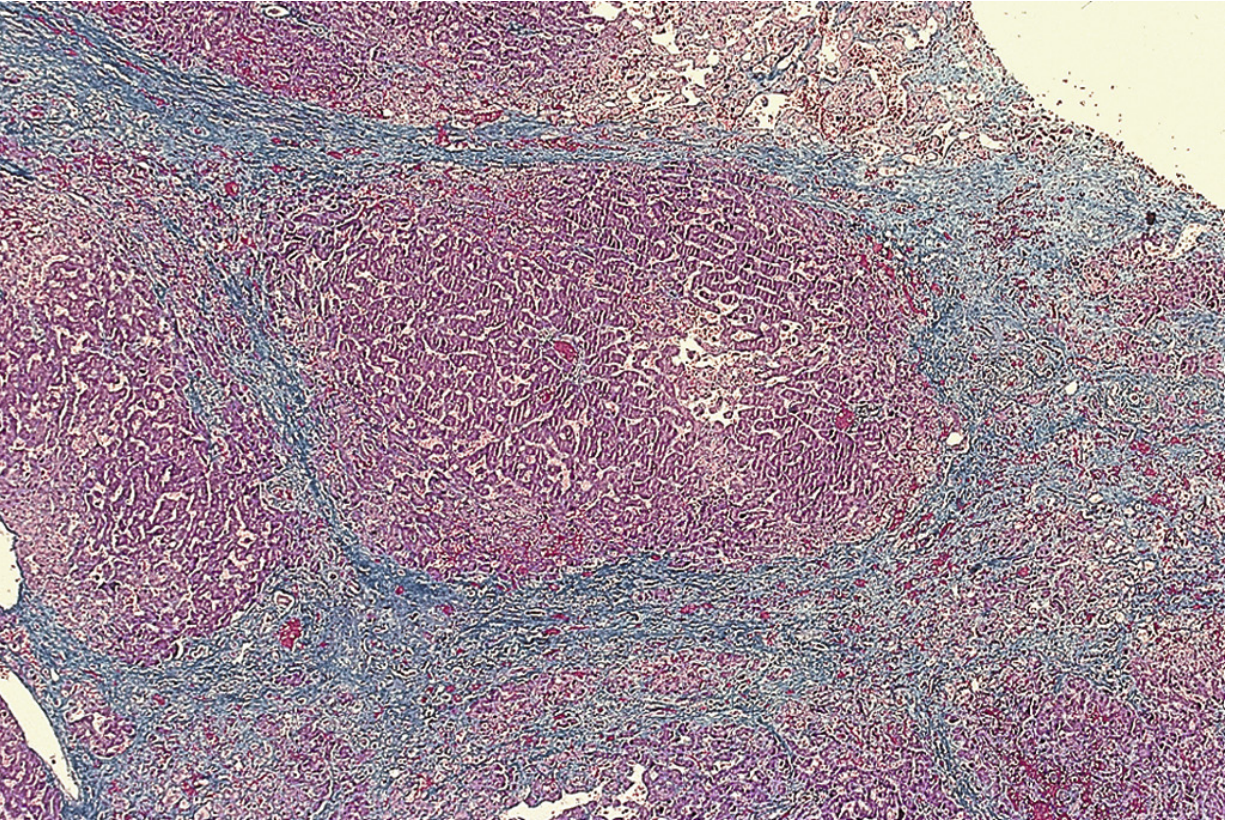
Mechanism: Right-heart failure → elevated CVP → hepatic venous hypertension → centrilobular sinusoidal congestion → hepatocyte atrophy in Zone 3 → perisinusoidal edema impairing O2 diffusion → "nutmeg liver"
Causes in this patient: Right heart failure from pacemaker-induced cardiomyopathy, cardiomyopathy, constrictive pericarditis, valvular heart disease
Clinical Features:
- Dull right upper quadrant ache
- Hepatomegaly (often tender)
- Hepatojugular reflux (compression over liver raises JVP)
- Pulsatile liver if tricuspid regurgitation is present
- Ascites (may need paracentesis if tense)
- Mild elevation of bilirubin (<3 mg/dL in most; jaundice in <10%)
- Prothrombin time prolonged in >75% - resistant to Vitamin K (hepatic dysfunction, not just coagulation factor deficiency)
- Spider angiomata and varices are usually absent
- Liver tests improve slowly with effective HF treatment
Investigations:
- LFTs: bilirubin (mild), ALP, GGT (elevated), ALT/AST (mild-moderate)
- INR/PT: prolonged
- Serum albumin: reduced in advanced disease
- USS/CT: hepatomegaly, dilated IVC + hepatic veins, ascites, portal hypertension signs
- CT portal phase: inhomogeneous hepatic enhancement ("nutmeg" pattern)
B. Ischemic Hepatitis ("Shock Liver")
Caused by systemic hypoperfusion superimposed on hepatic venous congestion (Yamada's Textbook of Gastroenterology). This is the acute liver injury component.
Key insight: Right-sided HF with hepatic venous congestion predisposes to ischemic hepatitis by lowering the threshold at which systemic hypotension triggers centrilobular necrosis. Even modest drops in BP (which alone would not cause ischemic hepatitis) can cause it in the background of hepatic congestion.
Causes of ischemic hepatitis in this patient:
- Low cardiac output (cardiogenic shock, severe HF)
- Arrhythmias (sustained VT/VF, complete AV block breakthrough)
- Myocardial infarction
- Cardiogenic shock
Diagnostic Features (Yamada's):
- Systemic hypotension + marked rapid elevation of serum aminotransferases (typically >1000 IU/mL) - rapidly reversible with treatment
- LDH markedly elevated with a low AST:LDH ratio - helps distinguish from viral hepatitis (in viral hepatitis, AST:LDH ratio is higher)
- Concomitant AKI almost always present (renal hypoperfusion)
- Classic histology: centrilobular necrosis
- Exclude other causes: viral hepatitis (HAV, HBV, HCV serology), drug/toxin-induced, autoimmune
Histopathology of Cardiac Cirrhosis (Sleisenger & Fordtran's, Masson trichrome):
Complete Liver Investigations:
| Test | Purpose |
|---|---|
| AST, ALT | Ischemic hepatitis: dramatically elevated (>1000); congestive: mildly elevated |
| ALP, GGT | Congestive hepatopathy: elevated (biliary pattern) |
| Total + direct bilirubin | Elevated in both; jaundice indicates severity |
| LDH | Markedly elevated in ischemic hepatitis; low AST:LDH ratio |
| PT/INR | Synthesisfunction marker; resistant to Vit K = hepatic origin |
| Serum albumin | Synthetic function; low = chronic/advanced |
| Serum ammonia | If encephalopathy suspected |
| Viral hepatitis serology | HAV IgM, HBsAg, anti-HCV, HCV RNA |
| Drug history | Paracetamol, statins, amiodarone, herbal supplements |
| Autoimmune panel | ANA, ASMA, AMA if other diagnoses unclear |
| USS hepatobiliary | Hepatomegaly, dilated hepatic veins/IVC, ascites, biliary disease |
| FibroScan (deferred) | Assess fibrosis burden if stable |
V. INTEGRATED MANAGEMENT STRATEGY
The primary treatment for all three organ complications is effective cardiac decongestion - reducing venous back-pressure and improving forward cardiac output.
1. Diuresis - Cornerstone of Therapy
Dose equivalency (NKF Primer on Kidney Diseases, 8e):
Furosemide 80 mg oral = Furosemide 40 mg IV = Torsemide 20 mg oral = Bumetanide 1 mg oral/IV
Strategy:
- IV route mandatory (oral absorption unreliable - furosemide bioavailability 10-90%, mean 50%)
- Start at double the home oral dose as IV furosemide
- Monitor urine output and urine Na+ at 1-2 hours post-dose; spot urine Na+ >50-70 mEq/L = adequate response
- DOSE trial: No significant difference between continuous infusion and bolus dosing; no need to routinely use infusion over bolus
- In CKD with GFR <30: bumetanide preferred - minimally affected by renal dysfunction, less ototoxicity
Diuretic Resistance (common with CKD background):
- Add metolazone (thiazide) for sequential/dual nephron blockade - monitor K+/Mg2+ closely
- Add spironolactone/eplerenone (MRA) - only if K+ <5.0 and creatinine stable
- SGLT2 inhibitor (e.g., empagliflozin) - proximal nephron blockade; emerging evidence in acute CRS (Systematic Review 2025, PMID 40435978)
- Ultrafiltration - precise, electrolyte-neutral fluid removal; preferred when diuretics fail
Goal of decongestion:
- Normalize JVP
- Clear pulmonary rales
- Resolve peripheral edema and hepatomegaly
- Reduce ascites
- Suppress BNP/NT-proBNP toward discharge
- Improve liver tests (will improve as congestion resolves)
2. Vasodilators
- IV nitroglycerin: first-line in hypertensive ADHF (SBP >140) + pulmonary edema
- Reduces preload and afterload; counteracts sympathetic/RAAS/AVP-mediated vasoconstriction
- IV nitroprusside: more potent; use with BP monitoring; caution in renal failure (thiocyanate toxicity)
- Avoid vasodilators if SBP <90 mmHg (worsens hepatic and renal perfusion)
3. Inotropes (Low-Output State / Cardiogenic Shock)
Per Harrison's 22E - indicated for hypoperfusion with end-organ dysfunction when diuretics insufficient:
| Agent | Notes |
|---|---|
| Dobutamine | First-line; reduces SVR; increases CO; arrhythmogenic; avoid if HR already high |
| Milrinone | PDE-3 inhibitor; greater SVR reduction; better on beta-blockers; dose-reduce in AKI (renally excreted); OPTIME-CHF: more arrhythmias and hypotension |
| Dopamine | Dose-dependent; "renal dose" dopamine has no benefit (ROSE-AHF) |
| Levosimendan | Calcium sensitizer; available outside USA; mixed trial results |
⚠️ Routine inotropes discouraged in ADHF - indicated only as bridge to LVAD/transplant or cardiogenic shock
4. Mechanical Circulatory Support (if refractory)
- Ultrafiltration / CRRT: preferred over intermittent HD in hemodynamically unstable patients (avoids BP swings that worsen hepatic and renal ischemia)
- IABP, Impella, VA-ECMO: for cardiogenic shock refractory to medical management
5. GDMT Continuation
| Drug Class | In-Hospital Action |
|---|---|
| Beta-blockers | Do NOT stop abruptly; reduce dose if low-output state |
| ACE-i/ARB/ARNI | Hold or reduce if AKI worsening or SBP <90; restart when stable |
| MRA | Hold if K+ >5.0 or rapidly rising creatinine |
| SGLT2 inhibitors | May continue or initiate in stable patients with HF |
| Digoxin | Continue with caution; reduce dose in AKI (renally cleared); monitor levels |
| Amiodarone | If on it, note hepatotoxicity potential - check LFTs against baseline |
VI. SPECIFIC HEPATIC MANAGEMENT
Since treatment of the heart treats the liver:
| Intervention | Rationale |
|---|---|
| Effective decongestion | Relieves hepatic venous congestion → LFTs improve with HF treatment |
| Avoid hepatotoxic drugs | NSAIDs, paracetamol overdose, statin (check level), amiodarone |
| Coagulopathy management | INR prolonged; vitamin K trial (1-3 doses); if resistant → hepatic synthetic failure |
| Nutrition support | Enteral preferred; avoid protein restriction unless encephalopathy present |
| Paracentesis | If tense ascites impairing breathing - therapeutic with albumin cover |
| Monitor for complications | Hepatic encephalopathy (raised ammonia, confusion), spontaneous bacterial peritonitis (if ascites) |
| Drug dosing adjustment | Reduce/avoid drugs with hepatic clearance: amiodarone, warfarin, statins, opioids |
In ischemic hepatitis specifically:
- Aminotransferases typically normalize within 1-2 weeks if systemic perfusion is restored
- Do NOT perform liver biopsy acutely - diagnosis is clinical
- Exclude viral hepatitis and drug causes with serology + drug history
- Acute liver failure workup (INR, ammonia, encephalopathy assessment) if rapidly deteriorating
VII. DRUG PRESCRIBING IN TRIPLE ORGAN DYSFUNCTION
This is where clinical judgment is most challenging:
| Drug | Kidney concern | Liver concern | Action |
|---|---|---|---|
| Furosemide | Impaired secretion in AKI → dose up | Hepatic edema impairs oral absorption | IV route; monitor response |
| LMWH (enoxaparin) | Accumulates in CKD → bleeding risk | Reduced clearance if severe liver failure | Use UFH; monitor anti-Xa if LMWH used |
| Milrinone | Renally excreted; half-life prolonged | Minimal hepatic metabolism | Dose reduce; monitor BP |
| ACE-i/ARB | Worsens AKI acutely | Caution in ascites (hepatorenal risk) | Hold until stable |
| Spironolactone | Hyperkalemia risk | Reduced hepatic metabolism | Hold if K+ >5.0 or severe liver failure |
| Amiodarone | Iodine load | Direct hepatotoxin; chronic use → cirrhosis | Check LFTs; consider discontinuation review |
| Statins | Generally safe | Can cause transaminase rise; avoid if ALT >3x ULN | Hold if severe acute liver injury |
| Morphine/Opioids | Accumulate in renal failure | Reduced hepatic metabolism; encephalopathy risk | Use cautiously; smallest effective dose |
| Contrast (CT) | Contrast nephropathy in AKI/CKD | Not an issue | Pre-hydrate; weigh risk/benefit; ISO-osmolar contrast |
| Metformin | Lactic acidosis risk in AKI | Lactic acidosis in hepatic failure | Hold immediately |
| NSAIDs | Worsens AKI | Worsens hepatic failure + coagulopathy | Contraindicated |
VIII. MONITORING TARGETS
| Parameter | Goal |
|---|---|
| SpO2 | ≥94-95% |
| Systolic BP | 90-140 mmHg |
| Heart rate (paced) | 60-90/min |
| Urine output | >0.5 mL/kg/hr |
| Net fluid balance | -500 to -1000 mL/day |
| Serum K+ | 3.5-5.0 mEq/L |
| Serum creatinine | Trend 12-hourly; acceptable mild rise during decongestion |
| LFTs (AST, ALT, bilirubin) | Trend; should normalize as cardiac function improves |
| PT/INR | Trend; improvement reflects hepatic recovery |
| BNP/NT-proBNP | Downtrend signals adequate decongestion |
| Lactate | Normalize (<2 mmol/L) - marker of end-organ perfusion |
| Blood glucose | Monitor closely - liver glycogen stores may be depleted |
IX. INDICATIONS FOR RENAL REPLACEMENT THERAPY
Consider CRRT (preferred over IHD in hemodynamic instability) if:
- Refractory volume overload
- K+ >6.5 mEq/L or ECG changes unresponsive to medical therapy
- Metabolic acidosis (pH <7.15)
- Uremic encephalopathy or pericarditis
- Inability to achieve adequate decongestion with diuretics/UF
CRRT also benefits the liver by removing inflammatory cytokines and ammonia, improving hepatic encephalopathy.
X. INDICATIONS FOR LIVER FAILURE WORKUP / HEPATOLOGY REFERRAL
Urgent hepatology referral if:
- INR >1.5 with encephalopathy (acute liver failure criteria - King's College criteria)
- Bilirubin rapidly rising
- Hepatic encephalopathy (confusion, asterixis, elevated ammonia)
- Does not improve with cardiac treatment (suggests alternative diagnosis)
Acute liver failure management adjuncts (if criteria met):
- N-acetylcysteine (improves microcirculatory flow; some evidence in non-paracetamol ALF)
- Lactulose + rifaximin for encephalopathy
- Vitamin K 10 mg IV x3 days (though likely resistant)
- Fresh frozen plasma only if bleeding (not for INR correction alone - distorts prognostication)
- CRRT + plasma exchange in severe cases
XI. DISPOSITION & TEAM
| Team | Role |
|---|---|
| Cardiology + EP/Device clinic | ADHF management, pacemaker interrogation, CRT evaluation |
| Nephrology | CKD/AKI management, RRT decisions |
| Hepatology | If liver injury severe or not improving |
| Intensivist | If hemodynamically unstable, cardiogenic shock |
| Setting | ICU/HDU given multi-organ dysfunction |
XII. KEY CLINICAL PITFALLS TO AVOID
- Withholding diuresis for fear of worsening AKI - venous congestion is the primary driver of AKI; aggressive decongestion improves survival even if creatinine rises transiently (Brenner & Rector)
- Attributing all liver injury to intrinsic hepatic disease - 95% of liver injury in ADHF is congestive/ischemic and will recover with cardiac treatment
- Using "renal-dose" dopamine - no benefit (ROSE-AHF)
- Giving vitamin K for coagulopathy without liver disease context - resistance to vitamin K confirms hepatic synthetic failure
- Not interrogating the pacemaker - device dysfunction may be the primary precipitant
- Stopping beta-blockers abruptly - worsens outcome in ADHF; reduce dose if needed
- NSAIDs for any pain - contraindicated in this triple-organ dysfunction setting
- Failing to check ammonia if patient is confused - hepatic encephalopathy can masquerade as "delirium" in elderly patients
Recent Evidence
- Congestive Hepatopathy Review (2024, PMID 38779242) - comprehensive review confirming treatment of underlying cardiac disease is the cornerstone; liver tests normalize with effective HF management
- Diuretic Resistance in Acute HF with Kidney Disease (2025, PMID 40435978) - SGLT2 inhibitors as adjuncts show promise; urine Na monitoring superior to weight for early diuretic response assessment
- ADHF Management Review (2025, PMID 41133931) - personalized approach to congestion; acetazolamide (ADVOR trial) aids decongestion but does not reduce readmissions
Sources: Harrison's Principles of Internal Medicine 22E (2025) | Brenner & Rector's The Kidney | NKF Primer on Kidney Diseases 8e | Yamada's Textbook of Gastroenterology 7e | Sleisenger & Fordtran's GI & Liver Disease
This is a shared conversation. Sign in to Orris to start your own chat.