
Pulmonary edema
"pulmonary edema"[MeSH Terms] AND management
pulmonary edema chest X-ray CXR radiograph
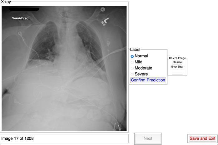
This diagnostic image illustrates a specialized medical data labeling interface designed for chest X-ray (CXR) classification. On the left, a semi-erect anteroposterior chest radiograph is displayed, showing a patient with visible medical support leads and tubing. The radiograph demonstrates diffuse bilateral lung opacities, commonly associated with conditions like COVID-19 pneumonia or pulmonary edema. The labeling panel on the right provides clinical severity classification options: Normal, Mild, Moderate, and Severe, with 'Normal' currently selected via a radio button. The interface includes interactive elements such as 'Confirm Prediction', an image resizing tool, and navigation controls ('Next', 'Save and Exit'). This platform is intended for medical education and research to establish ground truth datasets by capturing specialist interpretations of disease severity. Key concepts shown include thoracic radiology, disease severity grading, and the development of clinical decision support systems.
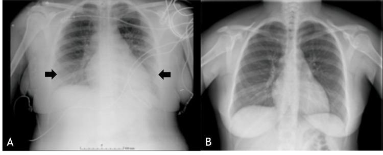
This comparison chart consists of two anteroposterior chest X-ray (CXR) images illustrating the progression and resolution of acute pulmonary edema. Image A, labeled as a postpartum clinical photograph from the postanesthesia care unit (PACU), demonstrates a significant bilateral pathology characterized by diffuse pulmonary infiltrates and marked hilar engorgement, indicated by black arrows. The cardiac silhouette appears widened, and peribronchial cuffing is suggestive of interstitial fluid accumulation. Monitoring leads and a central line are visible, indicating an acute clinical setting. In contrast, Image B shows the same patient five days later, following diuretic therapy. This follow-up radiograph reveals a complete resolution of the previous abnormalities; the lung fields are clear, the costophrenic angles are sharp, the hilar regions have returned to normal size, and the bronchovascular markings are within normal limits. The pair serves as a diagnostic comparison for identifying acute pulmonary congestion and its resolution over time in an obstetric or postoperative context.
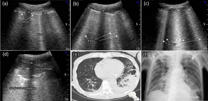
A multi-modal comparison of thoracic imaging findings in aspiration pneumonia, featuring point-of-care ultrasound (POCUS), computed tomography (CT), and chest X-ray (CXR). Panels (a)-(d) demonstrate a spectrum of lung ultrasound findings: (a) shows a single vertical B-line artifact originating from the pleural line; (b) displays multiple (≥3) B-lines; (c) shows confluent (≥5) B-lines creating a 'white lung' appearance; and (d) identifies sub-pleural consolidation (a hypoechoic area) and adjacent pleural effusion. Panel (e) is an axial CT scan of the chest showing significant consolidation and air bronchograms in the left lung, typical of aspiration pneumonia. Panel (f) is a posterior-anterior chest radiograph from the same patient, illustrating a loss of the silhouette of the left diaphragmatic arch behind the heart, indicating left lower lobe pathology. The collection serves as an educational comparison of diagnostic modalities for identifying pulmonary edema, interstitial syndrome, and consolidation in a clinical setting.
pulmonary edema pathophysiology alveolar interstitial fluid diagram
Pulmonary Edema
Definition
Pathophysiology
Jv = K × [(Pc - Pi) - σ(πc - πi)]
- Pc = pulmonary capillary hydrostatic pressure (avg 7 mmHg)
- Pi = interstitial hydrostatic pressure (-4 to -8 mmHg)
- πc = capillary oncotic pressure (~28 mmHg)
- πi = interstitial oncotic pressure (~14 mmHg)
- K = filtration coefficient; σ = reflection coefficient (albumin permeability)
The Pulmonary Edema Safety Factor
Classification and Causes
1. Hemodynamic (Cardiogenic) Pulmonary Edema
| Mechanism | Examples |
|---|---|
| Increased pulmonary venous pressure | Left ventricular failure (most common), mitral stenosis, left atrial obstruction |
| Volume overload | Fluid overload, large L-to-R shunts, severe anemia |
| Decreased oncotic pressure | Hypoalbuminemia, nephrotic syndrome, liver disease, protein-losing enteropathy |
| Lymphatic obstruction | Rare; post-transplant lymphangitis |
2. Noncardiogenic (Increased Permeability) Pulmonary Edema
| Type | Examples |
|---|---|
| Direct lung injury | Pneumonia (bacterial, viral), inhaled toxins (Cl₂, SO₂, O₂ in high concentration), aspiration (gastric contents), radiation, trauma |
| Indirect (systemic) injury | Sepsis/SIRS, pancreatitis, burns, extensive trauma, blood transfusion (TRALI) |
| Drugs/chemicals | Heroin, cocaine, methadone, bleomycin, amphotericin B, paraquat, kerosene |
3. Special/Mixed Types
| Type | Mechanism |
|---|---|
| High-altitude pulmonary edema (HAPE) | Hypoxic pulmonary vasoconstriction + microvascular leak |
| Neurogenic pulmonary edema | Massive sympathetic surge → severe pulmonary hypertension → alveolar-capillary disruption |
| Negative pressure pulmonary edema | Forceful inspiration against obstruction → markedly negative interstitial pressure → increased transmural capillary pressure |
| Re-expansion pulmonary edema | Rapid lung re-expansion after prolonged collapse |
| CKD/uremic pulmonary edema | Multifactorial: fluid overload + heart failure + uremic capillary injury + hypoalbuminemia - Murray & Nadel, p. 2962 |
Clinical Features
- Dyspnea - initially on exertion, then at rest; orthopnea, paroxysmal nocturnal dyspnea (PND)
- Tachypnea, tachycardia
- Cough - pink, frothy sputum in severe alveolar flooding
- Hypoxemia - often severe; central cyanosis
- Crackles (crepitations) - bilateral, basal initially, then spreading upward
- Wheeze - "cardiac asthma" from peribronchial edema
- Signs of underlying cause - raised JVP, S3 gallop, displaced apex in heart failure
Diagnosis
Chest X-Ray
- Kerley B lines - horizontal lines at lung bases (interstitial edema)
- Peribronchial cuffing
- Hilar enlargement/"bat-wing" pattern
- Bilateral diffuse alveolar opacities (alveolar flooding)
- Pleural effusions (cardiogenic)
- Cardiomegaly (in cardiogenic cases)
Other Investigations
- Lung ultrasound (POCUS): B-lines (≥3 per field = interstitial syndrome; "white lung" = severe alveolar edema) - highly sensitive and now preferred in acute settings
- BNP/NT-proBNP: elevated in cardiogenic; helps differentiate from noncardiogenic
- Pulmonary artery occlusion (wedge) pressure: >18 mmHg = cardiogenic (hemodynamic); normal = noncardiogenic
- Edema fluid protein ratio: low (<0.5) in cardiogenic; high (>0.7) in permeability edema
- ABG: hypoxemia ± hypocapnia (early), hypercapnia (late/severe)
- ECG, troponin, echo to assess cardiac cause
Management
Immediate/General Measures
- Sit patient upright (reduces preload, improves diaphragm mechanics)
- High-flow oxygen by mask (target SpO₂ ≥94%)
- Non-invasive ventilation (NIV): CPAP or BiPAP reduces preload, recruits alveoli, and reduces work of breathing. Strong evidence for benefit in cardiogenic pulmonary edema
- Intubation and mechanical ventilation if NIV fails or patient is obtunded
Cardiogenic Pulmonary Edema
| Drug | Action | Notes |
|---|---|---|
| Loop diuretics (furosemide IV 40-100 mg) | Reduces preload (immediate venodilation) + promotes diuresis | Drug of choice; also bumetanide, torsemide, ethacrynic acid |
| Nitrates (sublingual/IV nitroglycerin) | Venodilation → reduces preload; also afterload at higher doses | Start at 10-20 μg/min IV, titrate to effect |
| Morphine (cautiously) | Venodilation + anxiolysis | Use with caution - some evidence of harm in acute HF |
| ACE inhibitors | Reduce afterload | Useful in hypertensive pulmonary edema |
| Inotropes (dobutamine, milrinone) | Improve LV function | For cardiogenic shock/low-output failure |
| Nitroprusside | Mixed arterio/venodilation | For severe hypertension + pulmonary edema |
| IABP / Mechanical assist | Augment cardiac output | Acute ischemia/cardiogenic shock |
Noncardiogenic Pulmonary Edema (ALI/ARDS)
- Treat the underlying cause (antibiotics for sepsis, etc.)
- Lung-protective ventilation: tidal volume 6 mL/kg ideal body weight, PEEP titration
- Conservative fluid strategy (after resuscitation phase)
- Prone positioning in severe ARDS (PaO₂:FiO₂ <150)
- Neuromuscular blockade in severe cases
- Steroids (dexamethasone) in moderate-severe ARDS
Pulmonary Edema in CKD/ESRD
- Emergent dialysis or continuous venovenous hemofiltration (CVVH) is the definitive treatment - medical therapy alone is usually inadequate
- IV nitrates (sublingual nitroglycerin) for immediate preload reduction
- CPAP/BiPAP as adjunct
- Phlebotomy (150 mL) is safe in extreme/low-resource situations
- Loop diuretics only if residual renal function remains
- Peritoneal dialysis is too slow for acute pulmonary edema - Tintinalli's Emergency Medicine, p. 2756; Rosen's Emergency Medicine, p. 4854
High-Altitude Pulmonary Edema (HAPE)
- Immediate descent is the primary treatment
- Supplemental oxygen
- Nifedipine (pulmonary vasodilator)
- Portable hyperbaric chamber if descent delayed
- Phosphodiesterase-5 inhibitors (sildenafil, tadalafil) as prophylaxis
Pathological Stages
| Stage | Features |
|---|---|
| Stage 1 - Interstitial edema | Fluid in the interstitium; perivascular "cuffing"; Kerley B lines on CXR; mild hypoxemia |
| Stage 2 - Alveolar flooding | Fluid crosses the alveolar epithelium; alveolar opacities on CXR; severe hypoxemia |
| Chronic cardiogenic | "Heart failure cells" (hemosiderin macrophages), brown induration, fibrosis of alveolar walls |
Key Distinguishing Features: Cardiogenic vs. Noncardiogenic
| Feature | Cardiogenic | Noncardiogenic |
|---|---|---|
| Pulmonary wedge pressure | >18 mmHg | ≤18 mmHg |
| Edema fluid protein | Low (transudate) | High (exudate) |
| BNP | Elevated | Usually normal/low |
| CXR | Cardiomegaly + effusions common | No cardiomegaly; bilateral infiltrates |
| Response to diuretics | Good | Poor |
| Underlying cause | Heart failure, fluid overload | Sepsis, ARDS, toxin, trauma |
- Guyton and Hall Textbook of Medical Physiology, pp. 509
- Robbins, Cotran & Kumar Pathologic Basis of Disease, p. 2189
- Morgan and Mikhail's Clinical Anesthesiology (7e), pp. 2461-2464
- Murray & Nadel's Textbook of Respiratory Medicine, pp. 2962, 3109
- Tintinalli's Emergency Medicine, p. 2756
- Rosen's Emergency Medicine, pp. 4849-4855