
Anatomy of larynx
Reading File
Now let me search for larynx anatomy in the medical library.
Finding Sources
Finding Sources
Excellent - found dedicated sections. Let me read the most relevant sources in parallel.
Reading File
Reading File
Reading File
Good content. Let me read more from Scott-Brown's (the dedicated anatomy chapter) and Gray's Anatomy for deeper detail on cartilages, muscles, and nerves, plus fetch key images.
Reading File
Reading File
Loading Image
Excellent content. Let me read more of Gray's Anatomy (muscles, nerves, blood supply sections) and continue the Scott-Brown chapter on muscles and innervation.
Reading File
Reading File
Loading Image
Excellent material. Let me now read the intrinsic muscles table and innervation/blood supply sections from Scott-Brown and Gray's Anatomy.
Reading File
Reading File
I now have comprehensive material. Let me also read the intrinsic muscles table from Scott-Brown and fetch one more useful image.
Reading File
Loading Image
I have excellent, comprehensive material from Scott-Brown's Otorhinolaryngology and Gray's Anatomy for Students. I can now compile a thorough, well-illustrated answer.
Anatomy of the Larynx
The larynx is a hollow musculoligamentous structure with a cartilaginous framework that caps the lower respiratory tract. It serves two primary functions: acting as a valve (sphincter) to close the lower respiratory tract during swallowing, and as an instrument to produce sound (phonation).
1. Position and General Description
The larynx extends from the laryngeal inlet (at the tip of the epiglottis, level of C3) to the inferior border of the cricoid cartilage (level of C6). At rest, it lies anterior to the C3-C6 vertebrae - slightly higher in women. It is continuous below with the trachea and opens above into the pharynx, posterior and slightly inferior to the tongue.
The cavity of the larynx is divided into three anatomical regions by two pairs of mucosal folds:
| Region | Boundaries | Contents |
|---|---|---|
| Supraglottis | Epiglottis/aryepiglottic folds down to the apex of the laryngeal ventricle | Epiglottis, aryepiglottic folds, arytenoids, false vocal folds (vestibular folds), ventricle |
| Glottis | Upper border of ventricle apex to 1 cm below free edge of vocal folds | True vocal folds, anterior and posterior commissures |
| Subglottis | 1 cm below vocal fold free edge to inferior cricoid border | Transitions into trachea |
Note: The term "vocal cord" is anatomically imprecise - "vocal fold" is preferred, as these structures vibrate in a multiplanar (including vertical) fashion. - Scott-Brown's Otorhinolaryngology
2. The Cartilaginous Framework
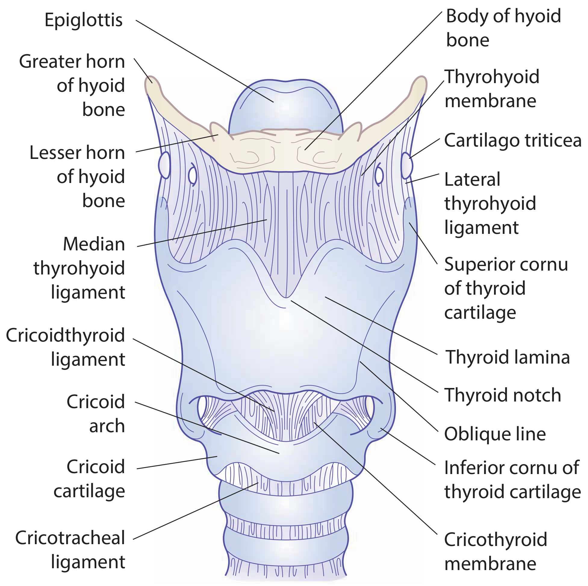
Framework diagram (Scott-Brown's):
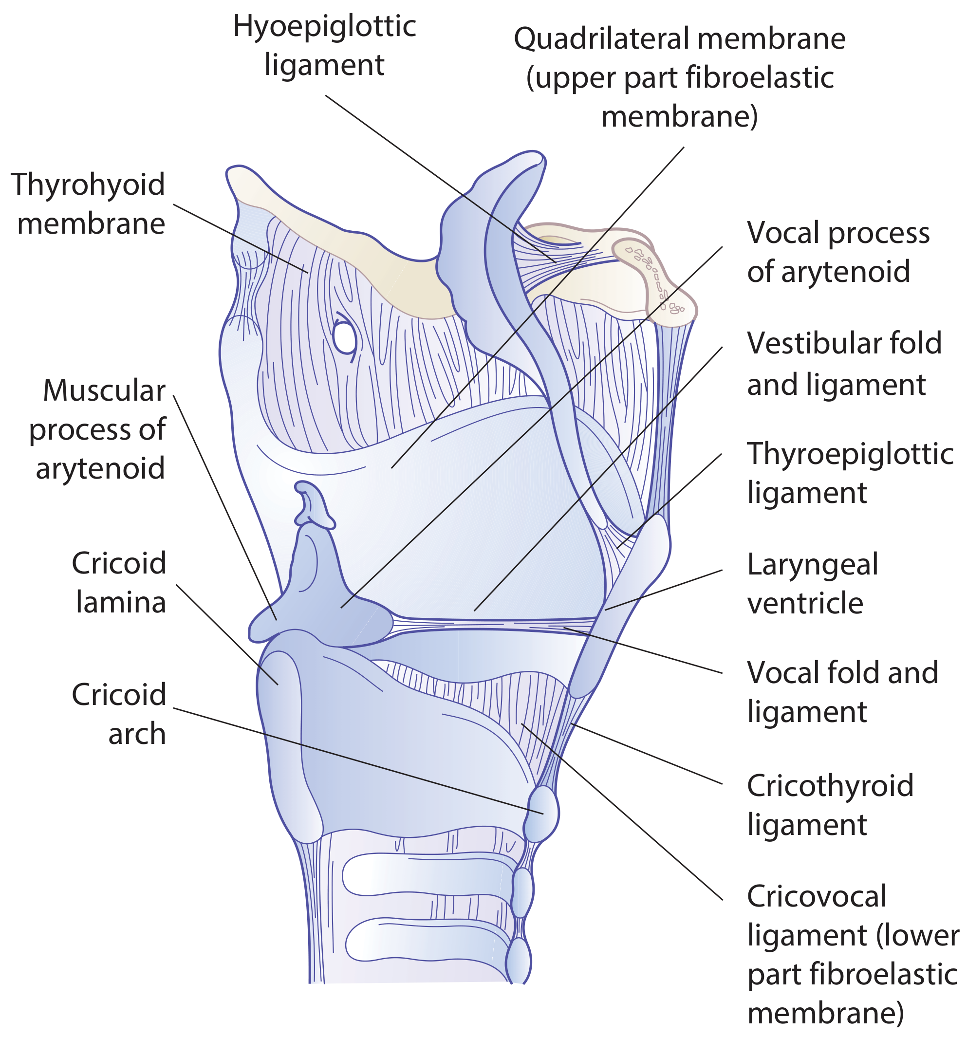
Sagittal section showing internal membranes and folds:
Unpaired Cartilages (3)
1. Cricoid Cartilage
- The most inferior laryngeal cartilage and the only one that completely encircles the airway
- Shaped like a signet ring: broad posterior lamina and narrow anterior arch
- Posterior lamina has two oval depressions (for posterior cricoarytenoid muscles) separated by a vertical ridge (for esophageal attachment)
- Articular facets: superolateral surface of lamina (for arytenoid cartilages) + lateral surface near base (for inferior horns of thyroid cartilage)
2. Thyroid Cartilage
- The largest laryngeal cartilage
- Two laminae fuse anteriorly to form the laryngeal prominence (Adam's apple)
- Fusion angle: ~90° in men, ~120° in women (hence more prominent in men)
- The superior thyroid notch is palpable above the prominence
- Posterior border of each lamina extends as superior horn (connected via lateral thyrohyoid ligament to hyoid's greater horn) and inferior horn (articulates with cricoid)
- Oblique line on the lateral surface is the attachment for sternothyroid, thyrohyoid, and inferior pharyngeal constrictor muscles
3. Epiglottis
- Leaf-shaped elastic cartilage (does not ossify)
- Attached below by the thyroepiglottic ligament to the back of the thyroid prominence, and superiorly by the hyoepiglottic ligament to the hyoid bone
- Connected to the tongue by the median and lateral glossoepiglottic folds; the valleculae are the recesses between these folds
Paired Cartilages (3 pairs)
Arytenoid cartilages - pyramid-shaped; sit on the sloping superolateral surface of the cricoid lamina. Each has:
- Vocal process (anteriorly) - attachment for vocal ligament
- Muscular process (posterolaterally) - attachment for most intrinsic muscles
- Movement at the cricoarytenoid joint (rotation + gliding) controls the rima glottidis
Corniculate cartilages - small, elastic; perch on the apex of each arytenoid; form the corniculate tubercles in the aryepiglottic folds
Cuneiform cartilages - small, elastic; lie within the aryepiglottic folds anterolateral to the corniculates; form the cuneiform tubercles
All cartilages except the epiglottis, corniculates, cuneiforms, and the apex/vocal process of the arytenoids are hyaline cartilage and begin to ossify around age 20. The remainder are elastic cartilage and do not ossify. - Cummings Otolaryngology
3. Ligaments and Membranes
The larynx is held together by an internal fibroelastic membrane divided into two parts:
| Membrane | Location | Free Border = Ligament |
|---|---|---|
| Quadrangular membrane (upper part) | Between epiglottis and arytenoid cartilages | Upper free border = aryepiglottic ligament; Lower free border = vestibular (false vocal fold) ligament |
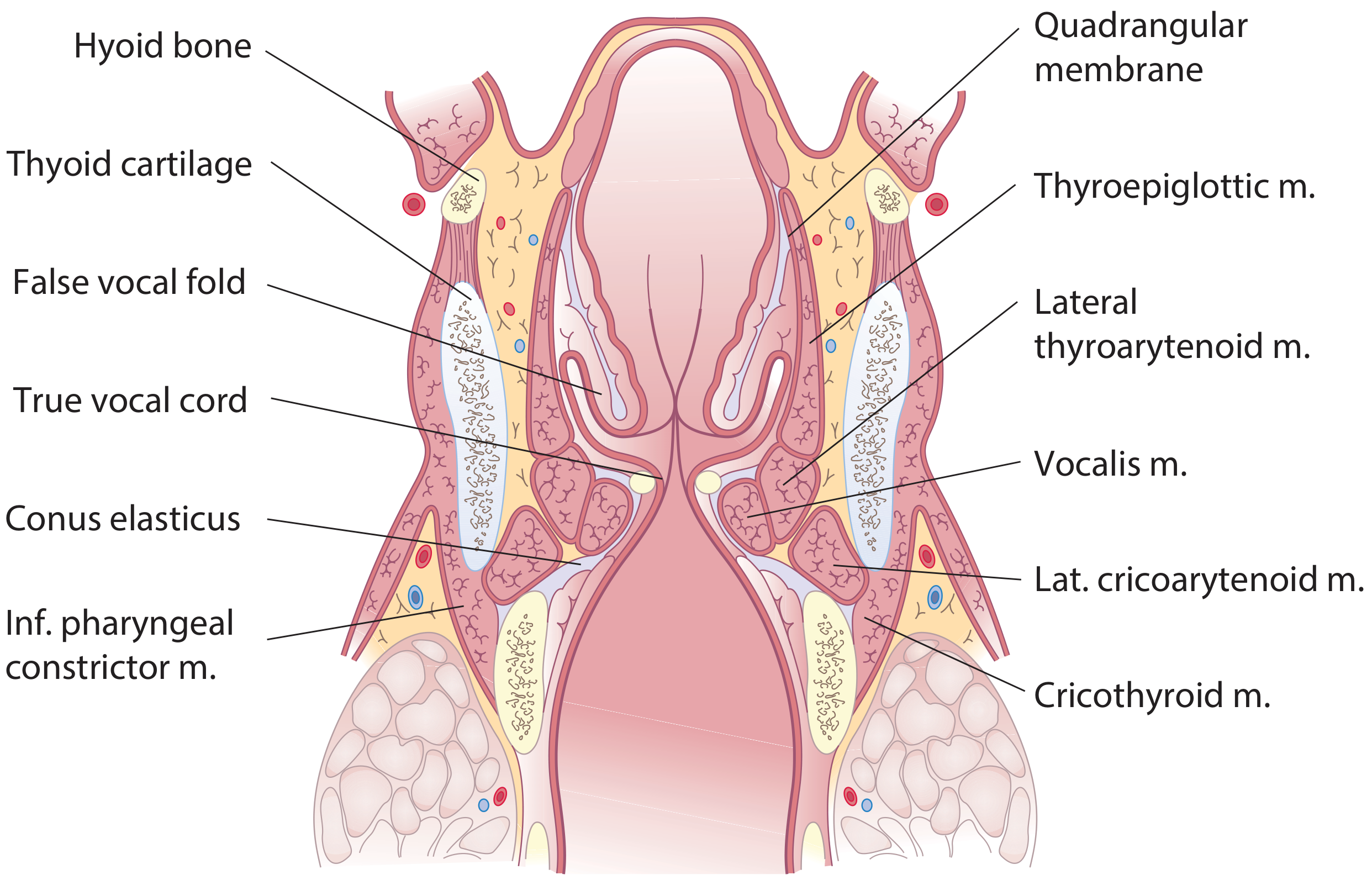
| Conus elasticus / Cricovocal membrane (lower part) | Arises from upper border of cricoid arch | Upper free border = vocal ligament (forms core of true vocal fold) |
External ligaments:
- Thyrohyoid membrane - connects thyroid cartilage to hyoid; pierced by the internal branch of the superior laryngeal nerve and superior laryngeal artery
- Cricothyroid ligament (median cricothyroid ligament) - the site of emergency cricothyrotomy
- Cricotracheal ligament - connects cricoid to the first tracheal ring
4. Intrinsic Muscles
Coronal section showing muscles:
Abductor (opens glottis)
| Muscle | Origin | Insertion | Action |
|---|---|---|---|
| Posterior cricoarytenoid (PCA) | Lower/medial surface of cricoid lamina | Muscular process of arytenoid | Only abductor - rotates arytenoids to separate vocal processes; also elevates vocal tip |
Adductors (close glottis)
| Muscle | Origin | Insertion | Action |
|---|---|---|---|
| Lateral cricoarytenoid (LCA) | Superior border of cricoid arch (lateral part) | Muscular process of arytenoid | Adducts and lowers vocal process - adducts and thins the vocal fold |
| Transverse arytenoid (unpaired) | Posterior surface/outer edge of one arytenoid | Same on other arytenoid | Adducts the arytenoids (closes posterior commissure) |
| Oblique arytenoids (paired) | Muscular process of one arytenoid | Apex of opposite arytenoid | Adduct and help close laryngeal inlet (fibers continue as aryepiglottic muscles) |
Tension Control
| Muscle | Action |
|---|---|
| Cricothyroid | Tilts thyroid forward / tilts cricoid backward → lengthens, tenses, and thins the vocal fold - the main pitch-raising muscle |
| Thyroarytenoid (vocalis) | Shortens and thickens the vocal fold; the medial vocalis fibers fine-tune vocal fold tension during phonation |
The cricothyroid is the only intrinsic muscle innervated by the external branch of the superior laryngeal nerve (not the RLN). All other intrinsic muscles are supplied by the recurrent laryngeal nerve (RLN). - Scott-Brown's Otorhinolaryngology
5. Extrinsic Muscles
These move the larynx as a whole (elevate or depress):
Infrahyoid (strap muscles - depress the larynx):
- Sternothyroid (ansa cervicalis C2,3) - inserts onto oblique line of thyroid
- Sternohyoid (ansa cervicalis C1,2,3) - depresses hyoid
- Thyrohyoid (hypoglossal, C1 root) - elevates larynx on fixed hyoid or depresses hyoid on fixed larynx
Suprahyoid (elevate the larynx):
- Mylohyoid, geniohyoid, stylohyoid, digastric, stylopharyngeus, palatopharyngeus
6. Laryngeal Ventricle and Saccule
Between the vestibular fold (false cord) above and the vocal fold (true cord) below, the mucosa bulges laterally to form the laryngeal ventricle on each side. An anterosuperior tubular extension - the laryngeal saccule - projects between the vestibular fold and thyroid lamina, containing mucous glands that lubricate the vocal folds. Abnormal dilatation of the saccule creates a laryngocele.
7. Nerve Supply
All laryngeal innervation is via branches of the vagus nerve (CN X):
| Nerve | Pathway | Motor supply | Sensory supply |
|---|---|---|---|
| Superior laryngeal nerve (SLN) - internal branch | Pierces thyrohyoid membrane with superior laryngeal artery | None | Mucosa above vocal folds (supraglottis + piriform fossa) |
| Superior laryngeal nerve (SLN) - external branch | Runs on inferior constrictor | Cricothyroid muscle only | None |
| Recurrent laryngeal nerve (RLN) - right | Loops under right subclavian artery; ascends in tracheoesophageal groove | All other intrinsic muscles | Mucosa below vocal folds (subglottis) |
| Recurrent laryngeal nerve (RLN) - left | Loops under aortic arch + ligamentum arteriosum; longer course | Same as right | Same as right |
Galen's anastomosis: The internal branch of SLN joins an ascending sensory branch of the RLN below the mucosa of the posterior larynx - a purely sensory communication.
8. Blood Supply and Lymphatics
| Vessel | Territory |
|---|---|
| Superior laryngeal artery (branch of superior thyroid artery from external carotid) | Upper larynx; enters with internal branch of SLN through thyrohyoid membrane |
| Inferior laryngeal artery (branch of inferior thyroid artery from thyrocervical trunk) | Lower larynx |
Lymphatic drainage follows the anatomical compartments:
- Supraglottis - drains to upper and middle deep cervical nodes (levels II, III) - rich lymphatic network, explaining higher rates of nodal metastasis in supraglottic cancers
- Glottis - the true vocal folds have virtually no lymphatics, which is why early glottic carcinoma rarely metastasizes to lymph nodes
- Subglottis - drains to paratracheal and lower deep cervical nodes (levels IV, VI)
9. Functional Summary
| Function | Mechanism |
|---|---|
| Respiration | PCA abducts vocal folds (rima glottidis opens, rhomboid shape) |
| Phonation | LCA + arytenoids adduct; air forces vibration; cricothyroid/vocalis adjust pitch and tension |
| Swallowing | Larynx elevates and moves forward; epiglottis swings down; rima glottidis and vestibule close |
| Effort closure | Complete glottic + vestibular closure to stabilize thorax (e.g., heavy lifting) |
| Cough reflex | Tight adduction followed by explosive release of subglottic pressure |
10. Clinical Correlates
- Cricothyrotomy: Emergency airway access through the median cricothyroid ligament - few structures between it and skin, making it the fastest surgical airway
- Tracheostomy: Performed below the cricoid when prolonged airway access is needed
- RLN injury: Unilateral = hoarseness/breathy voice (paralyzed vocal fold in paramedian position); Bilateral = stridor and respiratory distress
- SLN injury (external branch): Loss of cricothyroid function → inability to raise vocal pitch; common risk during thyroidectomy
- Laryngocele: Abnormal air-filled dilation of the laryngeal saccule; may be internal, external, or mixed
- Glottic cancer: Rarely metastasizes early due to poor lymphatics of the true vocal folds
Sources: Scott-Brown's Otorhinolaryngology Head & Neck Surgery; Gray's Anatomy for Students; Cummings Otolaryngology Head and Neck Surgery
This is a shared conversation. Sign in to Orris to start your own chat.