
Schannoma
I'll look up Schwannoma in the medical library for you right away.
Searching sources...
Reading File
Finding Sources
Finding Sources
Searching PubMed
schwannoma diagnosis treatment
Now let me read the most comprehensive sections - Harrison's (general overview), Andrews' Dermatology (skin schwannoma), and Campbell's Orthopaedics (surgical perspective) in parallel:
Reading File
Reading File
Reading File
Let me get the rest of the Campbell's surgical section and also fetch the key MRI image:
Reading File
Loading Image
Loading Image
Excellent - I now have rich, comprehensive information from multiple authoritative textbooks. Here is the full clinical overview:
Schwannoma (Neurilemmoma)
Definition & Origin
A schwannoma is a generally benign, encapsulated tumor arising from Schwann cells of peripheral, cranial, or spinal nerve sheaths. Also called a neurilemmoma, it is the most common benign peripheral nerve sheath tumor. Unlike neurofibromas, schwannomas do not infiltrate adjacent nerve fascicles - they push the nerve fibers aside, which is the key to nerve-sparing surgery.
- Harrison's Principles of Internal Medicine 22E, p. 765
- Andrews' Diseases of the Skin, p. 719
Epidemiology & Associations
| Feature | Detail |
|---|---|
| Sex | More common in women (peripheral type) |
| Usual presentation | Solitary, sporadic |
| NF2 (chromosome 22) | Bilateral vestibular schwannomas; multiple schwannomas |
| NF1 (chromosome 17q11.2) | Spinal nerve root schwannomas (less commonly) |
| Carney syndrome | Melanotic psammomatous schwannoma + spotty pigmentation + myxomas + endocrine overactivity |
Multiple schwannomas without NF features may be sporadic or familial (schwannomatosis).
Clinical Types & Locations
1. Vestibular Schwannoma (Acoustic Neuroma) - Most Common
- Arises from the vestibular portion of CN VIII
- Accounts for ~9% of all primary brain tumors
- Symptoms: Progressive unilateral hearing loss, tinnitus, dizziness; with large tumors - brainstem/cerebellar compression
- NF2 patients get bilateral vestibular schwannomas
2. Peripheral Schwannoma
- Solitary nodule, 3-30 mm in diameter
- Soft/firm, pale pink or yellowish
- Common sites: flexor surfaces of arms/wrists/knees, scalp, neck, tongue
- May or may not be painful; positive Tinel sign is characteristic
3. Plexiform Schwannoma
- Involves multiple fascicles; occurs as solitary or multiple lesions
- May be isolated or associated with NF1, NF2, or schwannomatosis
4. Ancient Schwannoma
- Long-standing schwannoma with nuclear atypia - a benign degenerative change, NOT malignant
- No mitotic figures; must be distinguished from malignant peripheral nerve sheath tumor (MPNST)
Imaging (MRI)
MRI is the investigation of choice:
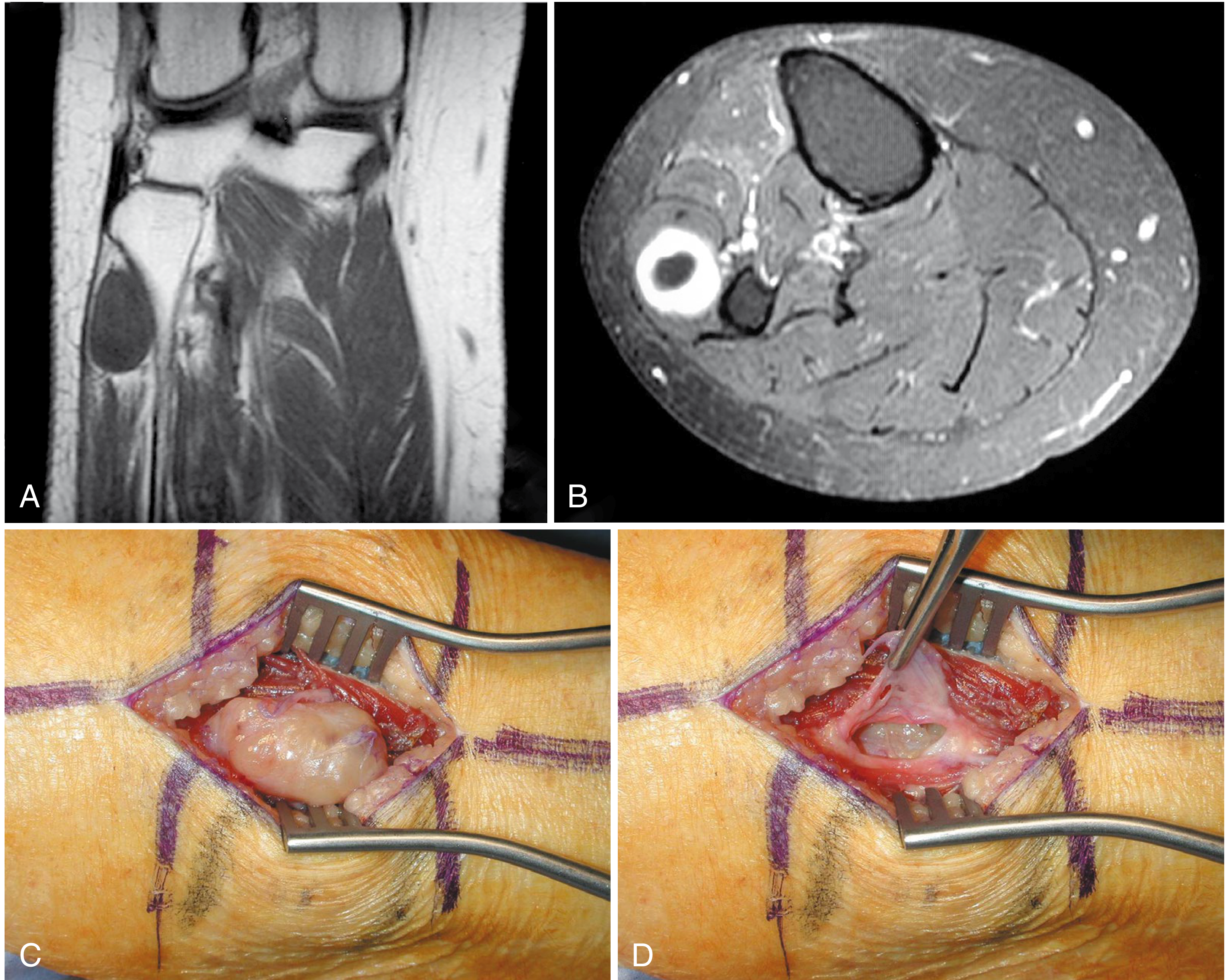
- Fusiform mass along the course of a major peripheral nerve
- "Split fat sign" on T1: a rim of fat surrounding the lesion
- "Target sign" on T2 (seen in ~50%): decreased central T2 signal with increased peripheral T2 signal - suggests benign diagnosis
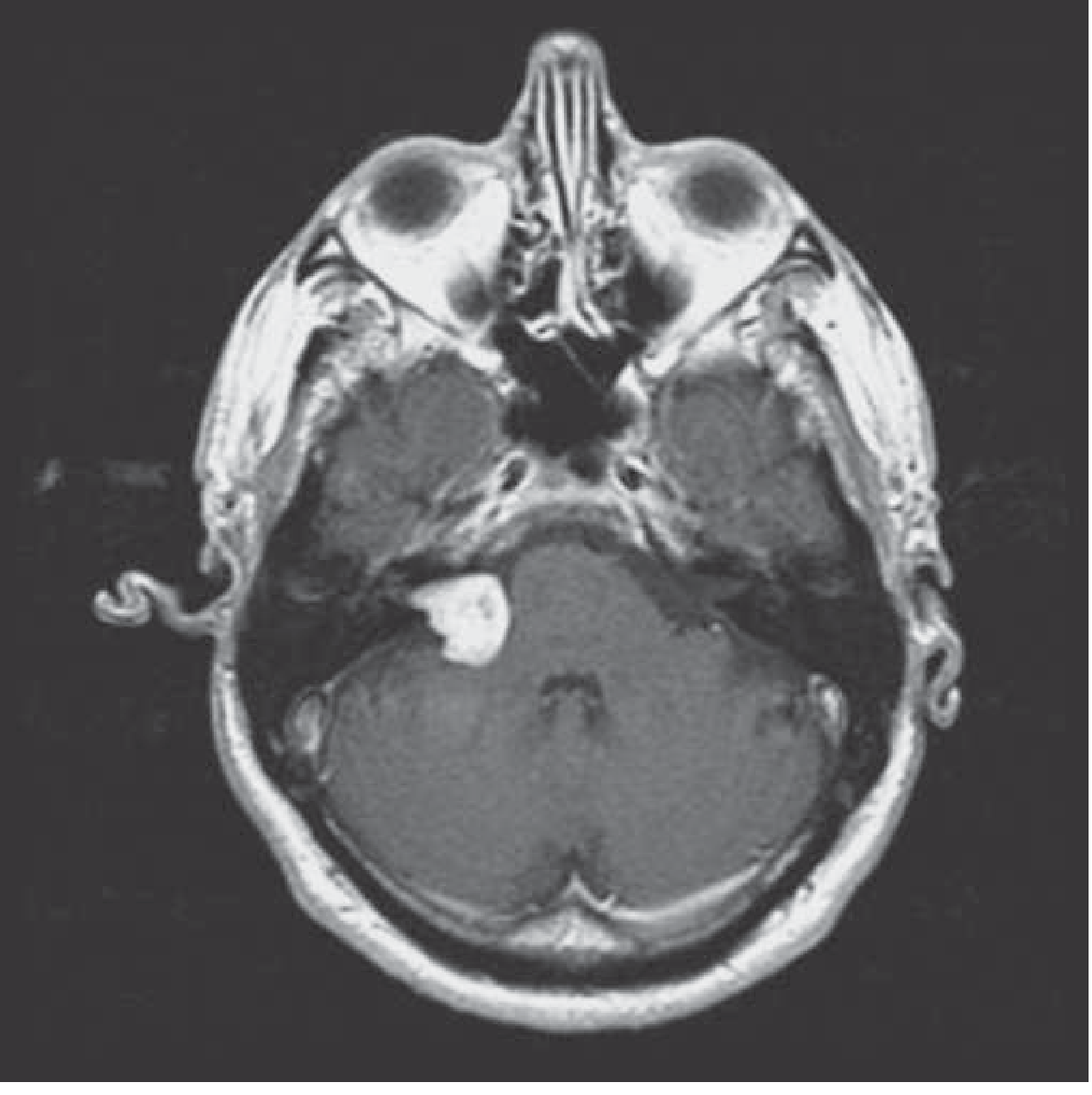
- Vestibular schwannoma: densely enhancing lesion, enlarges the internal auditory canal, extends into the cerebellopontine angle (CPA)
Postgadolinium MRI of right vestibular schwannoma (Harrison's, p. 765):
Peripheral schwannoma of peroneal nerve with intraoperative view (Campbell's Orthopaedics 15e):
Red flags for malignancy on imaging:
- Rapid size increase
- Peripheral enhancement pattern
- Perilesional soft-tissue edema
- Cystic change within the tumor
- Increased FDG uptake on PET scan
Histopathology - The Key Features
Schwannomas have two characteristic tissue patterns:
| Pattern | Description | Gross appearance |
|---|---|---|
| Antoni A | Compact spindle cells with marked nuclear palisading; Verocay bodies (two rows of palisading nuclei separated by fibrillary material) | Hard, firm |
| Antoni B | Loose, myxomatous, degenerative; ectatic/thick-walled blood vessels; cystic spaces | Soft, edematous |
Immunohistochemistry:
- S-100 positive (nuclear and cytoplasmic) - hallmark
- Also positive for vimentin and myelin basic protein (in Antoni A)
- EMA-positive capsule (derived from perineurium)
- Very few/no nerve fibers within the tumor bulk (a compressed nerve may be seen at one edge)
Treatment
Peripheral schwannomas:
- Surgical excision via longitudinal perineurial incision with careful blunt dissection
- Tumor can be shelled out without sacrificing nerve fascicles
- Excision is almost invariably curative
- Transient neurologic deficits occur in ~1/3 of cases post-resection
- Risk of neurologic injury is higher with tumors > 3 cm, age > 50, and longer symptom duration
Vestibular schwannomas:
- Very small, asymptomatic lesions: observe with serial MRIs
- Larger/symptomatic lesions: surgery or stereotactic radiosurgery (SRS)
- In patients with small tumors and intact hearing: early surgical intervention improves chance of hearing preservation
Malignant variant (MPNST - "malignant schwannoma"):
- Arises from long-standing neurofibromas in NF1 setting (NOT from benign schwannomas)
- Treatment: wide resection + radiotherapy
Key Differentials
| Feature | Schwannoma | Neurofibroma |
|---|---|---|
| Encapsulation | Yes | No |
| Nerve involvement | Displaces fascicles | Infiltrates fascicles |
| NF association | NF2 (also NF1) | NF1 (von Recklinghausen) |
| S-100 | Positive | Positive (less uniform) |
| Malignant potential | Very rare | Higher (plexiform type in NF1) |
| Surgery | Nerve-sparing excision possible | May require nerve sacrifice |
Recent Evidence (2024-2026)
Two recent systematic reviews are worth noting for vestibular schwannoma management:
- A 2024 network meta-analysis (PMID 39627752) compared treatment options for unilateral vestibular schwannoma across surgery, SRS, and observation.
- A 2026 systematic review and meta-analysis (PMID 41794963) in Journal of Neurology evaluated quality of life across all three management strategies (observation, SRS, microsurgery), providing patient-centred guidance.
Sources: Harrison's Principles of Internal Medicine 22E (p. 765) | Andrews' Diseases of the Skin (p. 719) | Campbell's Operative Orthopaedics 15e (p. 1262-1263)
This is a shared conversation. Sign in to Orris to start your own chat.