
Treatment of acute pancreatitis
"acute pancreatitis" AND management
"acute pancreatitis" AND treatment
PMID: 38857482
PMID: 40651900
PMID: 40085761
Treatment of Acute Pancreatitis
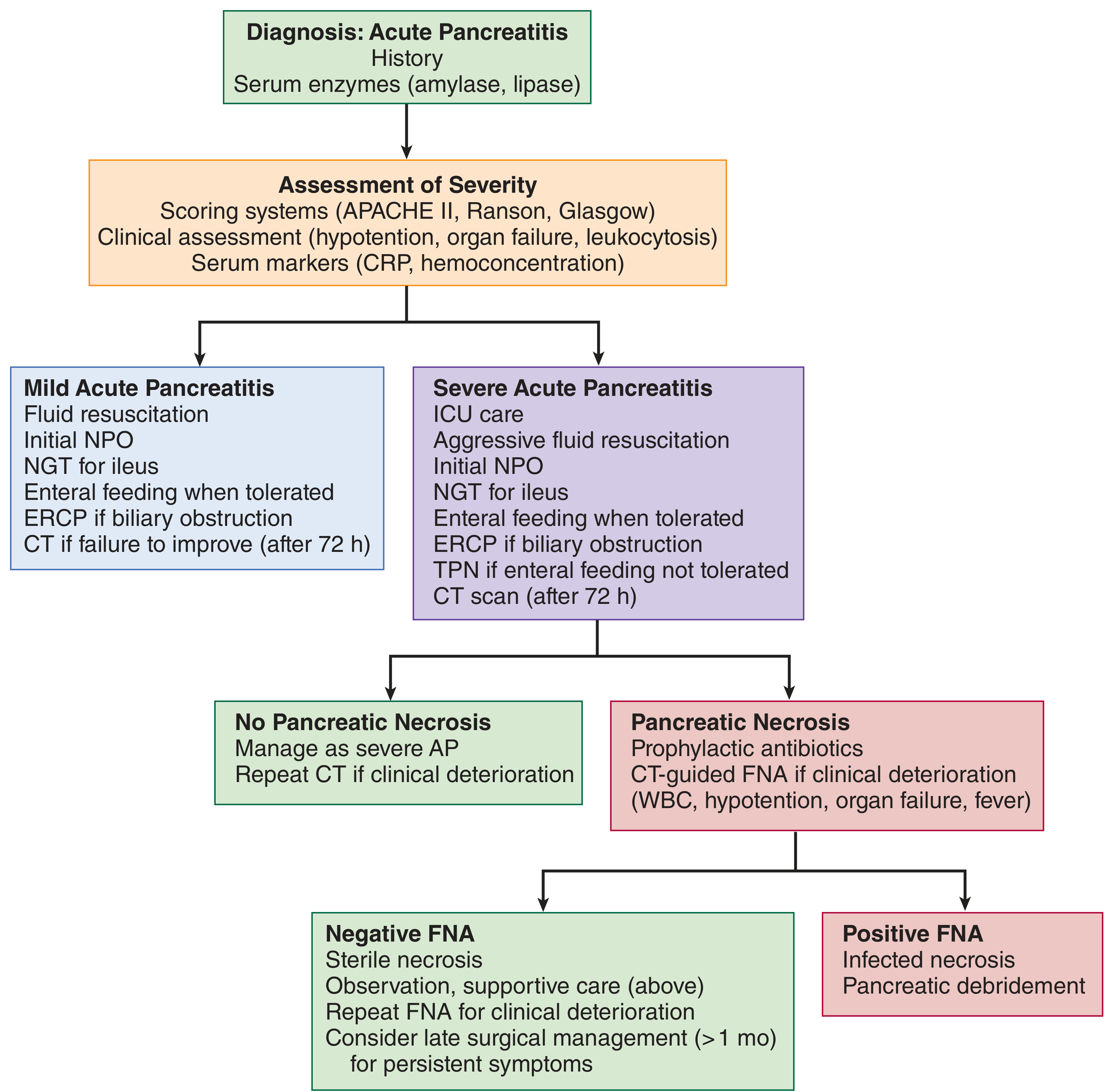
1. Diagnosis & Severity Assessment
- Characteristic epigastric pain (often radiating to the back)
- Serum lipase or amylase >3x upper limit of normal (lipase preferred - higher sensitivity/specificity)
- Characteristic findings on imaging (CT/MRI)
| Severity | Definition |
|---|---|
| Mild | No organ failure, no local/systemic complications |
| Moderately severe | Transient organ failure (<48 h) and/or local or systemic complications |
| Severe | Persistent organ failure (>48 h) - single or multi-organ |
| Critical | Persistent organ failure + infected pancreatic necrosis |
2. Fluid Resuscitation (MOST IMPORTANT EARLY INTERVENTION)
-
Lactated Ringer's (LR) is preferred over normal saline (NS)
- LR is more physiologic (balanced electrolyte solution)
- LR may provide anti-inflammatory effects via lactate-mediated mechanisms
- A 2025 meta-analysis of 6 RCTs (n=1,500 patients) showed LR vs. NS: significantly lower risk of moderate-to-severe AP (OR 0.48; 95% CI 0.34-0.67), shorter hospital stay, lower ICU admission rate (RR 0.42), and fewer local complications (Zhao et al., Int J Surg 2025, PMID 40085761)
-
Rate: Aggressive resuscitation - typically 250-500 mL/h during the first 12-24 hours in moderate-severe disease; careful goal-directed therapy to avoid overhydration
-
Titrate to: urine output >0.5 mL/kg/h, HR <100, MAP >65 mmHg, BUN trending down
3. Pain Management
- Adequate analgesia is essential; IV opioids (morphine, hydromorphone, fentanyl) are widely used and are safe
- NSAIDs and acetaminophen can supplement but are often insufficient alone in acute presentations
- There is no evidence that one analgesic is superior to another specifically for pancreatitis
- Epidural analgesia is an option in severe cases
4. Nutrition
- Mild AP: Oral diet should be restarted as soon as it is tolerated (often within 24-48 hours). There is no need to wait until pain resolves or amylase normalizes.
- Moderate-severe AP:
- Early enteral nutrition within 24-72 hours of admission is associated with decreased mortality, organ failure, and infectious complications vs. delayed nutrition
- Nasogastric (NG) or nasojejunal (NJ) tube feeding - RCTs show no significant difference in outcomes between gastric vs. jejunal delivery, so a nasogastric tube is acceptable
- NPO initially if there is ileus or if the patient cannot tolerate any intake
- TPN (parenteral nutrition) is reserved for cases where enteral feeding is not tolerated (refractory vomiting, intestinal obstruction, high-output fistula)
- Enteral nutrition maintains gut barrier function, reduces bacterial translocation, and reduces risk of infectious complications
5. Antibiotics
- Prophylactic antibiotics are NOT indicated in acute pancreatitis - this includes even severe or necrotizing pancreatitis without evidence of infection
- Harrison's (2025 ed.): "Prophylactic antibiotics are no longer recommended for severe acute pancreatitis"
- Antibiotics ARE indicated for:
- Documented infected pancreatic necrosis (confirmed by FNA or gas on CT, or clinical deterioration with fever/leukocytosis)
- Cholangitis (ascending cholangitis complicating biliary pancreatitis)
- Other clear sources of bacterial sepsis
- Preferred agents for infected necrosis: carbapenems (imipenem, meropenem) or fluoroquinolones + metronidazole (good pancreatic tissue penetration)
6. Role of ERCP
- ERCP is indicated in:
- Acute cholangitis complicating gallstone pancreatitis (urgent, within 24-72 hours)
- Biliary obstruction with evidence of persistent common bile duct stones
- ERCP is NOT indicated in mild biliary pancreatitis without cholangitis or persistent biliary obstruction (does not improve outcomes and risks complications)
- Rectal indomethacin (NSAIDs) or pancreatic duct stenting is used to prevent post-ERCP pancreatitis in high-risk patients
7. Management of Complications
Pancreatic Necrosis
| Type | Management |
|---|---|
| Sterile necrosis | Conservative - supportive care, antibiotics NOT required |
| Infected necrosis | Antibiotics (carbapenems preferred) + drainage/debridement |
- Minimally invasive approach preferred (step-up approach)
- CT/EUS-guided percutaneous or endoscopic drainage as first step
- Video-assisted retroperitoneal debridement (VARD) or endoscopic necrosectomy if drainage fails
- Open surgical necrosectomy - last resort (high morbidity/mortality)
- Intervention ideally delayed >4 weeks to allow "walled-off necrosis" (WON) to mature (better defined walls, safer to drain)
- FNA is now used less routinely; clinical judgment + CT findings guide decisions
Pseudocyst
- Most resolve spontaneously
- Drainage indicated if: symptomatic (pain, gastric outlet obstruction), infection, enlarging, or >6 weeks without resolution
- Endoscopic (EUS-guided cyst-gastrostomy or cyst-duodenostomy) is preferred over surgical drainage
Pancreatic Ascites/Pleural Effusion
- From ductal disruption; ERCP with stenting ± somatostatin analogue
8. Special Situations
Biliary/Gallstone Pancreatitis
- Cholecystectomy should be performed before discharge (or at least during the same hospitalization) in mild disease to prevent recurrence
- In severe disease, cholecystectomy is deferred until recovery
Hypertriglyceridemia-induced Pancreatitis
- Insulin infusion or plasmapheresis to rapidly lower triglycerides (target <500 mg/dL)
- Fibrates and dietary fat restriction for long-term prevention
Alcohol-induced Pancreatitis
- Alcohol cessation counseling and treatment (reduces recurrence and progression to chronic pancreatitis)
9. ICU Indications
- Persistent organ failure (renal, respiratory, cardiovascular)
- APACHE II score >8 or Ranson score >3 at 48 hours
- Clinical deterioration despite initial management
- Septic shock from infected necrosis
10. Summary of Key Guideline Recommendations (2024-2025)
- LR preferred over NS for fluid resuscitation
- Early enteral nutrition within 24-72 hours
- No prophylactic antibiotics in the absence of infection
- Minimally invasive, step-up approach for infected necrosis
- Cholecystectomy before discharge for biliary pancreatitis
- ERCP only for cholangitis/biliary obstruction
- Conservative management for sterile complications whenever possible
- Rosen's Emergency Medicine, 10th ed. - Chapter 77
- Maingot's Abdominal Operations - Chapter 55 (Complications of AP)
- Harrison's Principles of Internal Medicine, 22nd ed. (2025) - Chapter 7.39
- Washington Manual of Medical Therapeutics - Nutrition section
- ACG Guidelines 2024 (PMID 38857482)
- IAP/APA/EPC/IPC/JPS Revised Guidelines 2025 (PMID 40651900)
- Zhao et al., LR vs NS meta-analysis, Int J Surg 2025 (PMID 40085761)
Urinary tract infection in diabetic patients with history of chronic smoker and alcoholic
"urinary tract infection" AND "diabetes mellitus"
"urinary tract infection" AND diabetes AND management treatment
PMID: 34024733
UTI in Diabetic Patients with Chronic Smoking and Alcohol Use
1. Why This Patient is at Extreme Risk: Pathophysiology
Diabetes Mellitus
| Mechanism | Effect |
|---|---|
| Glucosuria | Elevated urinary glucose serves as a substrate for bacterial growth; impairs neutrophil phagocytosis and bactericidal function |
| Autonomic neuropathy | Causes dysfunctional voiding and urinary retention - prevents bacterial clearance through micturition |
| Reduced cytokine secretion | Decreased local urinary IL-6, IL-8, reducing neutrophil recruitment |
| Increased uroepithelial adhesion | Higher bacterial adherence to uroepithelial cells promotes colonization |
| Impaired cell-mediated immunity | Hyperglycemia generally suppresses phagocyte and T-cell function |
| Microangiopathy | Diminished vascularization reduces delivery of immune cells |
Chronic Alcohol Use
-
Impairs neutrophil maturation in the bone marrow and depresses cell-mediated immunity
-
Impairs neutrophil chemotaxis, phagocytosis, and intracellular killing
-
Alcohol-related liver disease (cirrhosis) results in hypocomplementemia, reduced opsonization, and impaired Kupffer cell function
-
Alcoholic patients are vulnerable to overwhelming gram-negative sepsis (E. coli bacteremia is common)
-
Malnutrition associated with alcoholism impairs zinc-dependent immune functions
-
Patients with liver disease/cirrhosis often develop spontaneous bacteremia predominantly with E. coli
-
Textbook of Family Medicine, 9th ed.; Goodman & Gilman's Pharmacological Basis of Therapeutics
Chronic Smoking
- Smoking impairs mucosal immunity - reduces secretory IgA in mucosal surfaces
- Reduces Langerhans cell populations and alters mucosal immune defense
- Promotes oxidative stress, impairing neutrophil function
- Smoking-related COPD/bronchiectasis increases risk for concurrent respiratory infections (Gram-negatives, including Pseudomonas)
- Nicotine causes bladder urothelial changes - increased bacterial adherence
- Carcinogens in cigarette smoke predispose to bladder cancer (which itself is a risk factor for complicated UTI and obstruction)
2. Classification: This is a COMPLICATED UTI
- Metabolic disease (diabetes)
- Immunocompromised state (alcohol-related immune deficiency + smoking)
- High likelihood of functional urinary tract abnormality (diabetic cystopathy, neurogenic bladder)
- Risk of resistant organisms
"Complicated urinary tract infection occurs in individuals with functional or structural abnormalities of the genitourinary tract... Complicated UTI: infection in the setting of metabolic, immunocompromised, functional, or anatomic abnormality."
- Brenner and Rector's The Kidney, 2-Volume Set; Smith and Tanagho's General Urology
3. Microbiology - Organisms to Expect
| Organism | Comments |
|---|---|
| E. coli | Still most common (45-58% even in diabetics) |
| Klebsiella pneumoniae | More common in diabetics than general population; gas-forming |
| Enterococcus | More prevalent in diabetics with asymptomatic bacteriuria |
| Staphylococcus aureus | Particularly important in diabetics - can cause renal carbuncle and urosepsis; not uncommon |
| Candida albicans / C. glabrata | Fungal UTI more common in diabetics, especially with poor glycemic control, catheterized patients, or prior antibiotics |
| Pseudomonas aeruginosa | Especially in smokers with structural lung disease, catheterized patients |
| Clostridium spp., Enterobacter | Gas-forming organisms in emphysematous infections |
4. Unique Complications in This Patient Population
Emphysematous Pyelonephritis (EPN)
- Life-threatening gas-forming necrotizing renal infection
- 95% of cases occur in diabetics (Kamei & Yamamoto, J Infect Chemother 2021)
- Gas in renal parenchyma or perirenal space on CT
- Organisms: E. coli (45-54%), Klebsiella (gas generated from glucose fermentation)
- High mortality: 7-20% with percutaneous drainage + antibiotics; up to 70% in fulminant disease
- Management: CT-guided percutaneous drainage + IV antibiotics + aggressive glycemic control; nephrectomy for non-responders
- Mortality rates: 50% with medical management alone vs. 13.5% with medical management + percutaneous drainage
Emphysematous Cystitis
- Less severe than EPN; gas in bladder wall
- 67% of cases have DM; E. coli (58%), Klebsiella (21%)
- Treatment: antibiotics + bladder drainage + glycemic control; surgical intervention only in 10%
- Overall mortality 7%
Renal Papillary Necrosis
- Seen particularly during acute pyelonephritis in diabetics
- Also with analgesic nephropathy, sickle cell disease, obstruction
- Presents as flank pain, hematuria, sloughing of papillae (visible on imaging as "ring shadow" on IVU or filling defects on CT)
Perinephric / Intrarenal Abscess
- More common in diabetics; higher risk with alcoholism-related immune depression
- Often requires CT-guided drainage + 4-6 weeks of antibiotics
Renal Carbuncle
- Due to hematogenous spread of Staphylococcus aureus (especially in diabetics)
- Treat with anti-staphylococcal antibiotics (nafcillin/oxacillin or vancomycin if MRSA suspected)
Xanthogranulomatous Pyelonephritis
- Chronic destructive renal infection; associated with urolithiasis and recurrent UTI
- Higher incidence in diabetics; usually requires nephrectomy
Urosepsis
- Diabetic patients have higher rates of bacteremia complicating pyelonephritis
- Gram-negative bacteremia (E. coli) common in alcoholics
- Combined patient is at very high risk for septic shock
5. Asymptomatic Bacteriuria (ASB) in Diabetics
-
Pregnancy
-
Pre-urological procedure
-
Renal transplant recipient (within first month)
-
Washington Manual of Medical Therapeutics; Smith and Tanagho's General Urology; Campbell Walsh Wein Urology
6. Diagnosis
Urine Studies
- Urinalysis: Pyuria (>5 WBC/hpf), bacteriuria, nitrite positive (Gram-negatives only), leukocyte esterase
- Urine culture with sensitivity: Mandatory in ALL complicated UTI - do NOT start empirical antibiotics without sending cultures first
- Threshold: ≥10⁵ CFU/mL symptomatic; ≥10² CFU/mL with catheter or suprapubic aspiration
Blood Tests
- CBC (leukocytosis, bandemia suggest severe infection)
- BMP/RFT (renal function - especially important given diabetic nephropathy risk)
- Blood glucose, HbA1c (glycemic control assessment)
- Blood cultures (if febrile or sepsis suspected - particularly in this patient)
- LFTs, coagulation profile (alcohol-related liver disease)
- CRP/procalcitonin (markers of severity)
Imaging
- Ultrasound KUB: First-line for obstruction, hydronephrosis, stones, perinephric abscess
- CT abdomen/pelvis (contrast-enhanced): Indicated if:
- Failure to respond to antibiotics after 48-72 hours
- Suspected EPN, abscess, papillary necrosis, or obstruction
- Gas on plain X-ray or U/S
- Sepsis with unknown source
7. Treatment
General Principles
- Always culture urine BEFORE starting antibiotics
- This patient requires hospitalization - oral outpatient therapy is NOT recommended for complicated UTI in a diabetic
- Strict glycemic control is essential - hyperglycemia impairs immune response and drives emphysematous infections
- No TMP-SMX as first choice in diabetics on oral hypoglycemic drugs - it potentiates hypoglycemia
- Alcohol withdrawal monitoring - a hospitalized chronic alcoholic must be monitored for withdrawal (CIWA protocol, thiamine supplementation)
- Avoid NSAIDs - nephrotoxic, especially with diabetic nephropathy
Antibiotic Treatment
| Setting | First Choice | Alternatives | Duration |
|---|---|---|---|
| Febrile complicated UTI / Pyelonephritis | IV Fluoroquinolone (ciprofloxacin 400 mg IV q12h) | IV 3rd-gen cephalosporin (ceftriaxone), aminoglycoside + pip/tazo | Minimum 2 weeks (febrile: continue 3-5 days after last fever) |
| Afebrile complicated UTI | Oral fluoroquinolone (after IV step-down) | Aminopenicillin + BLI (amoxicillin-clavulanate) | 2 weeks |
| Suspected/confirmed MRSA (renal carbuncle) | IV Vancomycin | Daptomycin | 4-6 weeks |
| Fungal UTI (Candida) | Fluconazole 200 mg daily (if susceptible) | Echinocandin (caspofungin) for fluconazole-resistant | 7-14 days |
| EPN/Perinephric abscess | IV carbapenem (meropenem/imipenem) + CT drainage | Pip-tazo | Until drainage complete + clinical improvement |
| Urosepsis | IV carbapenems (broad spectrum) | Consider antifungal coverage if poor glycemic control | Per clinical response |
- Smith and Tanagho's General Urology, Table 14-6; Kamei & Yamamoto, J Infect Chemother 2021 (PMID 34024733)
SGLT2 Inhibitors (Diabetic Patients on These Medications)
- SGLT2 inhibitors (dapagliflozin, empagliflozin, canagliflozin) significantly increase genital mycotic infections, but NOT the rate of UTI, pyelonephritis, or urosepsis significantly
- However, they promote glucosuria, which may lower threshold for candidal cystitis
- Consider temporarily holding SGLT2 inhibitors during acute illness (sick-day rule)
8. Special Considerations for the Alcoholic Patient
| Issue | Clinical Implication |
|---|---|
| Alcohol withdrawal | Monitor CIWA score; benzodiazepines if needed; Wernicke's prevention with thiamine 100 mg IV before any glucose |
| Liver disease/cirrhosis | Coagulopathy (avoid invasive procedures without correction), hypoalbuminemia, altered drug metabolism; higher infection severity |
| Malnutrition | Supplement thiamine, folate, zinc, B12 |
| Drug interactions | Metronidazole + alcohol = disulfiram-like reaction (avoid for fungal/anaerobic coverage) |
| Altered mental status | May mask typical UTI symptoms; have low threshold for sepsis workup |
| Thrombocytopenia | May complicate catheter placement or procedures |
9. Special Considerations for the Chronic Smoker
| Issue | Clinical Implication |
|---|---|
| Increased risk of resistant organisms | More likely colonized with Gram-negatives, including Pseudomonas |
| Bladder urothelial changes | Higher risk of urothelial malignancy - any unexplained hematuria needs cystoscopic evaluation |
| Impaired wound healing | Affects recovery from urological procedures |
| Concurrent respiratory infections | Common shared pathogens (E. coli, Klebsiella) may cause concurrent pneumonia + UTI |
| Smoking + alcohol + diabetes = triple risk | This combination is associated with worst UTI outcomes and highest risk for urosepsis |
10. Indications for ICU Admission / Urgent Intervention
- Signs of septic shock (hypotension, tachycardia, altered mental status, lactate >2 mmol/L)
- Emphysematous pyelonephritis (requires urgent CT + ICU-level care)
- Bilateral EPN or EPN with renal failure
- Urosepsis with DIC or multi-organ failure
- Failure to improve in 48-72 hours despite appropriate IV antibiotics
11. Prevention and Long-term Management
| Intervention | Rationale |
|---|---|
| Tight glycemic control (HbA1c <7%) | Directly correlates with reduced UTI risk |
| Alcohol cessation | Restores neutrophil function, improves liver function, reduces immunosuppression |
| Smoking cessation | Reduces urothelial changes, mucosal immune impairment |
| Regular post-void residual assessment | Detect diabetic cystopathy early; prevent urinary stasis |
| Adequate hydration | Dilutes bacteriuria, promotes voiding |
| Prophylactic antibiotics (if recurrent UTI, >3 per year) | Cephalexin 250 mg/day, ciprofloxacin 250 mg/day, or trimethoprim 100 mg/day (avoid TMP-SMX in those on sulfonylureas) |
| Methenamine hippurate | Non-antibiotic prophylaxis for recurrent UTI |
| Cranberry products | Modest evidence for prevention of recurrent UTI by reducing bacterial adhesion |
| Vaccinations | Pneumococcal, influenza, COVID-19 vaccines (diabetics at higher morbidity risk) |
| Avoid unnecessary urinary catheters | Major risk factor for hospital-acquired UTI |
- Smith and Tanagho's General Urology, 19th ed. - Chapter 14 (UTIs in Diabetes)
- Brenner and Rector's The Kidney, 2-Volume Set - Chapter 36 (UTI definitions, host defenses); Chapter emphysematous infections
- Harrison's Principles of Internal Medicine, 22nd ed. (2025) - Chapter on Diabetes Complications (Infections)
- Comprehensive Clinical Nephrology, 7th ed. - Diabetic patient with CKD and UTI
- Textbook of Family Medicine, 9th ed. - Acquired conditions predisposing to infection
- Washington Manual of Medical Therapeutics - Asymptomatic bacteriuria
- Kamei & Yamamoto, "Complicated UTIs with Diabetes Mellitus," J Infect Chemother 2021 (PMID 34024733)
Create a clinical decision-making flow chart for complicated UTI severity assessment
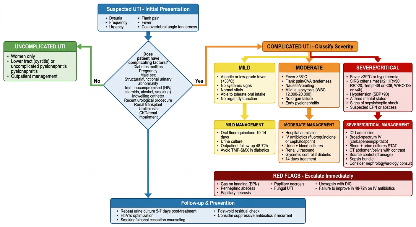
A detailed clinical decision-making flowchart for complicated UTI (urinary tract infection) severity assessment. The flowchart starts at the top with "Suspected UTI - Initial Presentation" and flows downward through the following decision nodes and pathways: 1. First box: "Suspected UTI - Initial Presentation" with sub-items: dysuria, frequency, urgency, flank pain, fever, costovertebral angle tenderness 2. Diamond decision: "Does patient have complicating factors?" with Yes/No branches - Complicating factors listed: Diabetes mellitus, Pregnancy, Male sex, Structural/functional urinary abnormality, Immunocompromised (HIV, steroids, alcohol, smoking), Indwelling catheter, Recent urological procedure, Renal transplant, Urolithiasis, CKD/renal impairment 3. No branch leads to "UNCOMPLICATED UTI" box (green): Women only, Lower tract (cystitis) or uncomplicated pyelonephritis, Outpatient management 4. Yes branch leads to "COMPLICATED UTI - Classify Severity" box (orange) 5. Severity classification splits into three columns: - MILD (yellow): Afebrile or low-grade fever (<38°C), No systemic signs, Normal vitals, Able to tolerate oral intake, No organ dysfunction - MODERATE (orange): Fever >38°C, Flank pain/CVA tenderness, Nausea/vomiting, Mild leukocytosis (WBC 12,000-20,000), No organ failure, Early pyelonephritis - SEVERE/CRITICAL (red): Fever >39°C or hypothermia, SIRS criteria met (≥2: HR>90, RR>20, Temp>38 or <36, WBC>12k or <4k), Hypotension (SBP<90), Altered mental status, Signs of sepsis/septic shock, Suspected EPN or abscess 6. Each severity level leads to management boxes: - MILD: Oral fluoroquinolone 10-14 days, Urine culture, Outpatient follow-up 48-72h, Avoid TMP-SMX in diabetics - MODERATE: Hospital admission, IV antibiotics (fluoroquinolone or cephalosporin), Urine + blood cultures, Renal ultrasound, Glycemic control if diabetic, 14 days treatment - SEVERE/CRITICAL: ICU admission, Broad-spectrum IV (carbapenem/pip-tazo), Blood + urine cultures STAT, CT abdomen/pelvis with contrast, Source control (drainage), Sepsis bundle, Consider nephrology/urology consult 7. Bottom section "RED FLAGS - Escalate Immediately" (dark red box): Gas on imaging (EPN), Perinephric abscess, Papillary necrosis, Fungal UTI, Urosepsis with DIC, Failure to improve in 48-72h on IV antibiotics 8. Final bottom box "Follow-up & Prevention": Repeat urine culture 5-7 days post-treatment, HbA1c optimization, Smoking/alcohol cessation counseling, Post-void residual check, Consider suppressive antibiotics if recurrent The chart uses color coding: green for uncomplicated, yellow for mild, orange for moderate, red for severe. Clean medical typography, professional clinical style, white background, clear arrows and connecting lines between all boxes. All text is clearly readable.
-
Entry point - Suspected UTI with presenting symptoms (dysuria, frequency, flank pain, fever, CVA tenderness)
-
First decision node - Identifies complicating factors (diabetes, pregnancy, male sex, immunosuppression from alcohol/smoking/HIV, structural anomalies, catheters, CKD, renal transplant)
-
Branching:
- No complicating factors → Uncomplicated UTI → outpatient oral therapy
- Complicating factors present → Complicated UTI → severity stratification
-
Three-tier severity classification:
- 🟡 Mild - Afebrile/low-grade fever, tolerating orals, no systemic signs → oral fluoroquinolone, outpatient with 48-72h follow-up
- 🟠 Moderate - Fever >38°C, pyelonephritis, nausea/vomiting, early leukocytosis → hospital admission, IV antibiotics, imaging
- 🔴 Severe/Critical - SIRS criteria, hypotension, sepsis/septic shock, suspected EPN → ICU, broad-spectrum carbapenems, CT imaging, source control
-
Red flags box - Triggers for immediate escalation: emphysematous pyelonephritis, perinephric abscess, papillary necrosis, urosepsis with DIC, failure to improve in 48-72h
-
Follow-up & prevention - Repeat cultures, HbA1c optimization, smoking/alcohol cessation, post-void residual monitoring, suppressive antibiotics if recurrent
Ureteric or reanl colic
"renal colic" AND management
PMID: 40084512
"ureteral stone" AND "medical expulsive therapy" alpha blocker
PMID: 39875883
Ureteric / Renal Colic
Terminology note: "Renal colic" is technically a misnomer. The correct term is ureteric colic - the pain arises from hyperperistalsis of the ureteric smooth muscle against an obstructing stone, not from the kidney itself. However, both terms are widely used in clinical practice. - Bailey and Love's Short Practice of Surgery, 28th ed.
1. Epidemiology & Stone Types
- Annual incidence: >1 per 1000 persons in industrialized nations; lifetime risk ~10% in women, ~15% in men
- Incidence peaks in the 4th-5th decades; prevalence has increased from 3% (1970s) to >5% (current), likely due to dietary and lifestyle changes
- Obesity (BMI >30) significantly increases stone risk
| Stone Type | Frequency | Key Features |
|---|---|---|
| Calcium oxalate | 70-80% | Most common; radio-opaque; associated with hypercalciuria, hyperoxaluria, hypocitraturia |
| Calcium phosphate | ~10% (often mixed with CaOx) | Radio-opaque; seen in renal tubular acidosis, hyperparathyroidism |
| Uric acid | <10% in USA; 70% in Middle East | Radiolucent on plain X-ray; associated with gout, low urine pH, dehydration |
| Struvite (infection stones) | 1-25% | Caused by urease-splitting bacteria (Proteus, Klebsiella); form staghorn calculi; always infected |
| Cystine | 1-2% | Genetic (cystinuria); very hard; do NOT respond to ESWL |
2. Clinical Presentation
Symptoms
- Ureteric colic: Sudden-onset, severe, colicky flank pain radiating to the groin, scrotum, or labia (following the path of the ureter)
- Pain is typically excruciating, unrelenting (patient cannot find a comfortable position - vs. peritonitis where patient lies still)
- Lower ureteric stones (near UVJ): cause urgency, frequency, dysuria mimicking cystitis
- Nausea and vomiting (reflex, very common)
- Haematuria: gross or microscopic in most cases
- Calculuria: passing of sand or gravel in urine
Radiation of Pain by Stone Location
| Stone Location | Pain Radiation |
|---|---|
| Pelviureteric junction (PUJ) | Loin/flank - deep, dull |
| Upper ureter | Flank → ipsilateral testicle/labium majus |
| Mid-ureter | Right: may mimic appendicitis; Left: mimics diverticulitis |
| Lower ureter / UVJ | Groin, scrotum/labia, inner thigh; irritative LUTS (urgency, frequency) |
Key clinical point
"Ureteric colic does NOT radiate to the chest or the back of the leg." - Bailey & Love
3. Differential Diagnosis
| Urinary Causes | Surgical Causes | Vascular |
|---|---|---|
| Clot colic (haemophilia, anticoagulation) | Acute appendicitis | Abdominal aortic aneurysm (must exclude!) |
| Papillary necrosis (DM, NSAIDs, sickle cell) | Ectopic pregnancy | Aortic dissection |
| Pyelonephritis | Ovarian torsion | |
| Retroperitoneal fibrosis | Acute intestinal obstruction | |
| Transitional cell carcinoma | Psoas abscess |
Critical: AAA must always be excluded in the middle-aged/elderly patient presenting with flank pain - especially if the pain is not colicky and there is a pulsatile mass.
4. Investigations
Emergency Setting
- Dipstick: haematuria (present in 85-90%, but can be absent), pyuria
- Mid-stream urine culture if pyuria/fever present
- Urine pregnancy test in women of reproductive age (mandatory - ectopic pregnancy)
- FBC (leukocytosis suggests infection), U&E/Cr (renal function), eGFR
- Serum calcium, phosphate, uric acid (initial metabolic screen)
- Serum lactate + blood cultures if sepsis suspected
| Modality | Role | Notes |
|---|---|---|
| CT KUB (non-contrast) | Investigation of choice | Detects both radio-opaque and radiolucent stones (except indinavir stones); 97% sensitivity, 96% specificity; also shows stone size, location, and signs of obstruction |
| USS (renal ultrasound) | Good first-line; preferred in pregnancy, children | Detects hydronephrosis and stones >5mm; misses many ureteric stones |
| Plain X-ray KUB | Detects radio-opaque stones (Ca, struvite) | Misses uric acid, cystine stones; limited value alone |
| IVU/IVP | Now largely replaced by CT | Still used in some centres; shows function and anatomy |
| MRI | Used in pregnancy when radiation avoided | Less sensitive for stones than CT |
Non-Emergency / Metabolic Evaluation
- 24-hour urine collection: volume, calcium, oxalate, uric acid, citrate, phosphate, creatinine
- Serum: calcium, phosphate, uric acid, PTH (if hypercalcaemia)
- Stone analysis (if passed or retrieved)
- Urine pH (low = uric acid; high = struvite/RTA)
5. Complications
| Complication | Description |
|---|---|
| Ureteric obstruction | With hydronephrosis and impaired renal function |
| Pyonephrosis | Infected obstructed kidney - surgical emergency |
| Urosepsis | Life-threatening infection from obstructed infected kidney |
| Calculous anuria | Bilateral obstruction or solitary kidney obstruction |
| Chronic renal failure | From recurrent obstruction + infection |
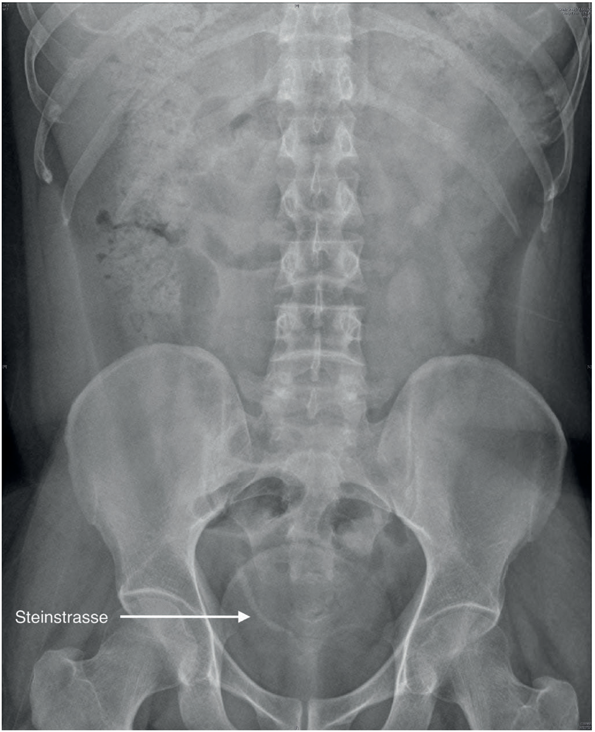
| Steinstrasse | "Street of stones" - linear row of stone fragments in ureter post-ESWL (see X-ray below) |
| XGP (Xanthogranulomatous pyelonephritis) | Rare; associated with Proteus stones; chronic destructive infection |
6. Management
6a. Emergency Management - Immediate Pain Relief
- Ketorolac 30 mg IV or diclofenac 75 mg IM
- NSAIDs reduce pain AND decrease ureteral/intrarenal pressure (by reducing prostaglandin-mediated ureteric spasm and urine production)
- A 2025 Cochrane systematic review (29 RCTs, 3,593 participants) confirmed NSAIDs significantly reduce renal colic pain vs. placebo (MD -3.84 cm VAS at 30 min); no one NSAID was clearly superior to another - (Afshar et al., Cochrane 2025, PMID 40084512)
- Oral route when tolerated; parenteral when nausea/vomiting prevents oral intake
- Morphine IV, pethidine, fentanyl
- Used when NSAIDs are contraindicated (renal impairment, peptic ulcer) or insufficient
- There is NO evidence that morphine causes more ureteric spasm than other opioids - it is safe and appropriate - Rosen's Emergency Medicine
- Antiemetics (ondansetron, metoclopramide) should accompany opioids
- NOT recommended as routine - evidence does not support their use for ureteric colic pain relief - Bailey & Love
- IV fluids for hydration but forced diuresis does NOT improve stone passage rates and may worsen pain
6b. Conservative Management (Watchful Waiting)
- Appropriate for small stones (<5 mm) that are likely to pass spontaneously
- Spontaneous passage rates:
- <4 mm stones: ~90% pass spontaneously
- 5-7 mm: ~50% pass spontaneously
-
7 mm: <25% pass; intervention usually required
- Stone <5 mm (distal ureteric)
- Well-controlled pain with oral analgesics
- No signs of infection
- Normal renal function
- Patient compliant with follow-up
6c. Medical Expulsive Therapy (MET)
- 0.4 mg once daily
- Causes smooth muscle relaxation of distal ureteric musculature
- Indicated for distal ureteric stones >5 mm
- Also used to assist passage of fragments following ESWL
- PDE-5 inhibitors (sildenafil/tadalafil): stone expulsion rate 2.7x better than alpha-blocker alone; also shorter expulsion time
- Corticosteroids: 2.7x higher stone expulsion rate
- Phytotherapy: 3.1x higher expulsion rate
6d. Surgical/Interventional Management
- Failure of conservative management (pain uncontrolled)
- Stone size >10 mm (unlikely to pass spontaneously)
- Obstructed infected kidney / pyonephrosis (EMERGENCY)
- Impaired renal function / solitary kidney
- Bilateral obstructing stones
- High-risk occupation (pilots, sailors, locomotive drivers)
- Patient preference
- Percutaneous nephrostomy (PCN): Radiologically guided drain into the renal pelvis - preferred in septic/unstable patients
- Retrograde ureteric stenting (JJ stent): Cystoscopy + fluoroscopy-guided placement; provides drainage past the obstruction
- Blood and urine cultures MUST be taken first; empirical broad-spectrum antibiotics started immediately
| Procedure | Indication | Stone Size |
|---|---|---|
| ESWL (Extracorporeal Shockwave Lithotripsy) | Renal stones <2 cm; proximal ureteric stones | <2 cm; NOT cystine (too hard) |
| Ureteroscopy (URS) ± laser lithotripsy | Ureteric stones; renal stones <2 cm | Any |
| Retrograde Intrarenal Surgery (RIRS) | Renal stones <2 cm; obesity; musculoskeletal deformities; bleeding disorders | <2 cm |
| Percutaneous Nephrolithotomy (PCNL) | Large renal stones, staghorn calculi | >2 cm |
| Open/laparoscopic surgery | Rare; failed endourology; anatomical anomalies | Complex cases |
- Non-invasive; introduced by Christian Chaussy in 1980
- Stone localised by fluoroscopy or ultrasound; focused shock waves fragment the stone
- Efficacy decreases with increasing stone burden
- Steinstrasse is a recognised complication (stone fragments queuing in ureter); usually pass spontaneously but may obstruct
- "Clinically insignificant residual fragments" (≤4 mm) may not clear in 20-40% of cases and form a nidus for regrowth
- Semirigid or flexible ureterorenoscopes (URSs)
- Working channels for laser/pneumatic lithotripsy, graspers, baskets
- Has largely replaced ESWL for many ureteric stones
- Complications: ureteric perforation, avulsion, retropulsion
- Involves creating a skin-to-pelvicalyceal tract (usually prone position)
- Fluoroscopy or USS guided; posterolateral calyx entry
- Complications: bleeding (may require embolisation), infection, pleural violation (supracostal approach)
7. Stone-Specific Management and Prevention
| Stone Type | Medical Treatment / Prevention |
|---|---|
| Calcium oxalate | High fluid intake (urine output >2.5 L/day); thiazide diuretics for hypercalciuria; potassium citrate for hypocitraturia; low oxalate diet; normal (not low) calcium diet |
| Uric acid | Urinary alkalinisation (potassium citrate, NaHCO3) to pH 6.5-7.0; allopurinol for hyperuricosuria; adequate hydration; low purine diet; stone can be dissolved medically |
| Struvite | Antibiotics targeting urease-splitting organisms; PCNL for large stones; acetohydroxamic acid (urease inhibitor) - rarely used |
| Cystine | Very high fluid intake (urine output >3 L/day); urinary alkalinisation; D-penicillamine or tiopronin (chelators); ESWL ineffective |
| Calcium phosphate | Treat underlying cause (RTA, hyperparathyroidism); potassium citrate |
General Prevention for All Stone Types
- Fluid intake: Target urine output >2.5 L/day (most important intervention)
- Avoid dehydration (hot climates, exercise, occupational fluid restriction)
- Dietary: reduce salt, limit animal protein; maintain normal calcium intake (low calcium diet WORSENS oxalate stones)
- Body weight control
- Treat underlying metabolic disorders
8. Indications for Hospital Admission
| Indication | Rationale |
|---|---|
| Uncontrolled pain despite parenteral analgesics | |
| Fever + obstruction (pyonephrosis/urosepsis) | Emergency - decompress urgently |
| Solitary kidney or bilateral obstruction | Renal failure risk |
| AKI or impaired renal function | |
| Vomiting preventing oral hydration/medications | |
| Diagnostic uncertainty (e.g., ? AAA) | |
| Stone >10 mm unlikely to pass spontaneously |
- Bailey and Love's Short Practice of Surgery, 28th ed. - Chapters 81 (Urinary symptoms) & 82 (Urolithiasis)
- Goldman-Cecil Medicine, International Edition - Chapter on Nephrolithiasis
- Rosen's Emergency Medicine, 10th ed. - Renal stone management
- Campbell Walsh Wein Urology - Stone surgery chapters
- Afshar et al., "NSAIDs for acute renal colic," Cochrane Database Syst Rev, 2025 (PMID 40084512)
- Taheri et al., "Combination MET for distal ureteral stone," BMC Urol, 2025 (PMID 39875883)
Antifungal cream
topical antifungal agents dermatophytosis treatment
Antifungal Creams (Topical Antifungal Agents)
1. Mechanisms of Action
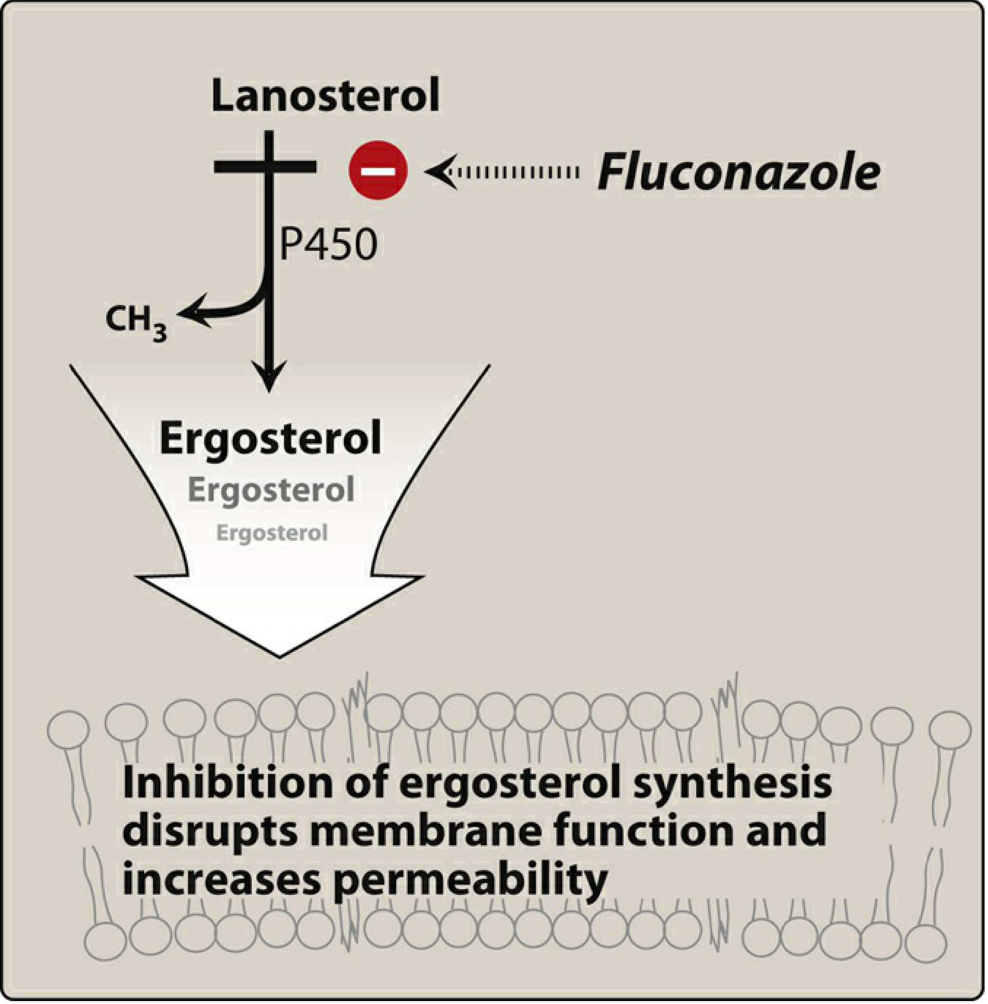
Ergosterol Synthesis Pathway and Drug Targets
| Drug Class | Target Enzyme | Effect |
|---|---|---|
| Azoles (imidazoles & triazoles) | 14-α-demethylase (CYP450) - blocks lanosterol → ergosterol | Ergosterol depletion + lanosterol accumulation → disrupted fungal membrane |
| Allylamines (terbinafine, naftifine) | Squalene epoxidase - earlier step in ergosterol synthesis | Ergosterol depletion + toxic squalene accumulation → fungicidal |
| Polyenes (nystatin, amphotericin B) | Bind directly to ergosterol in the membrane | Forms pores → K⁺ and small molecule leakage → cell death |
| Ciclopirox olamine | Chelates metal ions; disrupts membrane transport enzymes | Broad-spectrum fungistatic/fungicidal |
| Tolnaftate | Squalene epoxidase (weaker than allylamines) | Fungistatic vs. dermatophytes |
2. Drug Classes and Individual Agents
Class 1 - Azoles (Imidazoles - Topical)
| Drug | Brand Name | Formulations | Key Indications |
|---|---|---|---|
| Clotrimazole | Lotrimin, Mycelex | 1% cream, lotion, vaginal cream/tablet | Tinea corporis, tinea pedis, tinea cruris, tinea versicolor, cutaneous candidiasis, vulvovaginal candidiasis |
| Miconazole | Monistat, Micatin | 2% cream, lotion, powder, vaginal cream/suppository | Tinea infections, candidiasis, vulvovaginal candidiasis |
| Ketoconazole | Nizoral | 2% cream, shampoo, foam | Tinea infections, candidiasis, seborrheic dermatitis (unique indication) |
| Econazole | Spectazole | 1% cream | Tinea corporis, pedis, cruris, versicolor, candidiasis; once-daily dosing |
| Oxiconazole | Oxistat | 1% cream/lotion | Tinea infections; once daily; weaker anti-Candida activity |
| Sulconazole | Exelderm | 1% cream/solution | Tinea infections; weaker anti-Candida activity |
| Sertaconazole | Ertaczo | 2% cream | Tinea pedis |
| Luliconazole | Luzu | 1% cream | Tinea pedis, cruris, corporis |
| Efinaconazole | Jublia | 10% topical solution | Onychomycosis (toenails); 48 weeks; 15-18% cure rate |
| Butoconazole, Terconazole, Tioconazole | Various | Vaginal formulations | Vulvovaginal candidiasis only |
Class 2 - Allylamines and Benzylamines
| Drug | Brand Name | Formulations | Key Uses |
|---|---|---|---|
| Terbinafine | Lamisil | 1% cream, gel, solution | Tinea pedis (1 week!), tinea corporis, tinea cruris, tinea versicolor; also oral for onychomycosis |
| Naftifine | Naftin | 1-2% cream/gel | Tinea pedis, cruris, corporis; once-daily gel |
| Butenafine | Mentax | 1% cream | Tinea pedis (2 weeks daily), tinea cruris, tinea corporis; superior to azoles in clinical relapse rates |
Key advantage of terbinafine: Treatment course for tinea corporis/cruris can be shortened to 1 week (vs. 2-4 weeks for azoles). Clinical relapse rates are significantly lower. - Dermatology, 5th ed. (Bolognia et al.)
Resistance alert: In some geographic regions (notably India), Trichophyton species have developed increasing terbinafine resistance due to mutations in the squalene epoxidase gene. Local resistance patterns should guide choice.
Class 3 - Polyenes
| Drug | Brand Name | Formulations | Key Uses |
|---|---|---|---|
| Nystatin | Mycostatin, Nilstat | Cream, ointment, powder, oral suspension, vaginal tablet | Mucocutaneous Candida infections (skin, oral, vaginal, diaper rash); anogenital candidiasis; oral candidiasis (swish and swallow) |
| Amphotericin B | Fungizone (topical) | Topical cream/lotion | Cutaneous and mucocutaneous candidiasis; mainly a systemic drug |
Nystatin is not well absorbed from the GI tract when taken orally - this is used therapeutically to treat intestinal/anogenital candidiasis and candidal diaper dermatitis. - Dermatology, 5th ed.
Class 4 - Ciclopirox Olamine
- Spectrum: Broad - dermatophytes, Malassezia, Candida, actinomycetes, molds, and even some Gram-positive/Gram-negative bacteria
- Formulations: 0.77% cream, gel, lotion (Loprox); 8% nail lacquer (Penlac) for onychomycosis
- Indications: Tinea corporis, tinea pedis, tinea cruris, onychomycosis (nail lacquer), tinea versicolor, mucocutaneous candidiasis, seborrheic dermatitis
- More effective than azoles, allylamines, and benzylamines against Candida specifically
Class 5 - Miscellaneous Topical Antifungals
| Drug | Notes |
|---|---|
| Tolnaftate (Tinactin) | OTC; squalene epoxidase inhibitor; fungistatic vs. dermatophytes only; no anti-Candida activity |
| Undecylenic acid | OTC; weak; mainly for tinea pedis/cruris; inferior to azoles and allylamines |
| Haloprogin | Now rarely used; tinea infections |
| Tavaborole (Kerydin) | 5% solution; oxaborole; approved for toenail onychomycosis; 6-10% cure rate at 48 weeks |
3. Clinical Indications by Condition
| Condition | Pathogen | Preferred Topical | Duration |
|---|---|---|---|
| Tinea corporis (ringworm, body) | Trichophyton, Microsporum, Epidermophyton | Terbinafine (1 week) or azole (2-4 weeks) | 1-4 weeks |
| Tinea pedis (athlete's foot) | T. rubrum, T. mentagrophytes | Terbinafine or butenafine (1-2 weeks) | 1-4 weeks |
| Tinea cruris (jock itch) | T. rubrum, E. floccosum | Terbinafine or azole | 2-4 weeks |
| Tinea versicolor (pityriasis versicolor) | Malassezia furfur (Pityrosporum) | Ketoconazole shampoo/cream, selenium sulfide, or ciclopirox | 2-4 weeks |
| Cutaneous candidiasis | Candida albicans | Clotrimazole, miconazole, econazole, nystatin, or ciclopirox | 2-4 weeks |
| Vulvovaginal candidiasis | C. albicans | Clotrimazole, miconazole, butoconazole, tioconazole, terconazole (vaginal) | 1-7 days depending on formulation |
| Seborrheic dermatitis | Malassezia | Ketoconazole 2% cream or shampoo; ciclopirox | 2-4 weeks; maintenance |
| Onychomycosis (nail fungus) | T. rubrum, T. mentagrophytes | Efinaconazole 10% or ciclopirox 8% lacquer (topical); oral terbinafine preferred | 48 weeks topically |
| Oral candidiasis (thrush) | C. albicans | Nystatin suspension (swish and swallow); clotrimazole troches | 7-14 days |
| Diaper dermatitis (candidal) | C. albicans | Nystatin cream or miconazole/clotrimazole | 7-14 days |
4. When Topical Fails - Indications for Systemic Therapy
- Extensive skin involvement (>2 body areas)
- Tinea capitis (ALWAYS requires systemic - topical antifungals are ineffective for scalp ringworm)
- Tinea unguium / onychomycosis (nail - oral terbinafine or itraconazole preferred for higher cure rates)
- Fungal folliculitis / Majocchi granuloma (deep follicular involvement)
- Immunocompromised patient
- Failed adequate topical therapy
| Systemic Agent | Primary Use |
|---|---|
| Terbinafine oral | Onychomycosis (250 mg/day; 6 wks fingernails, 12 wks toenails), tinea capitis |
| Itraconazole | Onychomycosis (pulse therapy), dermatophytosis, blastomycosis, histoplasmosis, aspergillosis |
| Fluconazole | Vaginal candidiasis (150 mg single dose), systemic candidiasis, cryptococcal meningitis |
| Griseofulvin | Tinea capitis (especially Microsporum spp.); old drug, largely replaced |
5. Important Warnings and Precautions
Combination Antifungal-Corticosteroid Products (e.g., Clotrimazole/Betamethasone = Lotrisone)
These should be avoided. Use of potent corticosteroid combinations with antifungals frequently produces:
Resistance
- Azole resistance is increasing, especially in immunocompromised patients on prolonged therapy (mechanism: mutation in 14-α-demethylase gene; efflux pumps; reduced ergosterol)
- Candida auris - emerging multidrug-resistant pathogen; often azole-resistant; associated with healthcare settings
- Terbinafine resistance in Trichophyton spp. is rising in South Asia
Pregnancy
- Topical azoles (clotrimazole, miconazole) are generally considered safe
- Topical terbinafine: use with caution; avoid in first trimester if possible
- Oral antifungals should be avoided in pregnancy where possible
6. Quick Comparison - Which Cream for What?
| Agent | Best for Dermatophytes | Best for Candida | OTC Available | Duration |
|---|---|---|---|---|
| Terbinafine 1% | ✅✅ (superior) | ✅ (weak) | Yes | 1-2 weeks |
| Clotrimazole 1% | ✅ | ✅✅ | Yes | 2-4 weeks |
| Miconazole 2% | ✅ | ✅✅ | Yes | 2-4 weeks |
| Ketoconazole 2% | ✅ | ✅✅ | Rx only | 2-4 weeks; also seborrheic dermatitis |
| Nystatin | ❌ | ✅✅ | Rx only | 2-4 weeks |
| Ciclopirox | ✅ | ✅✅✅ | Rx only | 2-4 weeks; also nails, seborrheic dermatitis |
| Butenafine 1% | ✅✅ | ✅ (weak) | Yes | 2 weeks |
- Katzung's Basic and Clinical Pharmacology, 16th ed. - Chapter on Antifungal Agents / Topical Preparations
- Dermatology, 5th ed. (Bolognia et al.) - Chapter on Topical Antifungal Agents
- Andrews' Diseases of the Skin, Clinical Dermatology - Treatment of Tinea Corporis
- Lippincott Illustrated Reviews: Pharmacology - Antifungal Mechanisms
- Harrison's Principles of Internal Medicine, 22nd ed. (2025) - Candidiasis and Dermatophytosis
- Medical Microbiology, 9th ed. - Antifungal Agent Classes and Ergosterol Pathway