
Describe pump handle and bucket handle motion in brief
The pump handle and bucket handle motions describe how the ribs move during breathing.
Searching sources...
Reading File
Finding Sources
Reading File
Reading File
Loading Image
Reading File
Here is a clear description of both rib movements during breathing, sourced directly from Gray's Anatomy for Students and Medical Physiology:
Pump Handle Motion
The anterior ends of the ribs sit lower than their posterior ends (which articulate with the vertebral column). Because of this oblique orientation, when the ribs are elevated by the inspiratory muscles, the sternum is pushed upward and forward (superiorly and anteriorly). This resembles the arc traced by the handle of an old-fashioned water pump - the handle swings up and out from a fixed pivot point.
- Axis of movement: runs through the costovertebral joints (posterior)
- Dimension changed: anteroposterior (AP) diameter of the thorax
- Ribs mainly involved: upper ribs (ribs 1-6 / true ribs) and the sternum
- Muscles contributing: diaphragm, external intercostals, sternocleidomastoids (during forced inspiration)
Bucket Handle Motion
The middle (lateral) shafts of the ribs sit lower than both their anterior and posterior ends - the ribs curve downward in the middle like the arc of a bucket handle at rest. When these ribs are elevated, the midshafts swing upward and outward laterally, just like raising a bucket handle from the side of a bucket.
- Axis of movement: runs between the costovertebral and costochondral/sternocostal joints (front to back)
- Dimension changed: transverse (lateral) diameter of the thorax
- Ribs mainly involved: middle ribs (ribs 2-10), especially the lower true ribs and false ribs
- Muscles contributing: external intercostals (rostral and dorsal fibers), parasternal internal intercostals
Visual Summary
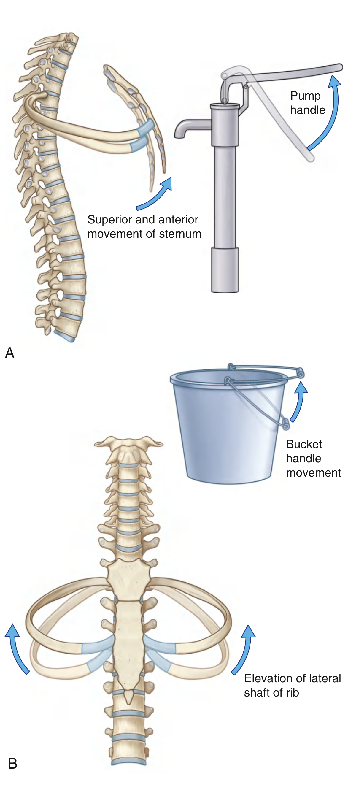
(A) Pump handle - superior and anterior movement of sternum. (B) Bucket handle - lateral elevation of rib shafts.
Key Comparison
| Feature | Pump Handle | Bucket Handle |
|---|---|---|
| Movement direction | Anterior + superior | Lateral (outward) |
| Diameter increased | Anteroposterior | Transverse |
| Pivot axis | Costovertebral joint | Costovertebral to sternocostal (front-to-back axis) |
| Ribs involved | Upper ribs + sternum | Middle and lower ribs |
| Analogy | Old water pump handle swinging up | Bucket handle lifting sideways |
Both movements occur simultaneously during normal inspiration, collectively expanding the thoracic volume and creating the negative intrathoracic pressure that draws air into the lungs.
- Gray's Anatomy for Students, p. 199
- Medical Physiology (Boron & Boulpaep), p. 888-889
Mention any four difference between ventricular and skeletal muscles.
Key differences between ventricular (cardiac) and skeletal muscle include: cardiac muscle is involuntary while skeletal is voluntary; cardiac cells are branched and interconnected via intercalated discs while skeletal fibers are long and unbranched; cardiac muscle cannot undergo tetanus while skeletal muscle can; and cardiac muscle has a much longer action potential with a plateau phase.
Searching sources...
Finding Sources
Reading File
Finding Sources
Reading File
Reading File
Reading File
Finding Sources
Reading File
Finding Sources
Difference between ventilatory and skeletal muscles
Before I dive in, a quick clarification - "ventilatory muscles" and "skeletal muscles" are actually the same tissue type (the diaphragm, intercostals, etc. are all skeletal muscle). You may have meant one of these:
Asking for Preferences
Which comparison did you intend?
Good choice - this is an interesting comparison. Respiratory muscles share the same basic skeletal muscle structure but have several unique adaptations that set them apart.
Searching sources...
Finding Sources
Finding Sources
Finding Sources
Finding Sources
Reading File
Reading File
Reading File
Reading File
Excellent - I now have comprehensive textbook data to provide a well-sourced answer. Here it is:
Ventilatory (Respiratory) Muscles vs. Other Skeletal Muscles
Ventilatory muscles - primarily the diaphragm, intercostals, scalenes, and abdominals - are anatomically skeletal muscles, but their adaptations to continuous, life-sustaining rhythmic work make them distinctly different from limb/peripheral skeletal muscles in several important ways.
1. Primary Load Encountered
| Feature | Ventilatory Muscles | Peripheral Skeletal Muscles |
|---|---|---|
| Load type | Resistive and elastic loads (airway resistance + lung/chest wall compliance) | Primarily inertial loads (moving limb mass against gravity) |
| Contraction pattern | Rhythmic and continuous (never truly rest during life) | Rhythmic only during movement; can rest completely |
"Limb muscles are essentially designed to produce movements and hence primarily work against inertial loads. Respiratory muscles mainly have to overcome resistive and elastic loads." - Fishman's Pulmonary Diseases, p. 97
2. Fiber Type Composition
| Feature | Ventilatory Muscles (Diaphragm) | Limb Skeletal Muscles (untrained) |
|---|---|---|
| Oxidative fibers (Type I + IIa) | ~80% | 36-46% |
| Type I (slow oxidative) fibers | ~55% | Lower proportion |
| Fast fatigable fibers (IIb/IIx) | Very few (~23% IIx; no IIb expressed in humans) | Greater proportion |
The diaphragm contains no type IIb fibers at all in humans - an important adaptation. The intercostals, abdominals, scalenes, and sternomastoids also contain at least 60% highly oxidative fibers.
3. Mitochondrial Density and Oxidative Capacity
| Feature | Ventilatory Muscles | Limb Skeletal Muscles |
|---|---|---|
| Volume density of mitochondria | ~2x greater than limb muscles | Baseline reference |
| Oxygen uptake capacity | Considerably greater | Lower |
| Capillary density | ~2x greater than limb muscle | Baseline reference |
| Maximal blood flow | Much greater | Lower |
This high oxidative capacity and vascular supply are what allow the diaphragm to contract ~20,000 times per day without fatigue under normal conditions.
4. Motor Unit Size and Force Development
| Feature | Ventilatory Muscles | Limb Skeletal Muscles |
|---|---|---|
| Innervation ratio | ~400 fibers/motor neuron (rat diaphragm) | ~2,500 fibers/motor neuron (humans, limb) |
| Motor unit force (fast units) | ~110 mN - considerably smaller than limb | Much larger forces |
| Recruitment principle | Size principle (smallest first) | Same principle, but wider force range |
Smaller motor units in respiratory muscles allow finer graded control of breathing depth and rate.
5. Force-Velocity Characteristics
The force-velocity curve of the diaphragm is intermediate between a pure type I (slow) muscle and a pure type II (fast) muscle - reflecting its mixed fiber composition. Limb muscles show a wider range depending on their function (e.g., soleus is nearly all type I; fast-twitch muscles are weighted toward type II).
6. Vital Dependence and Control
| Feature | Ventilatory Muscles | Peripheral Skeletal Muscles |
|---|---|---|
| Life-dependent | Yes - cannot stop even during sleep | No - voluntary use |
| Control | Both voluntary (cortex) and involuntary (medullary respiratory centers via phrenic nerve C3-C5) | Primarily voluntary |
| Fatigue consequences | Can cause respiratory failure, CO₂ retention, and death | Causes exercise limitation only |
Summary Table
| Property | Ventilatory Muscles | Other Skeletal Muscles |
|---|---|---|
| Primary load | Resistive + elastic | Inertial |
| Contraction rhythm | Continuous, lifelong | Intermittent |
| Oxidative fiber % | ~80% | 36-46% |
| Mitochondrial density | 2x greater | Baseline |
| Capillary density | 2x greater | Baseline |
| Motor unit size | Smaller (finer control) | Larger (greater force) |
| Fatigue resistance | Very high | Variable |
| Control type | Voluntary + involuntary | Primarily voluntary |
- Fishman's Pulmonary Diseases and Disorders, Chapter 3 - The Respiratory Muscles, pp. 96-98
This is a shared conversation. Sign in to Orris to start your own chat.