

What happened
folliculitis furuncle boil groin pubic area skin lesion
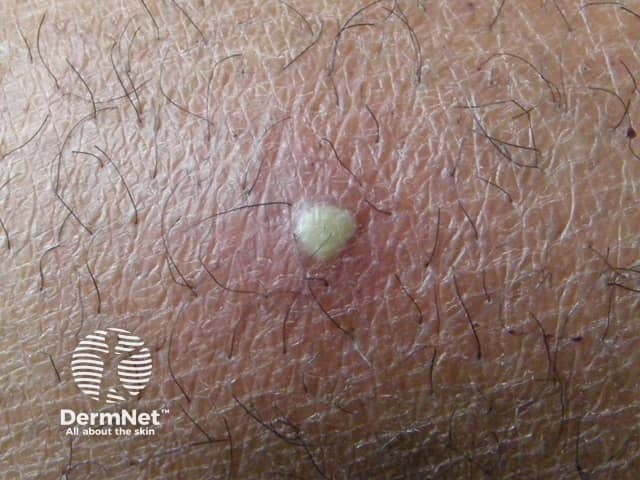
This dermatology clinical photograph documents a solitary boil (furuncle) on the cutaneous surface. Modality: Clinical photography; technique: macro close-up imaging of the skin with standard, noninvasive illumination. The lesion is a dome-shaped, erythematous nodule with a central yellowish-white pustular core representing purulent material within a hair follicle. Surrounding skin shows mild edema and perifollicular erythema. The lesion appears tender on palpation; no drainage is evident although the pus core is visible. The image captures typical signs of acute bacterial folliculitis progressing to furunculosis, most commonly caused by Staphylococcus aureus, with neutrophil-rich inflammatory infiltrate and abscess formation around the follicular unit. Clinically, this finding supports a diagnosis of furuncle rather than a simple acne lesion or epidermal cyst; differential diagnoses include carbuncle, abscess, or inflamed epidermoid cyst. Pathophysiology involves follicular occlusion, microbial colonization, and localized pus accumulation. Management implications include warm compresses, incision and drainage if fluctuance is present, and targeted antibiotics for extensive infection or cellulitis or systemic signs. This image is educational for medical trainees in recognizing cutaneous infections, dermatology case discussions, and demonstrations of localized skin abscesses and follicular infection. Useful for exam prep, clinical decision making, and patient education about boil treatment and outcomes globally.
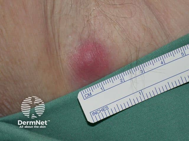
This is a clinical close-up photograph of a single left axillary skin lesion consistent with a boil (furuncle) or localized abscess. Modality: clinical photography; technique: macro close-up with standard color-calibrated lighting; scale is provided by a ruler for size estimation (~1.5–2 cm in diameter). The lesion is erythematous and tender with a raised, dome-shaped nodular configuration in the subcutaneous tissue of the axillary hair-bearing region. Surrounding perilesional erythema and mild edema reflect an acute inflammatory process. A central pus collection may be present; in some furuncles the area can be fluctuant, though drainage is not visible in this still image. The differential includes furuncle/abscess, folliculitis, hidradenitis suppurativa, and infected sebaceous cyst; HS typically shows recurrent painful nodules and sinus tracts in the axilla. Clinically, such lesions arise from bacterial infection of hair follicles, most often Staphylococcus aureus, with perifollicular inflammation. The image provides educational documentation for clinical scenarios, tele-dermatology triage, and surgical planning regarding drainage and antibiotic therapy. It supports visual recognition, teaching for medical students and residents, and correlation with histopathology or dermoscopy when available. Documentation of size, location, and morphology supports monitoring, patient education, and outcome assessment after drainage, incision, or antibiotic therapy, and aids research in cutaneous infectious disease.
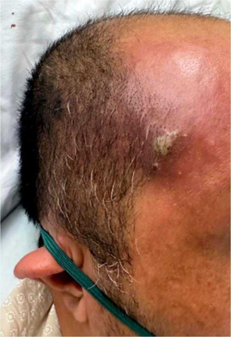
Clinical photograph showing a localized skin lesion, specifically a furuncle (boil), located on the right temporal/forehead region of a 44-year-old male. The lesion is characterized by an elevated, erythematous nodule approximately 1 cm in diameter, exhibiting central fluctuance and a small amount of yellowish-white purulent discharge (pus). There is significant surrounding perilesional erythema and soft tissue inflammation extending toward the scalp and forehead. The adjacent skin displays a slightly irregular texture with normal short hair distribution, including scattered graying. Clinically, such a lesion in a patient with poorly controlled diabetes mellitus can serve as a primary infectious nidus for hematogenous seeding, potentially leading to systemic complications such as septic pulmonary embolism (SPE). This image is used to demonstrate the morphology of a common cutaneous abscess and its potential role in the progression to sepsis.
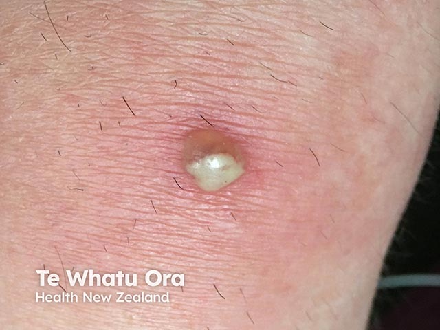
Imaging modality: Clinical photography of a cutaneous lesion. Anatomical location: left forearm skin, superficial dermis and hair follicle involvement. Perspective: close-up, frontal view of a solitary boil. Visual features: round, dome-shaped erythematous plaque with a central pustule and a creamy-white fluctuant core; surrounding perilesional induration; mild surrounding erythema; skin folds visible; no surrounding necrosis. Pathology inference: consistent with furuncle (boil) caused by Staphylococcus aureus infection of a hair follicle with inflammatory abscess formation within the dermis and subcutaneous tissue. In the clinical context, this lesion may arise in patients receiving Bruton tyrosine kinase inhibitors, which can disrupt B-cell function and predispose to bacterial skin infections; however, localization remains cutaneous and localized drainage is often therapeutic. Diagnostic significance: highlights a localized cutaneous infection; differential includes folliculitis, carbuncle, impetigo, abscess; MRSA risk; culture may guide antibiotics. Clinical correlation: correlate with systemic signs (fever, leukocytosis); assess need for wound drainage; versus incision and drainage; consider antibiotic therapy targeting Staphylococcus aureus; review BTK inhibitor therapy with treating oncologist for infection risk mitigation. Potential applications: educational dermatology cases, infection management training, BTK inhibitor adverse effect surveillance, antimicrobial stewardship. Image-guided documentation supports clinical decision-making, antibiotic stewardship, and comparative learning for dermatology and infectious disease education and training.
What's Happening: Most Likely a Furuncle (Boil) / Folliculitis
What Is It?
- Andrews' Diseases of the Skin describes it as: "an acute, round, tender, circumscribed, perifollicular staphylococcal abscess that generally ends in central suppuration."
- Friction from clothing and movement
- Moisture and sweating (apocrine glands)
- Shaving or hair removal (creates micro-breaks in skin)
- The perineum is a known S. aureus carrier site
Possible Causes
- Bacterial folliculitis / furuncle (most common) - S. aureus infection of a hair follicle
- Ingrown hair (pseudofolliculitis) - especially after shaving; the hair curls back into the skin causing a foreign body reaction that looks very similar
- Infected epidermoid/sebaceous cyst - blockage of a gland duct leading to abscess formation
What To Do
| Stage | Management |
|---|---|
| Early (no pus yet) | Warm compresses 3-4x/day to bring it to a head; topical antibiotic (mupirocin) |
| Fluctuant (has pus) | Needs incision and drainage (I&D) by a doctor - do NOT squeeze it yourself |
| Spreading redness/fever | See a doctor urgently - may need oral antibiotics (clindamycin, TMP-SMX, cephalexin) |
Warning Signs - See a Doctor Promptly If:
- The redness is spreading
- You develop fever or chills
- The lesion becomes very large (>2 cm) or fluctuant
- You have diabetes or a weakened immune system
- You've had multiple similar lesions (recurrent furunculosis)
Disclaimer: This is general medical information only, not a diagnosis. Please see a healthcare provider for proper evaluation and treatment.