
Pathology of polycystic kidney disease
I'll read the medical library skill first, then research polycystic kidney disease pathology.
Reading File
Searching sources...
Now let me search the medical library for polycystic kidney disease pathology in parallel.
Finding Sources
Finding Sources
Searching PubMed
"polycystic kidney disease"[MeSH Terms] AND pathology
Good results. Now let me read the key sections in parallel - Brenner and Rector's Kidney (most authoritative nephrology textbook) for both ADPKD and ARPKD, plus Goldman-Cecil and Comprehensive Clinical Nephrology:
Reading File
Reading File
Reading File
Excellent content. Let me now read more of Brenner's section on ADPKD (pathology specifically) and also the ARPKD section, plus fetch the polycystin protein diagram:
Reading File
Reading File
Loading Image
Now let me fetch the key ARPKD gross pathology images and read more on gross/microscopic pathology from Brenner's:
Reading File
Loading Image
Loading Image
I now have excellent content from Brenner's and Comprehensive Clinical Nephrology. Let me get a bit more on the gross and microscopic pathology of ADPKD, plus the molecular pathways:
Reading File
Reading File
I now have comprehensive content from Brenner and Rector's The Kidney. Let me fetch one more key image (the ADPKD gross specimen) and the polycystin structure diagram:
Loading Image
Loading Image
I now have very rich, authoritative content to compile a comprehensive answer. Let me now write the full response.
Pathology of Polycystic Kidney Disease (PKD)
PKD encompasses two major hereditary forms - autosomal dominant (ADPKD) and autosomal recessive (ARPKD) - which differ profoundly in genetics, protein products, gross morphology, microscopic features, and clinical course. Both are ciliopathies: the responsible proteins localize to primary cilia of renal tubular epithelium.
Comparison Overview
| Feature | ADPKD | ARPKD |
|---|---|---|
| Inheritance | Autosomal dominant | Autosomal recessive |
| Frequency | 1 in 400-1,000 | 1 in 10,000-40,000 |
| Gene/Protein | PKD1 (polycystin-1), PKD2 (polycystin-2) | PKHD1 (fibrocystin/polyductin) |
| Age of onset | 20s-30s (adult) | Perinatal/infantile |
| Cyst origin | Any tubular segment; majority collecting duct and distal nephron | Collecting ducts (fusiform dilation) |
| Renomegaly | Yes | Marked (up to 20x normal) |
| Liver involvement | Liver cysts (up to 90% of adults) | Congenital hepatic fibrosis (CHF) |
Source: Goldman-Cecil Medicine, Table 109, p. 1297
Part I: Autosomal Dominant PKD (ADPKD)
Genetics
ADPKD shows complete penetrance. Each child of an affected parent carries a 50% inheritance risk. At least 10% of cases arise from de novo mutation.
- PKD1 (chromosome 16p13.3): accounts for ~78% of pedigrees. Encodes polycystin-1 (PC1, ~4,303 amino acids). PKD1 mutations cause more severe disease; mean age at ESKD = 58.1 years.
- PKD2 (chromosome 4q21): accounts for ~15%. Encodes polycystin-2 (PC2, ~968 amino acids). Mean age at ESKD = 79.7 years.
- Rarer genes: GANAB and DNAJB11 (ER maturation defects) cause ADPKD-like phenotypes.
Individuals heterozygous for both PKD1 and PKD2 mutations have more severe disease than either alone. Inheritance of two fully inactivating alleles at either locus is lethal in utero.
Source: Brenner and Rector's The Kidney, p. 1983-1985
The Polycystin Proteins
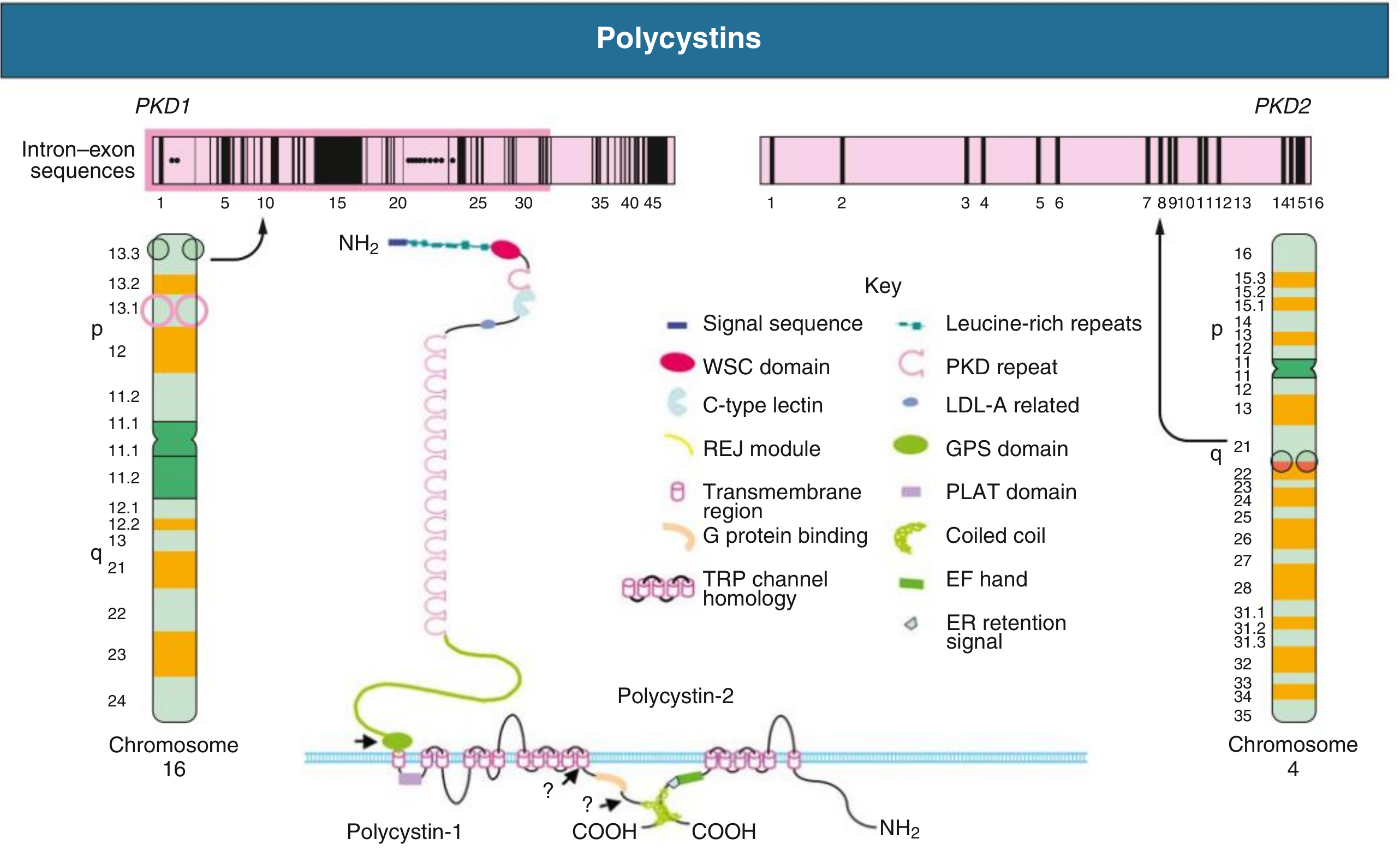
Fig. Polycystins: Gene locations and protein domain structures (Brenner and Rector's)
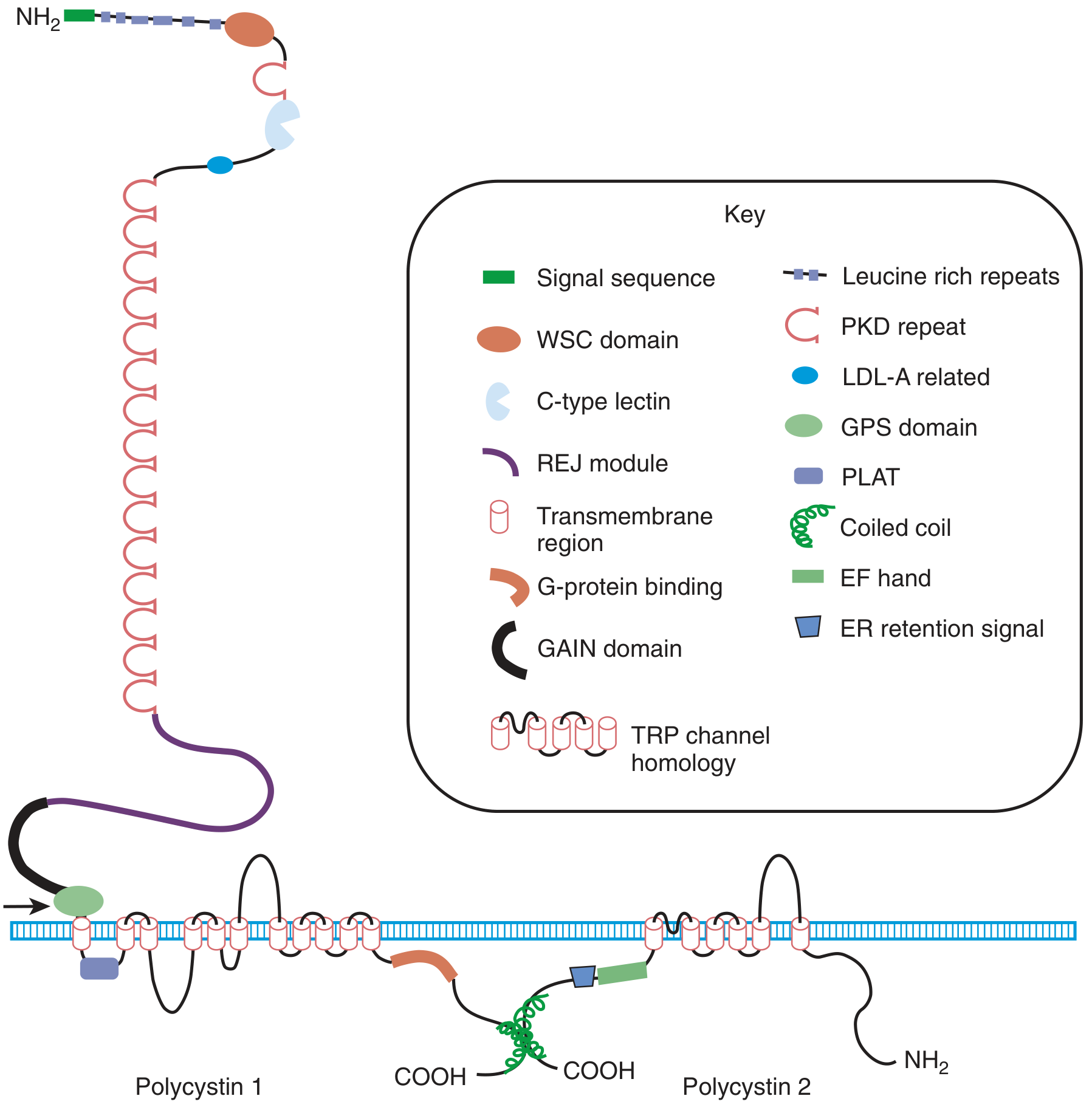
Fig. Detailed domain structure of PC1 and PC2 (Brenner and Rector's)
PC1 (~440 kDa): Large receptor/adhesion-molecule-like protein with:
- Extensive extracellular N-terminal region (leucine-rich repeats, C-type lectin, LDL-A, PKD repeats, REJ module, GAIN domain)
- 11 transmembrane domains
- Short intracellular C-terminal tail (G-protein binding, coiled-coil)
- Found in primary cilia, focal adhesions, desmosomes, adherens junctions, ER, and nucleus
PC2 (TRPP2, ~110 kDa): Transient receptor potential (TRP)-type calcium channel with:
- 6 transmembrane domains
- Short N-terminal cytoplasmic region
- C-terminal portion with EF-hand calcium-binding motif and ER retention signal
- Localized primarily in ER, but also in primary cilium, centrosome, and mitotic spindles
- The two proteins interact via coiled-coil domains in a 1 PC1 : 3 PC2 stoichiometry (cryo-EM data)
Both polycystins are found in high concentrations in urinary exosomes that can interact with primary cilia of neighboring cells.
Source: Comprehensive Clinical Nephrology 7e, p. 653
The "Two-Hit" Hypothesis
Although ADPKD is inherited as autosomal dominant (germ-line mutation in one allele), somatic inactivation of the second, normal allele is required for cyst formation - the "two-hit" model. This explains why only a minority of tubules (<1%) become cystic despite every cell carrying the germ-line mutation. Evidence includes:
- Loss of heterozygosity (LOH) detected in cyst-lining cells
- Cystic cells have reduced or absent PC1/PC2
- Experimental Pkd1 or Pkd2 knockout studies confirm two hits needed
Molecular Pathogenesis of Cyst Formation
Loss of polycystin function below a critical threshold disrupts:
- Primary ciliary signaling - PC1 and PC2 in cilia detect flow-induced bending; loss abolishes calcium influx response
- Intracellular calcium - Reduced resting [Ca²⁺], depleted ER calcium stores, impaired store-operated calcium entry
- Elevated cAMP - Reduced intracellular calcium disinhibits adenylyl cyclase (AC6) and impairs PDE activity → raised cAMP in kidneys, liver, and vasculature. cAMP activates:
- B-Raf/MEK/ERK proliferation pathway (via AMPK inhibition)
- PKA → phosphorylates CFTR and other chloride transporters → fluid secretion into cyst lumen
- mTOR pathway activation - mTORC1 is activated via PC1's interaction with TSC2. mTORC1 drives tubular cell hypertrophy and proliferation
- Planar cell polarity (PCP) loss - Normal tubulogenesis requires oriented cell division along the tubule axis; polycystin deficiency disrupts this, causing tubule dilation
- Increased proliferation - Cyst-lining cells show abnormally high rates of mitosis
- Abnormal fluid secretion - Instead of absorbing like normal tubular epithelium, cyst epithelium secretes Cl⁻ and fluid via CFTR-driven trans-epithelial transport, enlarging the cyst
- Extracellular matrix (ECM) remodeling - Altered collagen composition and increased matrix metalloproteinase activity promotes cyst expansion
Source: Brenner and Rector's The Kidney; Comprehensive Clinical Nephrology 7e
Gross Pathology of ADPKD
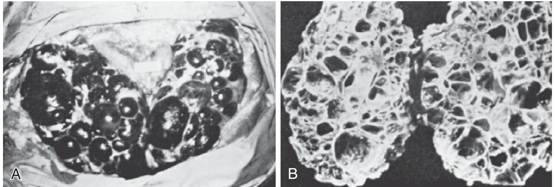
Fig. ADPKD - in situ (A) and cut section (B) showing diffuse, bilateral cysts (Brenner and Rector's)
Key gross features:
- Bilateral, diffuse cyst distribution (no dominant side)
- Kidneys can reach weights of 4,000 g or more per kidney at end-stage (normal ~150 g each)
- Kidney reniform shape may be maintained initially but lost at end-stage
- Cysts range from millimeters to several centimeters
- Cyst contents: clear serous fluid, or hemorrhagic (brown/red) - hemorrhage is a common complication
- At end-stage: only scant islands of normal parenchyma visible; fibrous tissue beneath capsule; fibrous bands encapsulating cysts on cut section
- Tubulointerstitial fibrosis and arteriolar sclerosis are cardinal end-stage features
The CT images below show typical radiologic appearance:
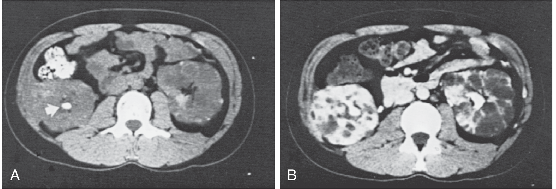
Fig. CT of polycystic kidneys - stone visible in right kidney (A), diffuse bilateral cysts after contrast (B) (Brenner and Rector's)
Microscopic Pathology of ADPKD
- Cyst origins: Cysts begin as outpouchings from preexisting tubules; most detach from the parent tubule once they exceed a few millimeters
- Segment of origin: The majority (especially large cysts >1 mm) stain for collecting duct markers; distal nephron/collecting duct origin predominates. Proximal tubule cysts occur early. Cysts can also arise from Bowman's capsule (parietal epithelium)
- Cyst epithelium: Not typical of mature, fully differentiated tubular epithelium - cells are partially dedifferentiated/immature; some show a flat, attenuated lining; others show cuboidal or low columnar cells
- Majority of cysts (75%) have high luminal Na⁺ (~plasma level) and leaky apical junctions, consistent with poorly differentiated/secretory epithelium; minority have low Na⁺ (more differentiated, absorptive-type)
- Interstitium: Progressive interstitial fibrosis; lymphocytic infiltration; arteriolar sclerosis
- Apoptosis is prominent in noncystic parenchyma adjacent to expanding cysts - this is the primary mechanism of progressive nephron loss and GFR decline
Extrarenal Pathology of ADPKD
Liver (most common extrarenal site):
- Up to 90% of adults have liver cysts
- Cysts are lined by biliary-type epithelium (single layer)
- Arise from proliferation/dilation of biliary ductules (biliary microhamartomas/von Meyenburg complexes) and peribiliary glands
- Macroscopic cysts usually do not communicate with the biliary system
- Clinically more severe in women (estrogen effect); can cause massive hepatomegaly
Intracranial aneurysms:
- Prevalence ~8% (higher in families with a history)
- Rupture is a major cause of mortality; risk increases with hypertension
Cardiovascular:
- Mitral valve prolapse (~25% of patients)
- Aortic regurgitation
- Left ventricular hypertrophy (often before hypertension)
Other:
- Pancreatic cysts (~10%)
- Seminal vesicle cysts (men)
- Arachnoid cysts
- Colonic diverticula
Source: Brenner and Rector's The Kidney, pp. 1993-1997
Part II: Autosomal Recessive PKD (ARPKD)
Genetics
- PKHD1 gene (chromosome 6p21.1-p12): one of the largest human genes (~470 kb, 67 exons)
- Encodes fibrocystin/polyductin (~460 kDa)
- No mutation hot spots; most mutations are private (single-family)
- Two truncating mutations → lethal neonatal phenotype; at least one missense/hypomorphic mutation required for survival
- Carrier frequency ~1 in 70; disease prevalence ~1 in 10,000-20,000
Fibrocystin (Polyductin)
- Single-pass transmembrane protein
- Large extracellular region with IPT/TIG (immunoglobulin-like) and PbH1 (parallel beta-helix) repeats
- Expressed in primary cilia (like polycystins), kidney, liver, and pancreas
- Expressed during embryogenesis in ureteric bud branches, intrahepatic/extrahepatic biliary ducts, and pancreatic ducts - explaining the renal and hepatic phenotype
Gross Pathology of ARPKD
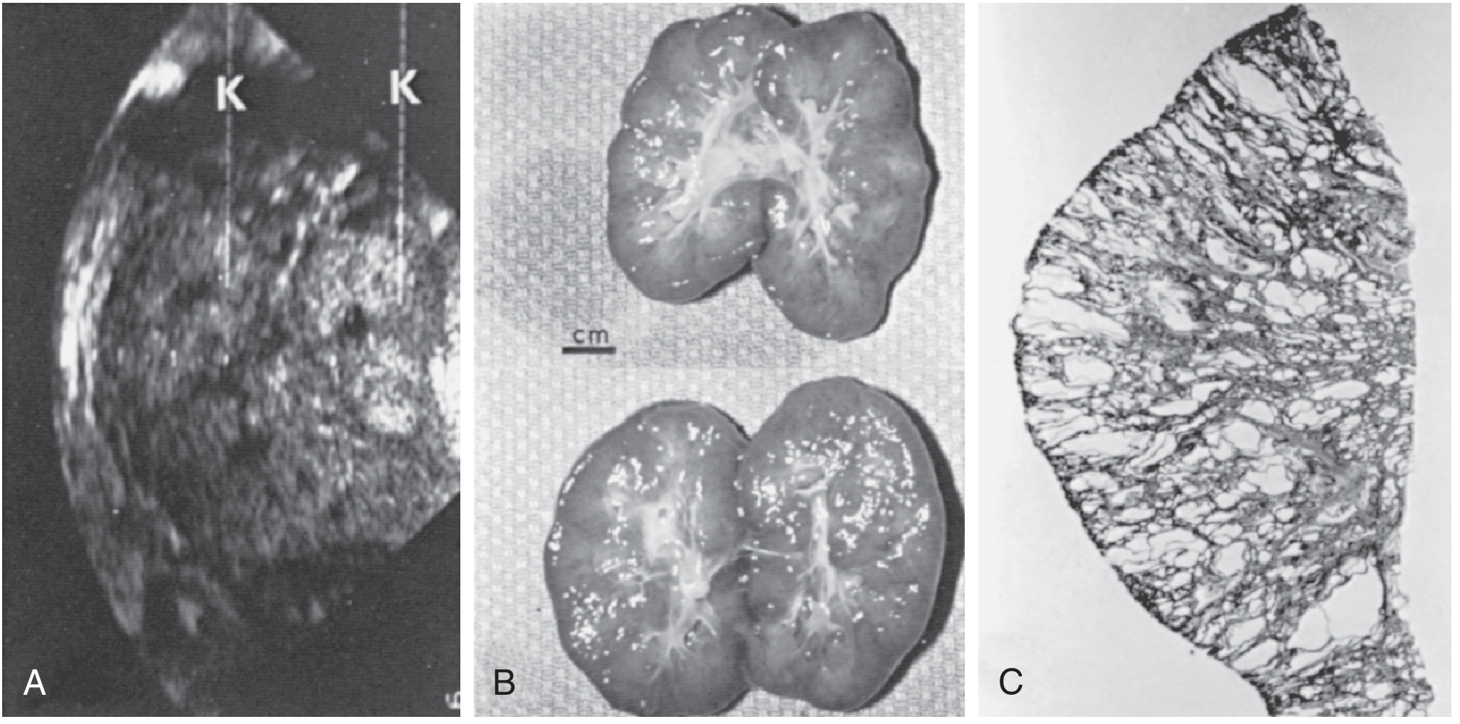
Fig. ARPKD in 32-week fetus - sonogram, gross, and microscopic sections showing radially oriented collecting duct cysts (Brenner and Rector's)
- Kidneys are symmetrically and bilaterally enlarged (combined weight up to 300 g vs. normal ~25 g in neonates - up to 20x normal)
- May cause dystocia due to size
- External surface: lobulated but smooth; kidney shape maintained
- Cut section: radially oriented, fusiform dilations of collecting ducts, 1-2 mm in diameter, throughout cortex and medulla - the classic "sponge-like" or "sunburst" pattern
- No discrete rounded cysts (unlike ADPKD) - the dilations are elongated/fusiform
- Almost 100% of collecting ducts are affected in severe perinatal cases
Microscopic Pathology of ARPKD
- Collecting ducts are lined by typical cuboidal epithelium (well-differentiated, unlike ADPKD)
- Glomeruli and proximal tubular elements appear histologically normal initially
- Proximal tubule cysts may occur in early fetuses (14-24 weeks) but resolve by 34 weeks
- In older children with milder renal involvement: medullary ductal ectasia is prominent; macroscopic cysts can develop
- Renal calcifications are common
Liver pathology (congenital hepatic fibrosis - CHF):
- Characteristic feature of ARPKD (inverse relationship with renal severity)
- Ductal plate malformation - failure of remodeling of primitive bile duct precursors
- Dense periportal fibrosis with proliferating, poorly formed bile ducts (biliary dysgenesis)
- Portal hypertension → esophageal varices, splenomegaly
- Caroli disease (non-obstructive intrahepatic bile duct dilation) variably present
Source: Brenner and Rector's The Kidney, pp. 2001-2003
Pathogenetic Summary: The Ciliopathy Concept
Both ADPKD and ARPKD proteins (polycystins and fibrocystin) localize to the primary cilium of renal tubular cells. These non-motile sensory organelles project into the tubular lumen and respond to fluid flow. Disruption of ciliary signaling is the unifying mechanism:
| Pathway | Effect |
|---|---|
| ↓ Intracellular Ca²⁺ | Disinhibits adenylyl cyclase → ↑ cAMP |
| ↑ cAMP | Activates PKA, CFTR → fluid secretion; activates B-Raf/MEK/ERK → proliferation |
| ↑ mTORC1 | Tubular cell hypertrophy and proliferation |
| ↓ Planar cell polarity | Abnormal cell division orientation → tubule dilation |
| ↑ Apoptosis (noncystic parenchyma) | Progressive nephron loss |
| ECM remodeling | Cyst expansion and fibrosis |
Key Differences: ADPKD vs. ARPKD at a Glance
| Feature | ADPKD | ARPKD |
|---|---|---|
| Cyst shape | Spherical, discrete | Fusiform/elongated dilations |
| Cyst epithelium | Partially dedifferentiated, flat-cuboidal | Well-differentiated cuboidal |
| % nephrons affected | <1% form cysts | ~100% of collecting ducts in severe cases |
| Parenchyma | Initially preserved; lost at end-stage | Normal-appearing between dilated ducts |
| Liver | Saccular cysts (biliary origin) | Portal fibrosis + Caroli disease |
| Dominant complication | Intracranial aneurysm, ESKD in adulthood | Pulmonary hypoplasia (neonatal), portal hypertension |
| Hypertension | Due to intrarenal RAAS activation and cyst compression | Present in severe cases |
Sources: Brenner and Rector's The Kidney, 2-Volume Set (primary); Comprehensive Clinical Nephrology 7th Ed.; Goldman-Cecil Medicine
This is a shared conversation. Sign in to Orris to start your own chat.