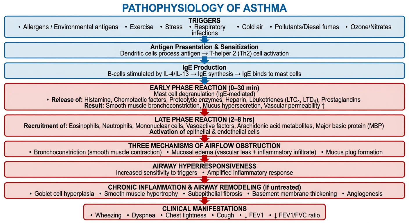
Flow chart for pathophysiology for asthma
A detailed medical flowchart showing the pathophysiology of asthma. The flowchart should flow top to bottom with clear arrows and boxes. Include these steps in order: 1. TRIGGERS box at top (Allergens / Environmental antigens, Exercise, Stress, Respiratory infections, Cold air, Pollutants/Diesel fumes, Ozone/Nitrates) 2. Arrow down to: Antigen Presentation & Sensitization (Dendritic cells process antigen → T-helper 2 (Th2) cell activation) 3. Arrow down to: IgE Production (B-cells stimulated by IL-4/IL-13 → IgE synthesis → IgE binds to mast cells) 4. Arrow down to: EARLY PHASE REACTION (0–30 min) box with: - Mast cell degranulation (IgE-mediated) - Release of: Histamine, Chemotactic factors, Proteolytic enzymes, Heparin, Leukotrienes (LTC4, LTD4), Prostaglandins - Result: Smooth muscle bronchoconstriction, Mucus hypersecretion, Vascular permeability ↑ 5. Arrow down to: LATE PHASE REACTION (2–8 hrs) box with: - Recruitment of: Eosinophils, Neutrophils, Mononuclear cells, Basophils - Release of: Cytokines (IL-3 to IL-6, TNF-α, IFN-γ), Vasoactive factors, Arachidonic acid metabolites, Major basic protein (MBP) - Activation of epithelial & endothelial cells 6. Arrow down to: THREE MECHANISMS OF AIRFLOW OBSTRUCTION box: - Bronchoconstriction (smooth muscle contraction) - Mucosal edema (vascular leak + inflammatory infiltrate) - Mucus plug formation 7. Arrow down to: AIRWAY HYPERRESPONSIVENESS box: - Increased sensitivity to triggers - Amplified inflammatory response 8. Arrow down to: CHRONIC INFLAMMATION & AIRWAY REMODELING box (if untreated): - Goblet cell hyperplasia - Smooth muscle hypertrophy - Subepithelial fibrosis - Basement membrane thickening - Angiogenesis 9. Arrow down to: CLINICAL MANIFESTATIONS box at bottom: Wheezing, Dyspnea, Chest tightness, Cough, ↓ FEV1, ↓ FEV1/FVC ratio Use a clean clinical white background, blue and red color scheme for the boxes, with clear bold text labels. Title at top: "PATHOPHYSIOLOGY OF ASTHMA". Make the chart professional and educational quality suitable for medical students.
Pathophysiology of Asthma
1. Triggers
2. Sensitization Phase
- Antigen presented by dendritic cells → Th2 cell activation
- Th2 cells release IL-4, IL-5, IL-13
- B-cells stimulated → IgE synthesis
- IgE binds to mast cells and basophils (sensitization complete)
3. Early Phase Reaction (0–30 min)
- Preformed mediators: Histamine, chemotactic factors, heparin, proteolytic enzymes
- Secondary mediators: Leukotrienes (LTC4, LTD4), prostaglandins, PAF
- Smooth muscle bronchoconstriction
- Mucus hypersecretion
- ↑ Vascular permeability → mucosal edema
4. Late Phase Reaction (2–8 hrs)
- Eosinophils, neutrophils, mononuclear cells, basophils
- Cytokines: IL-3 to IL-6, TNF-α, IFN-γ
- Arachidonic acid metabolites
- Major basic protein (MBP) from eosinophils → epithelial damage
- Activation of epithelial and endothelial cells amplifying the response
5. Three Mechanisms of Airflow Obstruction
| Mechanism | Cause |
|---|---|
| Bronchoconstriction | Smooth muscle contraction |
| Mucosal edema | Vascular leak + inflammatory infiltrate |
| Mucus plug formation | Goblet cell hypersecretion |
6. Airway Hyperresponsiveness
- Chronic inflammation lowers the threshold for bronchoconstriction
- Even mild stimuli trigger exaggerated responses
7. Chronic Inflammation → Airway Remodeling (if untreated)
- Goblet cell hyperplasia
- Smooth muscle hypertrophy
- Subepithelial fibrosis
- Basement membrane thickening
- Angiogenesis
- Leads to irreversible airflow limitation
8. Clinical Manifestations
- Wheezing, dyspnea, chest tightness, cough
- ↓ FEV₁, ↓ FEV₁/FVC ratio (< 0.75 in adults)
- Reversible with bronchodilators (unlike COPD)