
Diabetic foot which gangrene
diabetic foot gangrene types dry wet
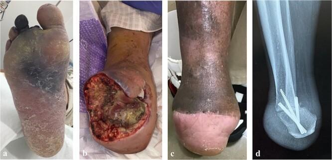
This composite series of four images documents the clinical progression and surgical management of a severe diabetic foot infection and necrotizing gangrene. Panel (a) is a clinical photograph of the plantar surface of the foot showing dry and wet gangrene with extensive tissue necrosis affecting the 2nd, 3rd, and 4th digits, along with surrounding erythema and purulent exudate indicative of advanced infection. Panel (b) shows the postoperative state after a Chopart joint transection, featuring a large, open granulating wound with exposed deep tissues. Panel (c) illustrates the healed outcome following a Pirogoff amputation, showing a shortened, weight-bearing stump with a stable skin graft or healed plantar flap. Panel (d) is a lateral plain radiograph of the same site, demonstrating the modified Pirogoff technique where the calcaneus is rotated and fused to the distal tibia. The radiograph confirms arthrodesis and stable internal fixation with three cannulated compression screws. This sequence is representative of salvage procedures for the diabetic foot to preserve functional weight-bearing length and avoid transtibial amputation.
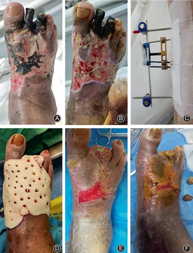
This series of clinical photographs (A-F) documents the surgical management and healing progression of a severely infected diabetic foot with localized gangrene. (A) Admission: Displays extensive dry and wet gangrene on the dorsal aspect of the foot, toes, and ankle with black necrotic eschar. (B) Post-debridement: Shows the removal of necrotic tissue and partial amputation of the second and third toes, revealing a raw, erythematous wound bed with viable granulation tissue. (C) mTTT Treatment: Illustrates the application of a metallic external fixator for transverse tibial bone transport to improve distal perfusion. (D) ABC Treatment: Shows the application of an Antibiotic-Loaded Bone Cement (ABC) spacer, appearing as a molded, perforated white material covering the wound surface for local infection control. (E) Discharge: Demonstrates significant wound contraction and the formation of a healthy pink epithelial layer. (F) Healing: Shows complete wound closure and re-epithelialization with residual hyperpigmentation and scarring. The sequence highlights a multidisciplinary approach combining surgical debridement, antibiotic spacers, and micro-tibia transverse transport (mTTT).
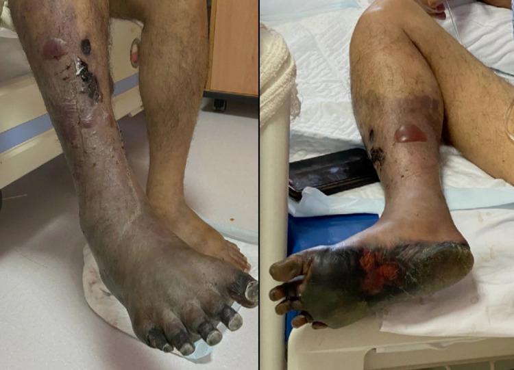
Clinical photograph of a right lower limb demonstrating concurrent dry and wet gangrene secondary to acute arterial insufficiency. The distal foot, including all phalanges, the dorsum, and the plantar surface, exhibits characteristic dry gangrene, visualized as mummified, shriveled, and dark black tissue with distinct demarcation. Proximal to the ankle, the lower leg shows signs of wet gangrene, characterized by significant edema, skin maceration, and the presence of multiple large, fluid-filled bullae and blisters. Notable tissue breakdown and serosanguinous discharge are visible on the pretibial and lateral surfaces of the leg. A clear line of demarcation is emerging below the knee, separating the necrotic, infected tissue from the viable proximal limb. This visual illustrates the progression of critical limb-threatening ischemia and the transformation from dry to infected wet gangrene, a surgical emergency requiring urgent intervention such as amputation to prevent systemic sepsis.
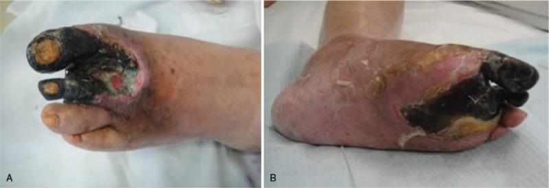
Clinical photographs (A and B) showing a severe diabetic foot ulcer with extensive necrosis and vascular occlusion in the left lower extremity. Image A (dorsal view) reveals significant tissue destruction of the first and second toes, characterized by dry gangrene (blackened, desiccated appearance) and a deep ulceration exposing underlying subcutaneous structures. The wound base contains yellow-green purulent exudate, suggesting secondary infection. Image B (plantar view) demonstrates a large, approximately 9cm x 5cm wound with a central area of black necrotic eschar and slough, involving two-thirds of the plantar surface. There is circumferential erythema (redness) and significant edema (swelling) extending from the wound margins toward the ankle. These clinical features illustrate Wagner Grade 4 diabetic foot complication, indicating critical limb ischemia and a high risk of amputation. Key educational concepts include the recognition of wet and dry gangrene, management of infected diabetic ulcers, and the visual assessment of peripheral arterial disease in chronic diabetic patients.
"diabetic foot" AND gangrene AND management
Diabetic Foot - Gangrene
Definition of Gangrene
Types of Gangrene in the Diabetic Foot
1. Dry Gangrene
- Occurs when tissues are desiccated by gradual slowing of the bloodstream
- Typically results from atheromatous (atherosclerotic) occlusion of arteries
- A clean line of demarcation forms between viable and dead tissue in a matter of days (when proximal blood supply is adequate)
- Separation occurs neatly with minimum infection
- If bone is involved, separation takes longer; the stump tends to be conical
2. Wet (Moist) Gangrene
- Occurs when superadded infection and putrefaction are present
- Crepitus may be palpated - due to gas-forming organisms - a hallmark feature in diabetic foot problems
- Must be considered a surgical emergency requiring urgent tissue debridement or amputation
- Infection and suppuration extend into neighbouring living tissue, pushing the line of demarcation more proximally than in dry gangrene
- Spread can occur as "skip areas" on the other side of the foot, heel, dorsum, or even the calf
Pathophysiology of Diabetic Gangrene
| Factor | Details |
|---|---|
| Ischaemia | Macrovascular atherosclerosis (typically affects crural vessels with relative sparing of pedal vessels) + microvascular dysfunction from increased circulatory shunting |
| Peripheral Sensorimotor Neuropathy (PSN) | Sensory loss in stocking distribution → trophic changes, unnoticed injury; motor involvement → muscle imbalance → abnormal pressure loading → callosities |
| Immunosuppression | Hyperglycaemia impairs immune function, predisposing to infection |
- Bailey and Love's Surgery, p.1031
Classification of Diabetic Foot Lesions
Wagner Classification (Foot Ulcers)
| Grade | Description |
|---|---|
| 0 | Skin at risk |
| I | Superficial ulcer |
| II | Exposed tendon and deep structures |
| III | Deep ulcers with abscess or osteomyelitis |
| IV | Partial gangrene |
| V | More extensive gangrene |
Brodsky Depth-Ischemia Classification
- 0 = At-risk foot, no ulceration
- 1 = Superficial ulceration, no infection
- 2 = Deep ulceration, tendons/joint exposed
- 3 = Extensive ulceration or abscess
-
A = Not ischemic
-
B = Ischemia without gangrene
-
C = Partial foot gangrene
-
D = Complete gangrene
-
Campbell's Operative Orthopaedics 15th Ed, p.5058
Treatment
- Arterial assessment and revascularisation - angioplasty or bypass surgery to improve distal circulation (enables more conservative amputation levels)
- Drainage of pus - urgent incision and drainage
- Liberal debridement - removal of all dead and infected tissue
- Antibiotics - broad-spectrum, covering gram-positives, gram-negatives, and anaerobes (diabetic foot infections are frequently polymicrobial)
- Primary amputation - indicated for:
- Life-threatening systemic sepsis
- When arterial supply cannot be improved
- Non-functional limb (contractures, etc.)
- Strict glycaemic control throughout
Key principle: amputation in the presence of poor circulation will fail - gangrene reappears at wound edges. Revascularisation must precede or accompany surgery.
Clinical Images
Key Points Summary
| Feature | Dry Gangrene | Wet Gangrene |
|---|---|---|
| Cause | Arterial occlusion, desiccation | Infection + arterial disease |
| Appearance | Mummified, blackened, shrunken | Swollen, moist, malodorous, blistered |
| Demarcation line | Clear, neat | Poorly defined, spreads proximally |
| Crepitus | Absent | May be present (gas-forming organisms) |
| Urgency | Less urgent | Surgical emergency |
| Risk | Dry can convert to wet | Systemic sepsis, death |
Give me one answer wheather it is dry or wet
Diabetic Foot Gangrene = WET Gangrene
- Diabetes causes immunosuppression (high tissue glucose impairs immunity) → predisposes to superadded infection and putrefaction → this is the defining feature of wet gangrene
- Gas-forming organisms (e.g. Clostridium, anaerobes) are common in diabetic foot infections → produce crepitus on palpation
- Infection spreads rapidly in subfascial planes → leads to fulminant foot sepsis
- It is therefore a surgical emergency
Bailey and Love's Surgery, 28th Ed: "Crepitus may be palpated as a result of infection by gas-forming organisms, commonly in diabetic foot problems, and should be considered a surgical emergency."
| Gangrene Type | Classic Cause |
|---|---|
| Dry | Atherosclerosis (old age, smoker) - no infection |
| Wet | Diabetes - infection + ischaemia |