
Polycythemia vera
"polycythemia vera"[MeSH Terms] AND management
polycythemia vera blood smear bone marrow JAK2
![This diagnostic visual presents an [18F]FDG PET/CT scan of a 59-year-old male, illustrating advanced polycythemia vera (PV) with transformation to myelofibrosis. Panel A is a Maximum Intensity Projection (MIP) showing widespread, intense metabolic activity throughout the axial and appendicular skeleton. High FDG uptake is particularly localized in the bone marrow of the trunk (spine, ribs, pelvis) and the proximal long bones (humeri and femurs), characteristic of panmyelosis. Panel B displays three transversal PET/CT fusion slices corresponding to the abdominal and pelvic levels. These slices reveal massive splenomegaly, with the spleen extending from the upper abdomen into the pelvis. The splenic tissue demonstrates uniform and significantly elevated metabolic activity, reflecting extramedullary hematopoiesis and high cellular turnover. The combination of intense skeletal uptake and hypermetabolic splenomegaly in the absence of nodal or solid organ malignancy is highly suggestive of a myeloproliferative neoplasm in the 'spent phase' or fibrotic transformation. The image serves as an educational reference for nuclear medicine and hematology-oncology in identifying metabolic patterns associated with JAK2-positive myelofibrosis.](https://cdn.orris.care/cdss_images/pmc_clinical_VQA_74f4452983353662bb06b37ba53e0735049ff71767119be5980ef9325d9b47e5.jpg)
This diagnostic visual presents an [18F]FDG PET/CT scan of a 59-year-old male, illustrating advanced polycythemia vera (PV) with transformation to myelofibrosis. Panel A is a Maximum Intensity Projection (MIP) showing widespread, intense metabolic activity throughout the axial and appendicular skeleton. High FDG uptake is particularly localized in the bone marrow of the trunk (spine, ribs, pelvis) and the proximal long bones (humeri and femurs), characteristic of panmyelosis. Panel B displays three transversal PET/CT fusion slices corresponding to the abdominal and pelvic levels. These slices reveal massive splenomegaly, with the spleen extending from the upper abdomen into the pelvis. The splenic tissue demonstrates uniform and significantly elevated metabolic activity, reflecting extramedullary hematopoiesis and high cellular turnover. The combination of intense skeletal uptake and hypermetabolic splenomegaly in the absence of nodal or solid organ malignancy is highly suggestive of a myeloproliferative neoplasm in the 'spent phase' or fibrotic transformation. The image serves as an educational reference for nuclear medicine and hematology-oncology in identifying metabolic patterns associated with JAK2-positive myelofibrosis.
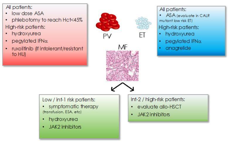
This infographic summarizes treatment recommendations for Myeloproliferative Neoplasms (MPN), specifically Polycythemia Vera (PV), Essential Thrombocythemia (ET), and Myelofibrosis (MF). The central panel illustrates the conditions: PV is shown with a cluster of erythrocytes; ET with light-colored thrombocytes; and MF with a histological bone marrow biopsy slide showing reticulin fibrosis. A red box for PV lists standard therapy (low-dose ASA, phlebotomy to Hct<45%) and high-risk options (hydroxyurea, pegylated IFṆ̑́, ruxolitinib). A blue box for ET outlines ASA use and high-risk treatments including hydroxyurea, pegylated IFṆ̑́, and anagrelide. The bottom green section displays management for MF stratified by risk: Low/Int-1 patients receive symptomatic therapy (transfusions, ESA), hydroxyurea, or JAK2 inhibitors; Int-2/High-risk patients are managed with JAK2 inhibitors and evaluation for allogeneic hematopoietic stem cell transplantation (allo-HSCT). This educational algorithm serves as a clinical guide for hematology-oncology professionals and students to differentiate subtype-specific therapeutic pathways based on patient risk stratification.
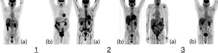
This diagnostic image displays a series of three whole-body Maximum Intensity Projection (MIP) 18F-FDG PET scans (1a, 2a, 3a) paired with age- and sex-matched controls (1b, 2b, 3b) for a comparative study of Polycythemia Vera (PV). The visual focuses on the metabolic activity of the bone marrow. Case 1a exhibits elevated, dispersed FDG uptake throughout the axial skeleton compared to control 1b, alongside incidental avid hepatic and esophageal lesions. Case 2a demonstrates diffuse heterogeneous uptake in both the axial and appendicular skeleton, notably more intense than control 2b, with focal uptake at the gastroesophageal (GE) junction and mediastinum. Case 3a shows more subtle visual differences from its control 3b, although the axial skeleton maintains increased metabolic activity. These images illustrate the hyper-metabolic state of bone marrow in myeloproliferative neoplasms and highlight the utility of functional imaging in identifying both disease-related marrow expansion and co-morbidities such as potential malignancies in secondary sites like the thyroid, lungs, or GE junction.
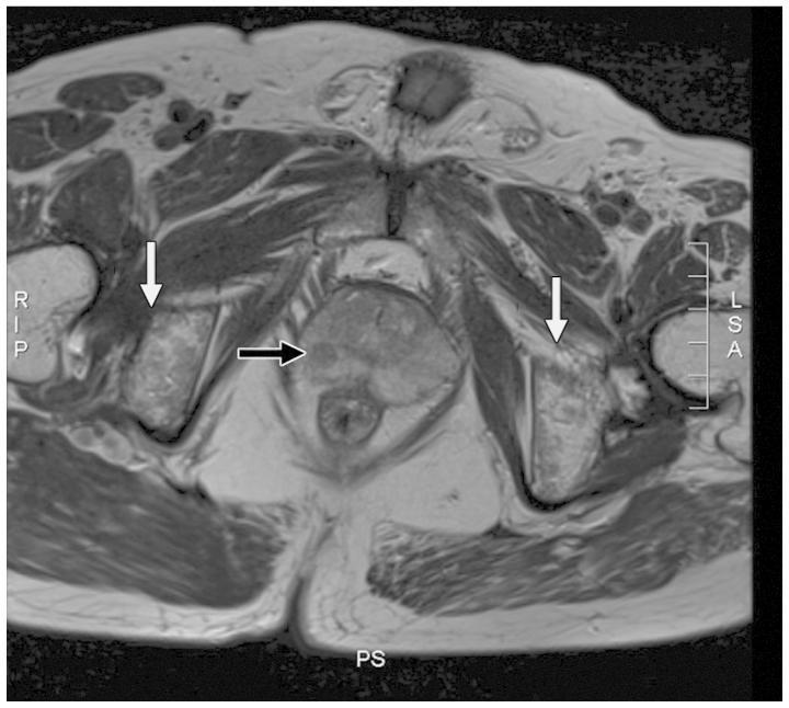
This diagnostic axial MRI image focuses on the pelvic region, specifically evaluating the prostate gland and surrounding musculoskeletal structures. Within the prostate, a black arrow highlights a small, localized nodule (approximately 5 mm) in the right peripheral zone characterized by abnormal signal intensity. The prostatic capsule appears continuous and intact, suggesting no extracapsular extension. Anatomically, the pelvic bone marrow (indicated by white arrows) exhibits a diffusely inhomogeneous signal intensity, which contrasts with the preserved integrity of the cortical bone structure. There is no evidence of localized lymphadenopathy in the immediate periprostatic area. This imaging is representative of clinical oncology and radiology, used to differentiate between localized primary prostatic adenocarcinoma and secondary hematologic or metastatic bone marrow involvement, such as that seen in myeloproliferative disorders like JAK2 mutation-positive polycythemia vera.
Polycythemia Vera (PV)
Definition and Overview
- Harrison's Principles of Internal Medicine 22E, p. 864
- Robbins, Cotran & Kumar Pathologic Basis of Disease
Pathogenesis
JAK2 Mutation
- Erythropoietin hypersensitivity and EPO-independent erythroid colony formation
- Increased Bcl-X_L expression (apoptosis resistance)
- Rapid terminal differentiation of erythroid progenitors
- Low serum EPO (contrast with secondary polycythemia where EPO is high)
- Harrison's 22E; Robbins & Kumar Basic Pathology; Robbins, Cotran & Kumar Pathologic Basis of Disease
Morphology
| Feature | Finding |
|---|---|
| Bone marrow | Hypercellular (panmyelosis) with residual fat; increased erythroid, granulocytic, and megakaryocytic precursors |
| Reticulin | Moderate-to-marked increase at diagnosis in ~10% of cases |
| Peripheral blood | Elevated RBC, often leukocytosis, large/abnormal platelets, basophilia |
| Late "spent phase" | Extensive marrow fibrosis, displaced hematopoiesis, prominent splenomegaly from extramedullary hematopoiesis |
- Robbins, Cotran & Kumar Pathologic Basis of Disease
Clinical Features
Symptoms from Hyperviscosity
- Headache, vertigo, tinnitus, visual disturbances, TIAs
- Systolic hypertension
- Fatigue, difficulty concentrating, lightheadedness
Characteristic Symptoms
- Aquagenic pruritus - itching after a warm bath/shower (highly characteristic)
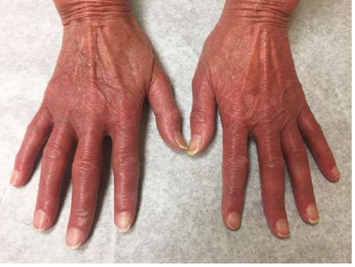
- Erythromelalgia - burning, erythema, and pain in the extremities due to platelet-mediated small vessel occlusion; responds dramatically to aspirin
Examination Findings
- Facial plethora, conjunctival injection, engorged retinal veins
- Splenomegaly (more frequent than hepatomegaly) - from congestion early, extramedullary hematopoiesis later
- Hypertension
Thrombosis and Bleeding
In a large international study of PV patients, arterial thrombosis, venous thrombosis, or major hemorrhage were noted prior to or at diagnosis in 16%, 7%, and 4% of patients, respectively.
- Cerebral, cardiac, and mesenteric vessels are most commonly involved
- Budd-Chiari syndrome (hepatic vein thrombosis) is particularly common in young women with PV - PV should be suspected in any woman who develops hepatic vein thrombosis
- Portal vein thrombosis is more common in male PV patients
- Bleeding can occur due to acquired von Willebrand disease (platelet-mediated proteolysis of high-MW vWF multimers when platelets >900,000/mL)
Metabolic
-
Hyperuricemia, secondary gout, uric acid stones
-
Gastrointestinal symptoms (epigastric discomfort, early satiety from splenomegaly)
-
Harrison's 22E, p. 863-864; Goldman-Cecil Medicine, p. 1751
Diagnosis
WHO 2022 Diagnostic Criteria for PV
- Hemoglobin >16.5 g/dL (men) or >16.0 g/dL (women), OR hematocrit >49% (men) or >48% (women), OR elevated red cell mass
- Bone marrow biopsy showing age-adjusted hypercellularity with trilineage proliferation (panmyelosis) including prominent erythroid, granulocytic, and pleomorphic megakaryocytic proliferation
- Presence of JAK2 V617F or JAK2 exon 12 mutation
- Subnormal serum EPO level
Key Diagnostic Notes
-
An elevated hemoglobin/hematocrit is not sufficient alone - plasma volume expansion in PV can mask the elevated red cell mass, especially in women. Red cell mass and plasma volume determinations may be necessary.
-
Normal arterial oxygen saturation + JAK2 mutation provides strong evidence for PV
-
Differential includes: secondary erythrocytosis (high altitude, COPD, high-O2-affinity Hgb), relative erythrocytosis (dehydration, diuretics), other MPNs
-
Harrison's 22E; Goldman-Cecil Medicine
Treatment
Risk Stratification
| Risk Category | Features |
|---|---|
| Low risk | Age <60 AND no prior thrombosis |
| High risk | Age ≥60 OR prior thrombosis |
Treatment Algorithm
- Phlebotomy - target hematocrit <45% (proven to reduce thrombotic risk in the CYTO-PV trial)
- Low-dose aspirin (75-100 mg/day) - reduces thrombotic risk; avoids in extreme thrombocytosis (>1,500,000/mL) due to bleeding risk
- Hydroxyurea (hydroxycarbamide) - first-line cytoreductive agent; reduces all blood cell lines, lowers thrombotic risk
- Pegylated interferon alpha (pegIFN-alpha) - alternative first-line, preferred in younger patients and pregnant women; can induce molecular remissions (JAK2 allele burden reduction)
- Ropeginterferon alfa-2b (Besremi) is an approved long-acting pegIFN with a 2025 systematic review/meta-analysis confirming efficacy and safety
- Ruxolitinib (JAK1/2 inhibitor) - for patients intolerant or resistant to hydroxyurea; also effective for pruritus and splenomegaly
- Busulfan - low-dose oral option especially in elderly patients
-
Pruritus: antihistamines, aspirin, interferon, ruxolitinib (most effective)
-
Hyperuricemia/gout: allopurinol
-
Erythromelalgia: aspirin (dramatic response)
-
Harrison's 22E; Goldman-Cecil Medicine
Disease Course and Complications
Transformation
- Prepolycythemic phase - subtle erythrocytosis, minimal symptoms
- Polycythemic phase - classic manifestations
- Spent phase / Post-PV Myelofibrosis - extensive marrow fibrosis, progressive anemia, massive splenomegaly; occurs in ~15-20% over 20 years
| Transformation | Frequency |
|---|---|
| Post-PV myelofibrosis | ~15-20% at 20 years |
| Transformation to AML | ~1-3% (higher with hydroxyurea treatment history, especially if alkylating agents used) |
- Robbins, Cotran & Kumar Pathologic Basis of Disease; Harrison's 22E
Key Distinguishing Features vs. Secondary Erythrocytosis
| Feature | PV | Secondary Erythrocytosis |
|---|---|---|
| Serum EPO | Low | Elevated |
| JAK2 mutation | Present (>95%) | Absent |
| Leukocytosis/thrombocytosis | Common | Absent |
| Splenomegaly | Present | Absent |
| Aquagenic pruritus | Characteristic | Absent |
| Oxygen saturation | Normal | Often low (hypoxic causes) |