
Chronic pancreatic in pregnancy
"chronic pancreatitis" AND pregnancy
PMID: 36498253
chronic pancreatitis pancreatic calcifications imaging
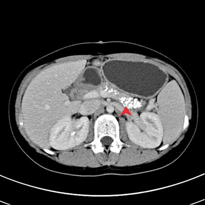
This diagnostic axial CT scan of the abdomen illustrates classic radiological findings of chronic pancreatitis. The image reveals numerous punctate, high-density areas representing parenchymal calcifications within the pancreas. A red arrow specifically highlights a dense cluster of these calcifications located in the head and neck region of the pancreas, anterior to the vertebral column. Surrounding anatomical structures include the liver in the right upper quadrant, the fluid-filled stomach positioned superiorly, the spleen, and both kidneys visualized posteriorly. The presence of extensive diffuse calcifications within the pancreatic parenchyma is a pathognomonic sign for end-stage chronic pancreatitis. This clinical imaging is essential for students and clinicians studying gastroenterology and radiology to identify permanent structural changes in the pancreas resulting from chronic inflammatory processes.
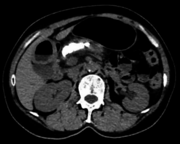
This axial non-contrast-enhanced computed tomography (CT) scan of the abdomen demonstrates classic imaging features of chronic pancreatitis. The primary finding is a series of dense, hyperattenuating (bright) focal areas within the pancreatic parenchyma, consistent with intraductal pancreatic stones or parenchymal calcifications. The largest calcification is visible in the region of the pancreatic head and body. The pancreas itself appears somewhat heterogeneous and atrophic. Surrounding anatomical structures are clearly visible, including the liver (right upper quadrant), the spleen (left upper quadrant), and bilateral kidneys. A cross-section of a lumbar vertebra is centered posteriorly. The stomach is visible anterior to the pancreas, appearing partially distended with air. No significant peripancreatic fluid collections or inflammatory stranding are immediately evident in this slice, suggesting a chronic rather than acute inflammatory state. This diagnostic image serves as a clinical example for medical students and gastroenterology fellows to identify obstructive pancreatic ductal lithiasis in the context of chronic pancreatitis.
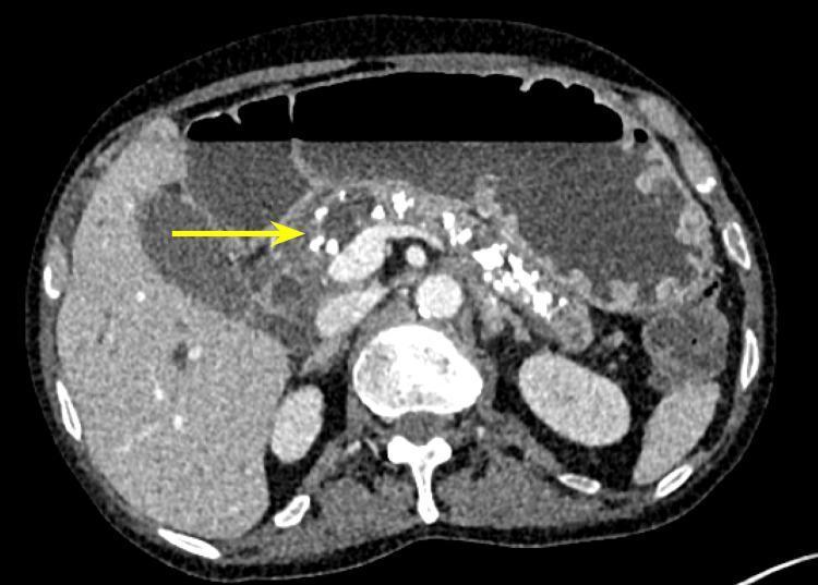
Axial contrast-enhanced CT scan of the abdomen demonstrating classic features of chronic pancreatitis. The primary diagnostic finding is the presence of multiple, coarse, hyperdense parenchymal calcifications (foci of high attenuation) distributed throughout the head, body, and tail of the pancreas. A yellow arrow points specifically to dense calcifications in the pancreatic head/groove region. The pancreas appears somewhat atrophic with irregular margins, indicative of long-standing inflammatory changes. Surrounding anatomical structures include the liver (right), spleen (left), a vertebra with its neural arch (center-bottom), and the stomach containing air-fluid levels (anterior). This imaging is characteristic of end-stage chronic pancreatitis, often associated with long-term alcohol use or metabolic disorders, and serves as a key diagnostic indicator for irreversible parenchymal damage.
Chronic Pancreatitis in Pregnancy
Overview
Pathophysiology
Etiology in Pregnancy
| Cause | Notes in Pregnancy |
|---|---|
| Gallstones / Cholelithiasis | Most common (~66%); estrogen increases bile lithogenicity |
| Alcohol | ~12%; abstinence is essential |
| Idiopathic | ~17% |
| Hypertriglyceridemia | ~4%; elevated by estrogen and progesterone; can precipitate acute flares on top of CP |
| Genetic (CFTR, PRSS1, SPINK1 mutations) | More common in younger patients with "idiopathic" CP |
| Autoimmune pancreatitis | Rare; steroid-responsive |
| Medications | Various; drug history essential |
| Acute fatty liver of pregnancy (AFLP) | Rare but carries particularly poor prognosis when associated with pancreatitis |
- Yamada's Textbook of Gastroenterology; Creasy & Resnik's Maternal-Fetal Medicine, p. 1598
- Elevated estrogen increases cholesterol saturation of bile → gallstone formation
- Progesterone causes gallbladder hypomotility → bile stasis
- Elevated triglyceride levels (physiologic in pregnancy) can reach pancreatitis-inducing thresholds, especially in women with underlying lipid disorders
Clinical Features
-
Presentation is not significantly altered by pregnancy
-
Most common in the third trimester and puerperium
-
Symptoms: epigastric pain (may radiate to flanks/back/shoulders), nausea, vomiting, abdominal tenderness
-
Some patients present with nausea/vomiting as the only complaint
-
Associated findings: mild fever, leukocytosis, adynamic ileus on imaging
-
Chronic CP features in pregnancy also include: steatorrhea, weight loss, malabsorption, and pancreatogenic diabetes (type 3c)
-
Creasy & Resnik's Maternal-Fetal Medicine, p. 1599
Diagnosis
Laboratory
| Test | Finding | Notes |
|---|---|---|
| Serum lipase | Elevated (>3× ULN) | Sensitivity 94%, specificity 96% for acute pancreatitis |
| Serum amylase | Elevated (>3× ULN) | Less specific; also elevated in cholecystitis |
| Serum amylase | May be low in CP | Chronic destruction of acinar tissue reduces amylase output |
| Blood glucose | Elevated | Indicates endocrine involvement |
| Serum calcium | Decreased | Saponification / severity marker |
| Triglycerides | Elevated | May be the trigger |
Imaging
- Ultrasound - first-line; safe in pregnancy; excellent for gallstone detection, less reliable for pancreatic parenchyma
- MRCP - preferred when ultrasound is indeterminate; confirms diagnosis, identifies ductal changes, pseudocysts, choledocholithiasis; avoids radiation; performed successfully during pregnancy
- CT - reserved for severe cases where fetal radiation risk is justified; identifies necrosis, pseudocysts, calcifications
- ERCP - therapeutic only (not diagnostic) during pregnancy; used for bile duct stone removal
Management
General Principles (same as non-pregnant)
- Bowel rest ± nasogastric decompression during acute flares
- IV fluid resuscitation with electrolyte replacement
- Analgesia - Meperidine (pethidine) is traditionally preferred over morphine as it does not constrict the sphincter of Oddi; however, opioids should be used with caution in pregnancy
- Fetal monitoring - continuous during acute flares
- Positioning - lateral decubitus to avoid IVC compression
Specific Considerations in Pregnancy
| Issue | Management |
|---|---|
| Gallstone-related pancreatitis | ERCP for common duct obstruction; early laparoscopic cholecystectomy advocated in all trimesters (70% relapse rate if deferred) |
| Hypertriglyceridemia-induced | Fat restriction, nutritional supplements, plasma exchange, insulin infusion, heparin (limited pregnancy data) |
| Exocrine insufficiency | Pancreatic enzyme replacement therapy (PERT) with each meal (e.g., pancrelipase); safe in pregnancy |
| Pancreatogenic diabetes | Insulin is the treatment of choice in pregnancy (oral agents generally avoided) |
| Pain management (chronic) | Conservative first; neuropathic agents (e.g., pregabalin) require careful risk-benefit assessment in pregnancy |
| Nutritional support | Enteral nutrition preferred over TPN; fat-soluble vitamin supplementation (A, D, E, K) and fat-restricted diet |
| Pseudocysts | Most observed; spontaneous resolution ~30-40%; intervention if symptomatic (laparoscopic, endoscopic, or percutaneous drainage reported) |
| Alcohol cessation | Mandatory; results in pain relief in 60-75% |
- Creasy & Resnik's Maternal-Fetal Medicine, p. 1599-1600; Schwartz's Principles of Surgery
Endoscopic / Surgical Options
- ERCP with sphincterotomy during pregnancy limits radiation by using fluoroscopy sparingly; can prevent recurrent biliary pancreatitis and allow cholecystectomy to be deferred
- However, sphincterotomy alone carries a 25-50% risk of gallstone complications within 2 years
- Surgical drainage procedures (Puestow, Frey, Berne) are rarely performed during pregnancy but may be necessary in refractory cases
Maternal and Fetal Outcomes
-
For mild disease responsive to conservative management: prognosis for mother and fetus is excellent
-
For severe pancreatitis (necrotizing, multiorgan failure): significant maternal and fetal morbidity/mortality
-
Perinatal mortality has decreased to ~3.6% with modern neonatal care; maternal mortality <1%
-
Risk of preterm labor is increased during acute flares
-
CP patients may have pre-existing malnutrition that compounds fetal growth risk
-
Pancreatic necrosis can progress to SIRS and multiorgan failure
-
50-70% of untreated biliary pancreatitis will have recurrent episodes if managed conservatively without addressing the biliary cause
-
Yamada's Textbook of Gastroenterology; Creasy & Resnik's Maternal-Fetal Medicine
Multidisciplinary Care
- Maternal-fetal medicine / Obstetrics
- Gastroenterology / Hepatology
- Surgery (HPB surgeon)
- Dietetics/Nutrition
- Endocrinology (if pancreatogenic diabetes)
- Neonatology (for preterm risk)
Key Points Summary
| Most common cause in pregnancy | Gallstones |
| Most dangerous cause | Hypertriglyceridemia (+ AFLP) |
| Preferred imaging | Ultrasound first, then MRCP |
| Preferred analgesia | Meperidine (avoids sphincter of Oddi spasm) |
| Ranson criteria | Not validated in pregnancy |
| Serum amylase in chronic CP | May be LOW (acinar destruction) |
| Cholecystectomy timing | All trimesters if gallstone-related (70% relapse without it) |
| Enzyme replacement | Safe in pregnancy |
| Diabetes treatment | Insulin only |
Pancreatogenic diabetes
"pancreatogenic diabetes" OR "type 3c diabetes" AND "chronic pancreatitis"
PMID: 36219100
PMID: 41357647
pancreatic islet cells diabetes insulin glucagon pathology diagram
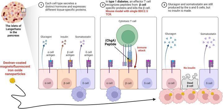
This pathophysiology diagram illustrates the cellular mechanisms of Type 1 Diabetes (T1D) within a pancreatic islet of Langerhans. The diagram is divided into three sequential panels. Panel 1 shows healthy endocrine cells: alpha (α) cells secreting glucagon, beta (β) cells secreting insulin, and delta (δ) cells secreting somatostatin, each expressing specific surface antigens. Panel 2 depicts an 'immune attack' phase where an autoreactive cytotoxic T cell recognizes a Chromogranin A (ChgA) peptide presented on the β-cell surface. Panel 3 illustrates the outcome: selective β-cell destruction ('Dead β cell') and the absence of insulin production, while α and δ cells remain intact and functional. The diagram also highlights the role of diagnostics, showing dextran-coated magnetofluorescent iron oxide nanoparticles being internalized by infiltrating macrophages and monocytes (insulitis). These nanoparticles serve as contrast agents for MRI to visualize microvascular leakage and inflammation in the pancreas. This educational tool demonstrates the transition from normal hormonal homeostasis to autoimmune-mediated islet dysfunction.
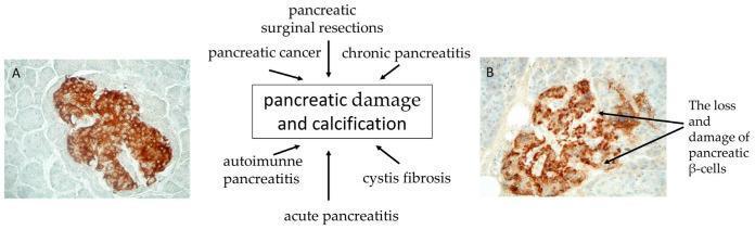
This medical figure presents a side-by-side comparison of insulin immunohistochemistry (IHC) in pancreatic islet cells, integrated with a central conceptual diagram. Image A (210× magnification) shows a normal pancreatic islet characterized by a strong, dense, and uniform brown chromogen reaction within the β-cells, indicating high insulin content and intact cellular architecture. Image B (265× magnification) depicts an islet from a patient with Type 3c Diabetes Mellitus (T3cDM), displaying a diffuse and reduced staining intensity. Arrows in Image B specifically highlight the loss and damage of pancreatic β-cells, resulting in fragmented islet morphology. Between the two micrographs, a central flowchart identifies etiologies leading to 'pancreatic damage and calcification'—including acute and chronic pancreatitis, pancreatic cancer, surgical resections, autoimmune pancreatitis, and cystic fibrosis—which ultimately contribute to the β-cell depletion visualized in Image B. This visual aid is designed for pathology and endocrinology education, illustrating the histological manifestation of pancreatogenic diabetes.
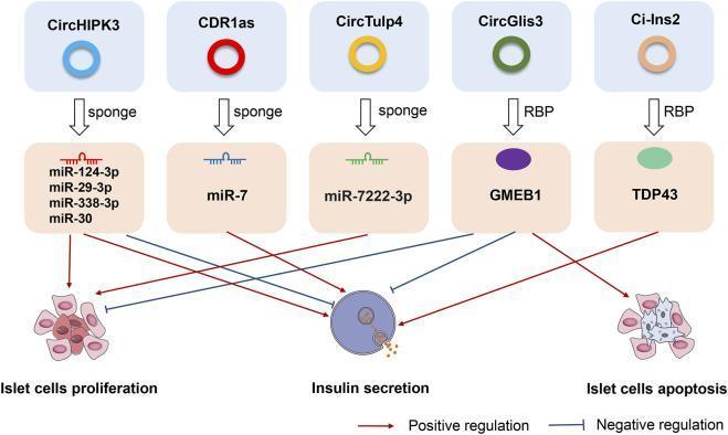
A medical pathophysiology diagram illustrating the regulatory roles of circular RNAs (circRNAs) in pancreatic islet β cells. The diagram shows five specific circRNAs: CircHIPK3, CDR1as, CircTulp4, CircGlis3, and Ci-Ins2. These molecules act through two main mechanisms: miRNA sponging and interaction with RNA-binding proteins (RBPs). CircHIPK3, CDR1as, and CircTulp4 act as 'sponges' for various microRNAs, including miR-124-3p, miR-29-3p, miR-338-3p, miR-30, miR-7, and miR-7222-3p. These pathways positively regulate islet cell proliferation and insulin secretion, with the exception of miR-7 which negatively regulates proliferation. CircGlis3 and Ci-Ins2 interact with RBPs GMEB1 and TDP43, respectively. The diagram indicates that GMEB1 negatively regulates insulin secretion and proliferation while promoting islet cell apoptosis. In contrast, TDP43 positively regulates insulin secretion. Red arrows denote positive regulation (stimulation), while blue T-bar lines represent negative regulation (inhibition). The illustration concludes at the functional level, showing impacts on islet cell proliferation, insulin secretion, and apoptosis, which are critical factors in diabetes mellitus research.
Pancreatogenic Diabetes (Type 3c Diabetes Mellitus / T3cDM)
Definition
It is estimated to affect 5-10% of all patients with diabetes but is frequently misdiagnosed as Type 2 DM. - Vonderau & Desai, JAAPA 2022 (PMID 36219100)
Causes (Etiologies)
| Cause | Notes |
|---|---|
| Chronic pancreatitis | Most common cause overall |
| Pancreatic adenocarcinoma | T3cDM occurs in ~30% of pancreatic cancer patients |
| Pancreatectomy / surgery | Distal pancreatectomy and Whipple > drainage procedures; worst after total pancreatectomy |
| Cystic fibrosis | CFRD (CF-related diabetes) is a form of T3cDM |
| Hemochromatosis | Iron deposition damages islets |
| Fibrocalculous pancreatopathy | Tropical pancreatitis; geographic variant |
| Autoimmune pancreatitis | Type 1 AIP in particular |
- Robbins, Cotran & Kumar Pathologic Basis of Disease; Goldman-Cecil Medicine; Schwartz's Principles of Surgery
Pathophysiology
Islet Pathology
-
Islets are relatively resistant to destruction in chronic pancreatitis - acinar tissue and fibrosis predominate first
-
Islets become smaller, isolated from their vascular network by surrounding fibrosis
-
With progressive disease, endocrine failure eventually follows exocrine failure
-
Frank diabetes is seen in ~20% initially, but impaired glucose metabolism is detectable in up to 70% of CP patients
-
In alcoholic CP, diabetes develops in 83% within 25 years; >50% ultimately need insulin
-
Point prevalence of diabetes in CP: ~40%; cumulative lifetime prevalence: >80%
-
Yamada's Textbook of Gastroenterology; Sleisenger & Fordtran's GI and Liver Disease
The Three-Hormone Deficiency
| Hormone | Cell | Loss in T3cDM | Consequence |
|---|---|---|---|
| Insulin | Beta (β) cells | Decreased (but sufficient to prevent ketosis) | Hyperglycemia |
| Glucagon | Alpha (α) cells | Decreased | Impaired hypoglycemia counterregulation → prolonged/severe hypoglycemia |
| Pancreatic Polypeptide (PP) | PP cells (ventral pancreas) | Decreased | Hepatic insulin resistance; paradoxical reduced hepatic insulin sensitivity |
The Paradox of Dual Insulin Sensitivity
- Enhanced peripheral sensitivity to insulin (muscle and adipocytes) - due to insulin deficiency upregulating insulin receptors, as in Type 1 DM
- Decreased hepatic sensitivity to insulin (hepatic insulin resistance) - mediated by PP deficiency
- This results in hepatic glucose overproduction when insulin is insufficient, but severe peripheral hypoglycemia when insulin is barely in excess
Incretin Deficiency
-
GIP and GLP-1 (primary incretin hormones) mediating postprandial insulin secretion are also impaired in T3cDM
-
Likely due to impaired nutrient absorption from coexisting exocrine pancreatic insufficiency (EPI)
-
Reduced incretin response = reduced postprandial insulin secretion on top of already-depleted beta cells
-
Yamada's Textbook of Gastroenterology
Comparison: T3cDM vs T1DM vs T2DM
| Feature | T1DM | T2DM | T3cDM |
|---|---|---|---|
| Cause | Autoimmune beta-cell destruction | Insulin resistance + relative deficiency | Exocrine pancreatic disease |
| Insulin levels | Absent | Elevated initially, then declines | Low (but enough to prevent ketosis) |
| Glucagon levels | Elevated | Elevated | Decreased |
| PP levels | Normal | Normal | Decreased |
| Insulin resistance | Peripheral resistance (upregulated receptors) | Generalized resistance | Paradoxical: peripheral sensitivity ↑, hepatic resistance ↑ |
| Ketoacidosis | Common | Rare | Rare (residual insulin prevents ketosis) |
| Hypoglycemia risk | Moderate | Low | High (no glucagon counterregulation) |
| Brittle diabetes | Possible | Rare | Characteristic |
| Steatorrhea / malabsorption | Absent | Absent | Common (coexisting EPI) |
| Retinopathy / neuropathy | Common | Common | Occurs at similar frequency |
| Ketoacidosis / nephropathy | Common | Common | Relatively uncommon |
- Sleisenger & Fordtran GI and Liver Disease; Schwartz's Principles of Surgery
Clinical Features
- Hyperglycemia - often persistent, poorly controlled
- Hypoglycemic episodes - frequent, prolonged, severe (hallmark)
- Weight loss and malnutrition (coexisting exocrine insufficiency)
- Steatorrhea / fat malabsorption
- Abdominal pain (if due to chronic pancreatitis)
- Microangiopathic complications (retinopathy, neuropathy) - occur at similar frequency as in T1DM
- Nephropathy and ketoacidosis - relatively uncommon
Diagnosis
Diagnostic Criteria for T3cDM (requires ALL major criteria + at least one minor criterion)
- Exocrine pancreatic insufficiency (EPI) confirmed by low fecal elastase or 72-h fecal fat
- Abnormal pancreatic imaging (CT/MRI/MRCP/EUS showing structural changes)
- Absence of T1DM autoantibodies (anti-GAD, anti-islet cell antibodies)
- Impaired beta-cell function (low C-peptide, insulin)
- No insulin resistance (normal or low HOMA-IR)
- Impaired incretin secretion (low GIP/GLP-1)
- Low serum levels of fat-soluble vitamins (A, D, E, K)
- Reduced PP response to mixed meal
Management
General Principles
- Glycemic control
- Prevention of hypoglycemia
- Treatment of coexisting exocrine insufficiency (PERT)
- Nutritional support
Step-by-Step Pharmacological Approach
| Step | Agent | Role | Notes |
|---|---|---|---|
| 1st line (mild hyperglycemia) | Metformin | Reduces hepatic glucose production | Convenient oral route; theoretically chemoprotective against PDAC; use cautiously if malnutrition |
| 2nd line | Insulin | Primary treatment for uncontrolled hyperglycemia | Required in ~50% of CP-related T3cDM; start at low doses; frequent monitoring essential |
| Adjuncts | Thiazolidinediones (pioglitazone) | Improve hepatic insulin sensitivity | PP-deficiency-mediated hepatic resistance; limited by osteoporosis risk in CP patients |
| Emerging | GLP-1 agonists (exenatide, liraglutide), DPP-4 inhibitors (sitagliptin, linagliptin) | Incretin enhancement | Previously avoided (feared AP/PDAC risk); evidence now clarifies GLP-1 agonists do NOT promote AP or PDAC - can be considered |
- Yamada's Textbook of Gastroenterology
Insulin Therapy - Special Considerations
- Start low, titrate slowly - risk of severe hypoglycemia
- Target HbA1c carefully (7-8% may be safer than strict <7%)
- Insulin pump therapy has been studied; addition of subcutaneous PP infusion reduced insulin requirements
- Avoid long-acting insulin as the sole agent without rapid-acting cover at meals
- Always co-prescribe pancreatic enzyme replacement therapy (PERT) - uncontrolled EPI worsens glycemic variability by impairing nutrient absorption
PERT and Nutrition
- All T3cDM patients with coexisting EPI need PERT with every meal (pancrelipase)
- PERT improves glucose variability by restoring predictable nutrient absorption
- Annual screening for fat-soluble vitamin deficiencies (A, D, E, K) - deficiencies are common (Vitamin D deficient in ~58%)
- Fat-restricted diet is generally recommended; adequate protein and calories to prevent sarcopenia
Experimental / Future
- Pancreatic Polypeptide (PP) analogs / PP receptor agonists - currently under investigation to specifically correct hepatic insulin resistance in T3cDM - Schwartz's Principles of Surgery, 11e
Natural History and Complications
- Progression: From impaired glucose tolerance → overt diabetes over years to decades (median 11-26 years from CP onset depending on etiology)
- Alcoholic CP: ~20 years median; late-onset idiopathic CP: ~12 years
- Pancreatic cancer risk: T3cDM in chronic pancreatitis carries significant risk of PDAC; new-onset T3cDM in a patient without prior CP diagnosis should prompt pancreatic cancer screening
- Microangiopathy (retinopathy, neuropathy) comparable to T1DM
- Malnutrition-related complications
Key Summary Points
| Point | Detail |
|---|---|
| Most common cause | Chronic pancreatitis |
| Most misdiagnosed as | Type 2 DM |
| Defining feature | Triple hormone deficiency: insulin + glucagon + PP |
| Why hypoglycemia is dangerous | No glucagon counterregulation |
| Why ketoacidosis is rare | Sufficient residual insulin |
| Hepatic insulin resistance | PP deficiency-mediated |
| 1st line treatment | Metformin (mild) → Insulin (uncontrolled) |
| Always co-prescribe | PERT (pancreatic enzyme replacement) |
| Lifetime DM prevalence in CP | >80% |
| PP analog research | Under active investigation |