
Diabetic Retinopathy.
diabetic retinopathy fundus photograph
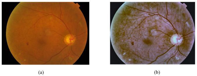
Diagnostic Image: Fundus Photographs. This figure shows two side-by-side retinal fundus images illustrating Diabetic Retinopathy (DR) manifestations and the effect of digital preprocessing. Image (a) is the original raw fundus photograph with a standard reddish-orange hue, showing the optic disc and retinal vasculature. Pathological findings include red lesions (microaneurysms and hemorrhages) appearing as dark spots, and bright lesions representing hard exudates (yellowish spots) and soft exudates/cotton wool spots (fluffy white areas). Image (b) shows the same retina after preprocessing to enhance contrast and sharpen clinical features. In the processed version, the overall tone shifts to a muted brownish-purple, making the vascular network more distinct and the lesions more defined. The preprocessing helps differentiate between red lesions and bright exudative lesions, which are critical for DR staging. This comparison demonstrates the role of image processing in improving the detectability of subtle retinal pathology for diagnostic AI models or clinical review.
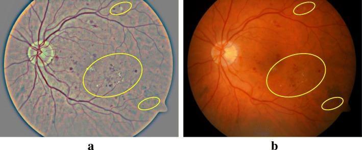
This clinical comparison shows side-by-side retinal fundus photographs demonstrating the effect of contrast enhancement (CE) on the visualization of diabetic retinopathy (DR). Image (a) is the original fundus photograph, presenting a somewhat washed-out, grayish-blue tone with low contrast between retinal features. Image (b) shows the result of the image enhancement technique, featuring a more natural orange-red retinal hue with significantly improved contrast and sharpness. Both images utilize yellow elliptical annotations to highlight specific pathological signs of DR, including hard exudates (bright, yellowish spots) and microaneurysms or small hemorrhages (dark red spots). The optic disc is visible on the left side of each image, with retinal vessels radiating outward. The enhancement in image (b) allows for better singularization and identification of lesions, particularly small microaneurysms and subtle exudates that are less distinct in the raw image. This demonstrates the clinical utility of computational image processing in improving the diagnostic sensitivity of fundus imaging for automated or manual screening.
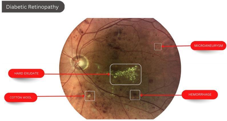
This clinical fundus photograph illustrates the classic ocular manifestations of diabetic retinopathy. The image displays a posterior view of the retina with several labeled pathological lesions. Key clinical findings identified include: 1) Hard Exudates: clustered, yellowish-white lipid deposits with distinct borders located in the macular region; 2) Microaneurysms: small, punctate red dots representing localized capillary dilatations; 3) Retinal Hemorrhages: larger, dark red blotches indicating blood leakage within the retinal layers; and 4) Cotton Wool Spot: a fluffy, pale-white lesion with indistinct margins signifying an area of nerve fiber layer infarction. The background shows an orange-red retinal hue with visible major retinal arteries and veins emanating from the optic disc. This visual is designed for educational indexing in ophthalmology to demonstrate non-proliferative diabetic retinopathy features and their typical distribution patterns within the fundus.
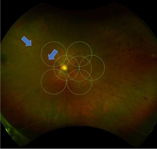
This ultra-widefield (UWF) pseudocolor fundus photograph illustrates advanced diabetic retinopathy in a human eye. The central retina is overlaid with a standardized template of seven overlapping dashed circles, representing the Early Treatment Diabetic Retinopathy Study (ETDRS) 7-standard fields used for clinical grading. The primary pathology demonstrated is peripheral retinal vessel whitening, indicated by blue arrows in the superotemporal quadrant. This whitening signifies vascular sheathing or sclerosis, often associated with chronic ischemia. The background fundus shows a variegated orange-red appearance with visible intraretinal hemorrhages and microvascular abnormalities characteristic of proliferative diabetic retinopathy (PDR). The image highlights the diagnostic utility of UWF imaging in identifying peripheral vascular changes that occur beyond the traditional 30-to-45-degree field of view of conventional fundus photography. These peripheral findings are clinically significant as they may correlate with areas of capillary non-perfusion and increased risk of disease progression.
"diabetic retinopathy"[MeSH Terms] AND treatment
Diabetic Retinopathy
Definition and Epidemiology
Pathogenesis
- Pericyte loss: The number of pericytes relative to endothelial cells diminishes - this is the earliest and most specific structural change in retinal capillaries.
- Basement membrane thickening: Retinal blood vessel basement membranes thicken, similar to changes elsewhere in diabetic microangiopathy.
- Blood-retinal barrier breakdown: VEGF (originally called "vascular permeability factor") increases vascular permeability, causing macular edema. This is a major cause of visual loss.
- Microaneurysm formation: Pericyte dropout leads to outpouching of capillary walls.
- Microvascular occlusion: Leads to retinal ischemia and nonperfusion.
- VEGF upregulation: Retinal nonperfusion/hypoxia upregulates VEGF, driving intraretinal angiogenesis and eventually neovascularization that breaches the internal limiting membrane (proliferative DR). - Robbins, p. 1802-1808; Kanski's, p. 522
Risk Factors
| Factor | Detail |
|---|---|
| Duration of diabetes | Most important predictor; DR rarely occurs in the first 5 years of T1DM |
| Glycemic control | HbA1c directly correlated with DR risk |
| Hypertension | Accelerates progression |
| Nephropathy | Renal disease and DR are closely linked; renal transplantation may improve retinopathy |
| Dyslipidemia | Hyperlipidemia increases exudate formation; fenofibrate may reduce progression |
| Pregnancy | ~5% with mild DR and ~1/3 with moderate DR progress to PDR during pregnancy |
| Drugs | Pioglitazone associated with worsening macular edema |
| GLP-1 agonists | Rapid glycemic improvement with GLP-1 RAs (e.g., semaglutide) can transiently worsen DR, especially with marked HbA1c reduction |
Classification (ETDRS / International Scale)
Non-Proliferative Diabetic Retinopathy (NPDR)
| Stage | Features | Follow-up |
|---|---|---|
| No DR | Normal fundus | 12 months |
| Very mild NPDR | Microaneurysms only | 12 months |
| Mild NPDR | Microaneurysms + any of: hemorrhages, exudates, cotton-wool spots | 6-12 months |
| Moderate NPDR | More than mild but less than severe; may include cotton-wool spots, venous beading, IRMA | 4-6 months |
| Severe NPDR (PPDR) | 4-2-1 rule: >20 intraretinal hemorrhages in all 4 quadrants, OR venous beading in ≥2 quadrants, OR prominent IRMA in ≥1 quadrant | 3-4 months; consider PRP |
Proliferative Diabetic Retinopathy (PDR)
- Vitreous or preretinal hemorrhage
- NVD >1/3 disc area
- NVE >1/2 disc area when associated with vitreous hemorrhage
- Any NVI (neovascularization of the iris) or NVA (angle)
Clinical Signs
Background / NPDR Signs
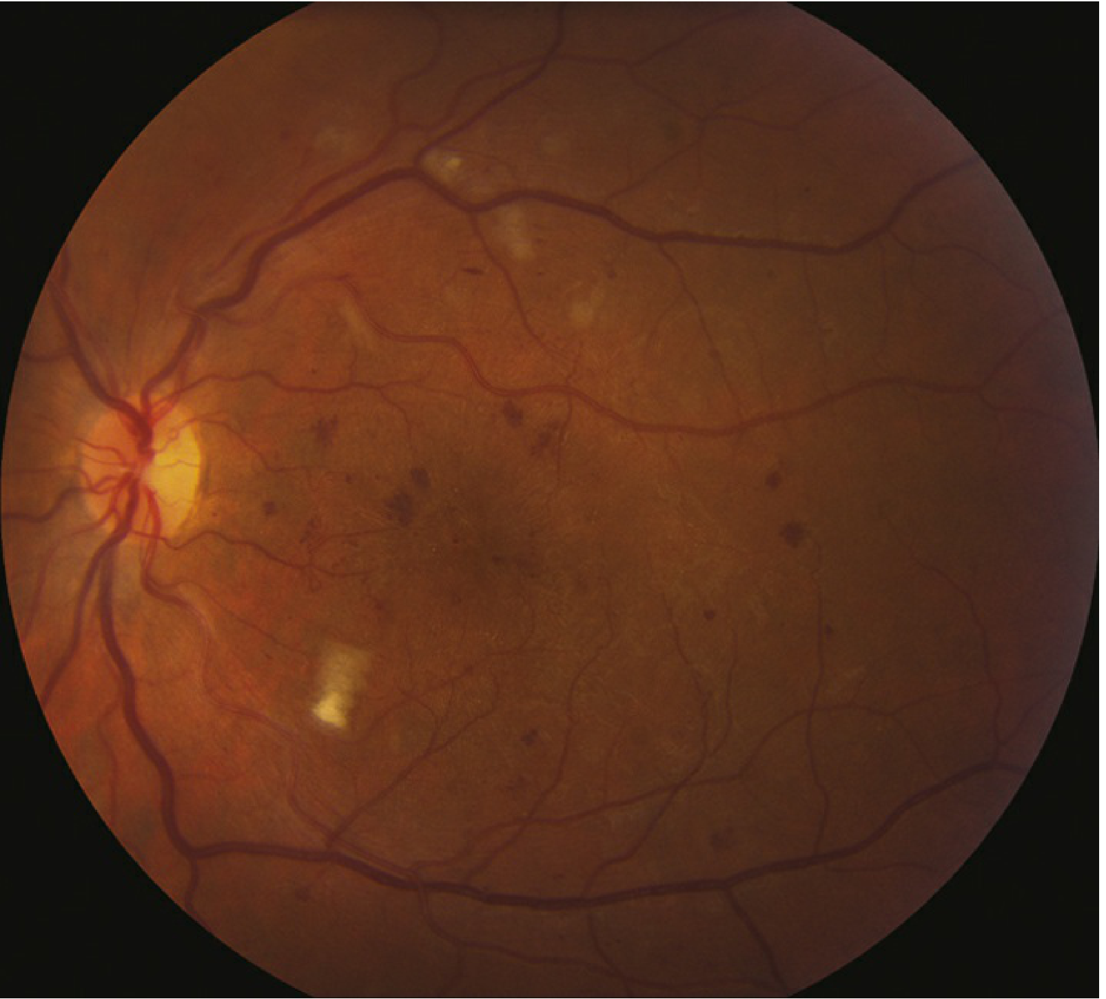
- Microaneurysms: First clinical sign - tiny red dots, hyperfluorescent on FA (hyperfluorescent leaking spots). Located at sites of pericyte dropout.
- Dot and blot hemorrhages: Intraretinal hemorrhages in the middle retinal layers ("blot") and deeper dark round hemorrhages representing hemorrhagic retinal infarcts.
- Hard exudates: Waxy yellow lesions with distinct margins in clumps or rings at the posterior pole, composed of lipoproteins and lipid-filled macrophages in the outer plexiform layer. Often surrounding leaking microaneurysms.
- Cotton-wool spots (CWSs): Fluffy white areas - nerve fiber layer infarcts due to precapillary arteriolar occlusion.
- Venous beading: Sausage-like irregularities in venous caliber - marker of severe ischemia.
- IRMA (Intraretinal microvascular anomalies): Dilated pre-existing capillaries acting as shunts around areas of nonperfusion; no leakage on FA.
Diabetic Macular Edema (DME)
PDR Signs
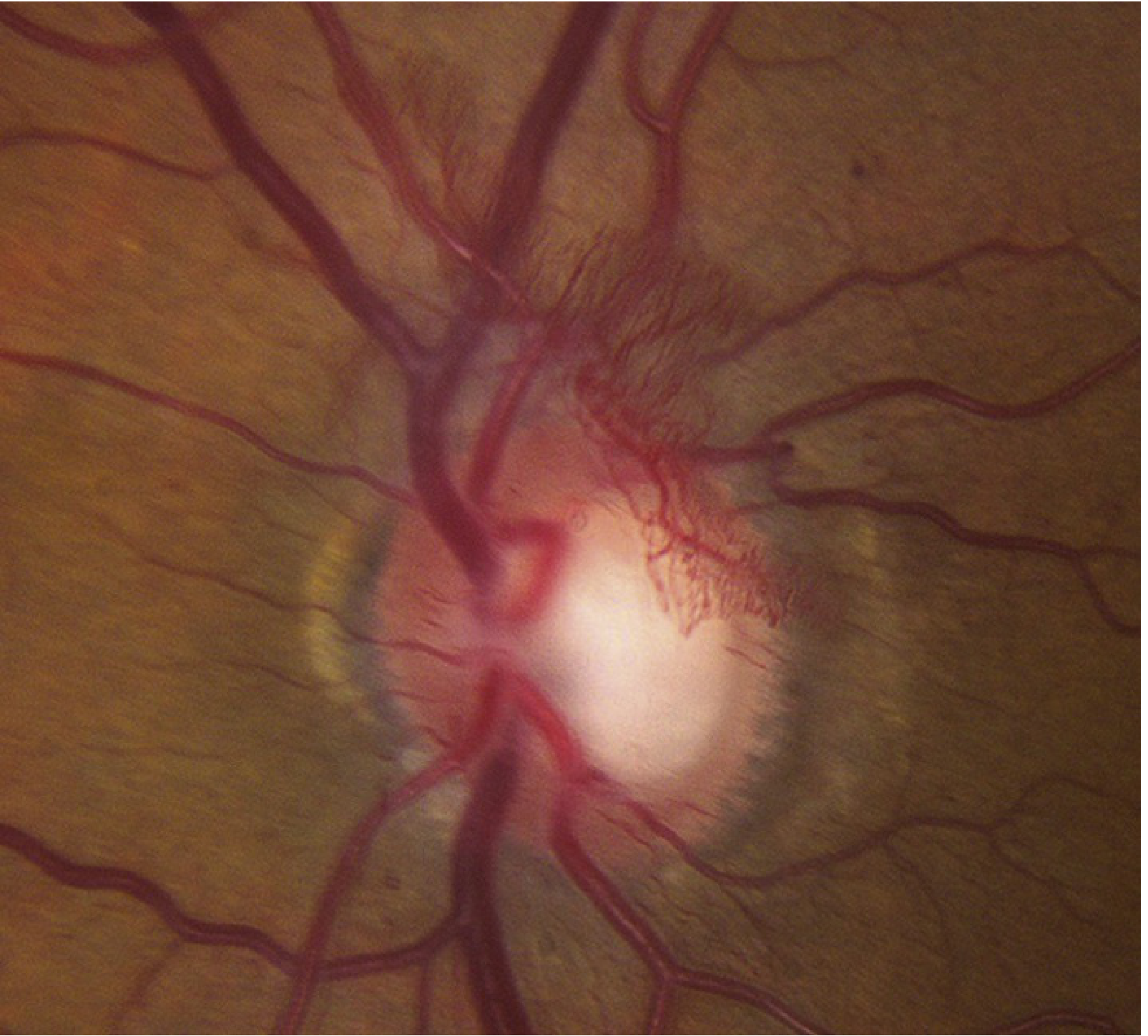
- NVD: New vessels at or within 1 disc diameter of the disc - high-risk because of proximity to vitreous base.
- NVE: New vessels elsewhere in the retina.
- NVI / NVA: Neovascularization of iris/angle (rubeosis iridis) - risk of neovascular glaucoma.
- Vitreous hemorrhage: From fragile new vessels; may cause sudden visual loss.
- Tractional retinal detachment: Fibrovascular membrane contraction.
Fundus Images
Differential Diagnosis
| Condition | Key Distinguishing Features |
|---|---|
| CRVO | Optic disc swelling, dilated tortuous veins, flame hemorrhages, typically unilateral, sudden onset |
| BRVO | Hemorrhages along a vein, do not cross the horizontal raphe |
| Hypertensive retinopathy | Flame hemorrhages, AV nicking, microaneurysms rare, arteriolar narrowing |
| Ocular ischemic syndrome | Midperipheral hemorrhages, no exudates, pain, mid-dilated pupil |
| Radiation retinopathy | History of radiation, microaneurysms rare |
| Sickle cell retinopathy | Peripheral "sea fan" neovascularization |
Investigations
- Dilated fundoscopy: Essential - nondilated exams by primary care are inadequate.
- Fundus photography: Allows remote reading; validated screening tool.
- Fluorescein angiography (FA): Detects microaneurysms (hyperfluorescent dots), areas of nonperfusion, leakage, and IRMA.
- Optical coherence tomography (OCT): Gold standard for detecting and quantifying macular edema; identifies structural layers of retinal pathology.
- B-scan ultrasound: When dense vitreous hemorrhage obscures fundal view - to rule out tractional retinal detachment.
Screening Recommendations
- Type 1 DM: First screen at 5 years after diagnosis; then annually.
- Type 2 DM: Screen at time of diagnosis (hyperglycemia often present for years before recognition); then annually.
- Pregnancy: Screen prior to and during pregnancy; high-risk patients require up to 2-weekly review. - Goldman-Cecil Medicine, p. 1392; Harrison's 22E
Treatment
1. Systemic Control (Prevention and Slowing Progression)
- Glycemic control: Intensive control delays development and slows progression. Note: rapid normalization (e.g., with GLP-1 agonists, pancreatic transplant) can transiently worsen DR in first 6-12 months.
- Blood pressure control: Especially with renin-angiotensin system blockers (ACE inhibitors/ARBs).
- Lipid control: Fenofibrate reduces progression of retinopathy and decreases hard exudates.
- Aspirin: Does not alter the natural history of DR.
2. Anti-VEGF Therapy (Intravitreal injections)
- Agents: ranibizumab, bevacizumab, aflibercept, faricimab
- DME: Anti-VEGF is first-line for center-involving DME affecting or threatening the fovea.
- PDR: Can be used as an alternative to PRP, especially when: (a) DME is coexistent, (b) view is limited by vitreous hemorrhage, (c) patient wishes to avoid laser. However, anti-VEGF without PRP requires close follow-up - patients lost to follow-up have worse outcomes.
3. Laser Photocoagulation
- NVD >1/4 to 1/3 disc area
- Any NVD + preretinal or vitreous hemorrhage
- NVE >1/2 disc area + preretinal or vitreous hemorrhage
- Any NVI or NVA
4. Vitrectomy
- Dense, nonclearing or recurrent vitreous hemorrhage
- Tractional retinal detachment involving or threatening the macula
- Macular epiretinal membrane or vitreomacular traction
- Dense premacular hemorrhage
- Chronic DME unresponsive to other treatment
- Severe fibrovascular proliferation unresponsive to laser/anti-VEGF
Prognosis
- With effective screening and treatment (ETDRS protocols + anti-VEGF), severe visual loss can be reduced to <5%.
- Once advanced retinopathy is present, glycemic control has limited impact on the retina.
- Appropriate ophthalmologic care can prevent most blindness from DR.
- Tractional retinal detachment and neovascular glaucoma carry a poor visual prognosis if not treated promptly.
Key Clinical Pearls
- DR is largely asymptomatic even in severe PDR - hence the absolute necessity of regular screening.
- The 4-2-1 rule defines severe NPDR (the "preproliferative" stage with high risk of progression to PDR).
- DME can occur at any stage of DR, including mild NPDR.
- Anti-VEGF is preferred over focal laser for center-involving DME.
- GLP-1 receptor agonists (semaglutide) can worsen DR with rapid glycemic improvement - ophthalmology review before initiating is prudent.
- Aspirin and anticoagulants do NOT need to be stopped for intravitreal injections.
- In pregnancy with PDR, treat aggressively; DME often resolves spontaneously postpartum.