
Total Abdominal Hysterectomym
Reading File
Total Abdominal Hysterectomy (TAH) is the surgical removal of the uterus (and optionally the cervix, ovaries, and fallopian tubes) through an abdominal incision. It is one of the most commonly performed gynecological surgeries.
Searching sources...
Finding Sources
Finding Sources
I have two excellent primary sources: Sabiston Textbook of Surgery and Schwartz's Principles of Surgery. Let me read both in parallel.
Reading File
Reading File
Excellent content from both textbooks. Let me also fetch the key surgical steps images and look for indications/complications content.
Reading File
Finding Sources
Loading Image
Now let me fetch the complications content and the cardinal ligament division image.
Reading File
Loading Image
Finding Sources
I now have comprehensive textbook content. Let me compile a thorough, well-organized answer.
Total Abdominal Hysterectomy (TAH)
Definition
A total hysterectomy is surgical removal of both the uterine corpus and the cervix. When performed through an abdominal incision, it is called a Total Abdominal Hysterectomy (TAH). It is one of the most commonly performed major surgical procedures - the abdominal approach accounts for approximately 60% of all hysterectomies (vs. 14% laparoscopic and 26% vaginal) in the United States.
A subtotal (supracervical) hysterectomy removes only the corpus, leaving the cervix behind. TAH removes both.
Types of Hysterectomy (by extent)
| Type | Structures Removed |
|---|---|
| Subtotal / Supracervical | Uterine corpus only (cervix left in) |
| Total (TAH) | Uterine corpus + cervix |
| TAH + BSO | Uterus + cervix + bilateral salpingo-oophorectomy (tubes + ovaries) |
| Radical (Wertheim's) | Uterus + cervix + parametria + upper vagina + pelvic nodes (for cervical cancer) |
Indications
- Uterine leiomyomas (fibroids) - most common benign indication
- Abnormal uterine bleeding refractory to conservative management
- Endometriosis / adenomyosis (especially with bilateral oophorectomy if severe)
- Uterine prolapse (often via vaginal route, but TAH if added pathology)
- Endometrial carcinoma (TAH + BSO is standard surgical staging)
- Ovarian carcinoma staging (TAH + BSO + omentectomy + lymph node dissection)
- Uterine leiomyosarcoma (TAH + bilateral oophorectomy)
- Intractable postpartum hemorrhage / placenta accreta (emergency cesarean hysterectomy)
- Cervical carcinoma (early stages)
Preoperative Preparation
- Informed consent including risks of bleeding, infection, urinary tract injury, bowel injury, and loss of fertility
- Urine culture, CBC, coagulation profile, blood typing
- Bowel preparation (individualized)
- Prophylactic antibiotics (cephalosporin) within 60 minutes of incision
- DVT prophylaxis (compression stockings ± LMWH)
- Foley catheter placement
Patient Positioning & Incision
The patient is placed in the dorsal supine position (modified lithotomy if combined vaginal access is needed). The incision is either:
- Pfannenstiel (low transverse) - preferred for benign disease; better cosmesis, lower wound complications
- Vertical midline - chosen for malignancy, large pathology, need for full abdominal exploration, or re-operative cases
Choice of incision depends on indication, body habitus, prior surgical history, and size of uterus/pathology.
Step-by-Step Surgical Technique
(Based on Sabiston Textbook of Surgery and Schwartz's Principles of Surgery)
1. Abdominal Entry & Exploration
- Enter the abdomen through the chosen incision
- Inspect the upper abdomen for evidence of extrapelvic disease
- Place a suitable self-retaining retractor (e.g., Balfour)
- Grasp the uterus at each cornu with clamps (Kocher or Lahey) and elevate into the incision
2. Round Ligament Transection
- Identify and divide the round ligament bilaterally
- This opens the anterior and posterior leaves of the broad ligament
3. Broad Ligament Incision & Ureter Identification
- Extend the peritoneal incision from the round ligament laterally past the ovarian hilum (if oophorectomy planned) or medially below the utero-ovarian ligament (if ovaries are conserved)
- Bluntly open the retroperitoneal space and identify the ureter on the medial leaf of the broad ligament - this is critical to prevent ureteric injury
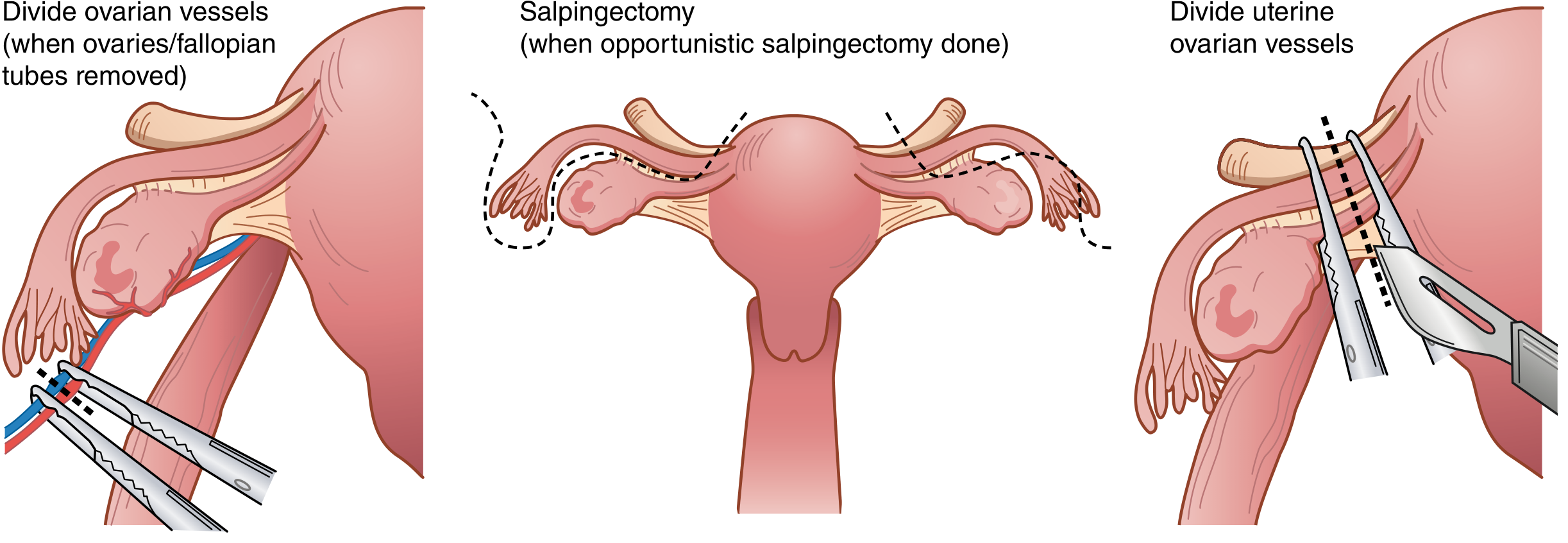
4. Adnexal Management
If ovaries/tubes are to be removed (BSO):
- Incise posterior leaves of broad ligament
- Double-clamp the infundibulopelvic (IP) ligament containing the ovarian vessels with curved Heaney or Zeppelin clamps
- Divide between clamps with curved Mayo scissors; doubly ligate pedicles
If ovaries are to be conserved:
- Create an opening below the utero-ovarian ligament and fallopian tube
- Clamp, divide, and doubly ligate the utero-ovarian vessels with Kelly or Heaney clamps
Opportunistic salpingectomy (removal of tubes only, conserving ovaries) is increasingly performed to reduce future ovarian cancer risk.
5. Bladder Mobilization
- Sharply dissect the bladder off the anterior surface of the uterus and cervix using Metzenbaum scissors or electrocautery
- Retract the bladder inferiorly below the level of the cervix
6. Uterine Vessel Ligation
- Skeletonize the uterine vessels along the lateral uterus by incising the remaining posterior broad ligament leaves
- Place a curved Heaney or Zeppelin clamp across the uterine vessels at the level of the internal cervical os
- Cut and suture-ligate the pedicle
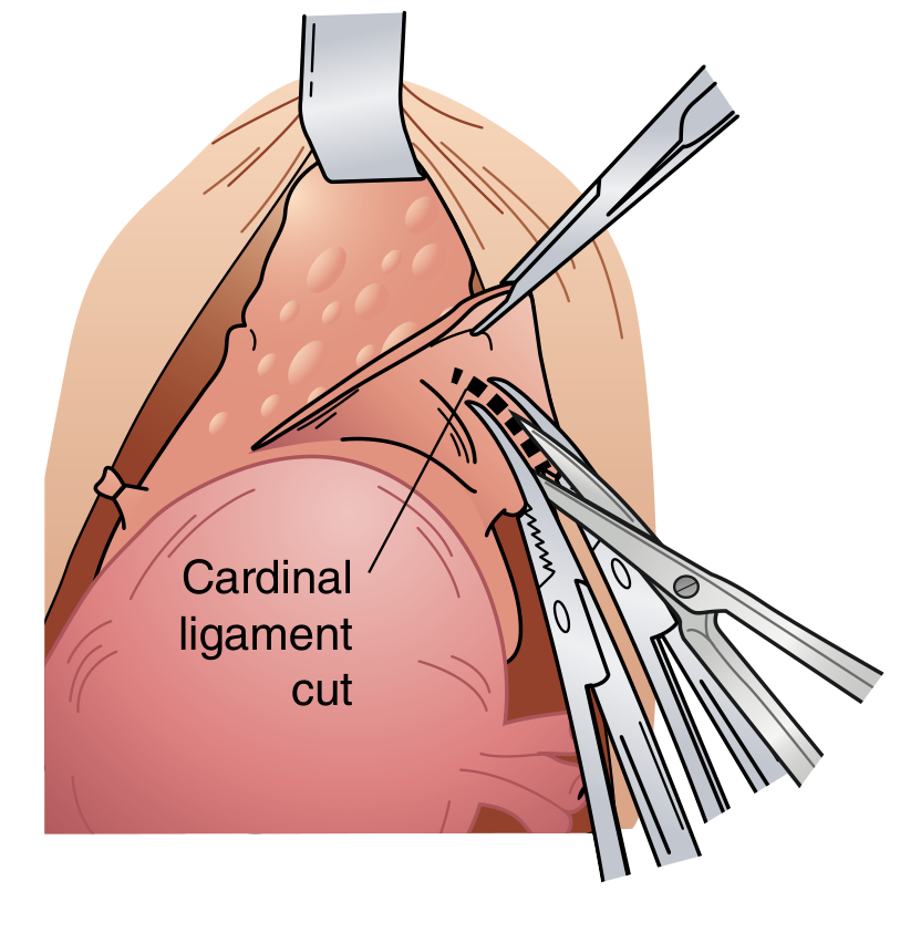
7. Cardinal & Uterosacral Ligament Division
- Serially clamp, cut, and ligate the cardinal ligaments (Mackenrodt's ligaments) bilaterally in steps
- Divide the uterosacral ligaments
- Continue until both sides are clear at the level of the external cervical os
8. Colpotomy & Specimen Removal
- Place curved clamps across the vagina just below the cervix bilaterally
- Cut with curved scissors just above the clamps to amputate the cervix from the vagina
- The specimen (uterus + cervix ± adnexa) is removed
9. Vaginal Cuff Closure
- Place Heaney transfixion stitches at each lateral vaginal angle - importantly, incorporating the ipsilateral uterosacral ligament for apical support (prevents vault prolapse)
- Close the remainder of the vaginal cuff with running or interrupted absorbable sutures (e.g., Vicryl 0 or 1)
- An alternative: direct sharp colpotomy around the cervix with Allis clamps placed on the vaginal edges for visualization before closure
10. Closure
- Pelvic reperitonization is not necessary
- Hemostasis confirmed
- Abdominal wall closed in layers (fascia, subcutaneous tissue, skin)
Key Anatomical Danger Points
| Structure at Risk | During Which Step | Prevention |
|---|---|---|
| Ureter | Broad ligament dissection, uterine vessel ligation | Retroperitoneal identification before clamping |
| Bladder | Anterior dissection, colpotomy | Sharp dissection under direct vision; mobilize completely below cervix |
| Uterine/ovarian vessels | Pedicle ligation | Double clamping and suture ligation |
| Bowel (sigmoid, rectum) | Posterior dissection, closure | Careful retraction, identify anatomy |
The ureter is at highest risk near the infundibulopelvic ligament and near the uterine artery (where it passes under the uterine artery - "water under the bridge"). Always identify it before clamping.
Complications
(Tintinalli's Emergency Medicine)
Infection rate: 3-10% (higher with abdominal vs. vaginal route). Overall complication rate is significantly higher for malignant vs. benign indications (19.4% vs. 7.9%). Risk factors include obesity, diabetes, and prolonged operative time.
| Category | Specific Complications |
|---|---|
| Infectious | Wound infection, infected vaginal cuff hematoma/cellulitis/abscess, ovarian abscess, UTI |
| Hemorrhagic | Intraoperative hemorrhage, postoperative hematoma |
| Urological | Bladder injury, ureteral injury, urinary retention |
| GI | Ileus, bowel injury, bowel obstruction |
| Wound | Dehiscence, evisceration |
| Thromboembolic | DVT, pulmonary embolism, phlebitis |
| Pulmonary | Atelectasis, pneumonia |
| Long-term | Vaginal vault prolapse, pelvic floor dysfunction, premature menopause (if BSO in premenopausal patient) |
Variants & Related Procedures
- TAH + BSO: adds bilateral salpingo-oophorectomy; standard for endometrial cancer, ovarian cancer, or peri/postmenopausal patients with benign disease
- TAH + BSO + Staging laparotomy: for gynecologic malignancies - includes omentectomy, peritoneal washings, and lymph node dissection
- Radical (Wertheim) hysterectomy: for cervical carcinoma - includes parametrectomy and upper vaginectomy
- Laparoscopic hysterectomy (TLH): same steps but using ports, energy devices, and a uterine manipulator with colpotomizer cup; avoids laparotomy
- Robotic hysterectomy: similar to laparoscopic but higher cost; no proven clinical benefit over laparoscopic for benign conditions
Postoperative Care
- Ambulation within 24 hours (DVT prevention)
- Foley catheter removal by postoperative day 1
- Diet advancement as tolerated
- Pain management (multimodal - NSAIDs + opioid-sparing)
- Pelvic rest for 6-8 weeks (no intercourse, tampon use, or heavy lifting)
- Discharge typically on postoperative day 2-4
- If premenopausal with BSO: hormone replacement therapy counseling
Sources:
- Sabiston Textbook of Surgery, pp. 2799-2800 (Chapter 120 - Steps to completing hysterectomy)
- Schwartz's Principles of Surgery, 11th Edition, p. 1825 (Chapter 41 - Procedures for Abnormal Uterine Bleeding)
- Tintinalli's Emergency Medicine, Chapter 105 - Complications of Major Gynecologic-Abdominal Surgery
This is a shared conversation. Sign in to Orris to start your own chat.