
Infertility
"infertility" AND "diagnosis" AND "management"
infertility causes diagram male female
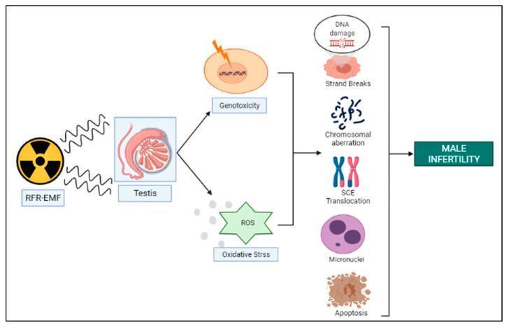
This pathophysiology diagram illustrates the proposed mechanisms of male infertility induced by Radio Frequency Electromagnetic Field (RFR-EMF) exposure. The process begins with RFR-EMF radiation affecting the testis, which triggers two primary pathological pathways: Genotoxicity (represented by cellular nuclear damage) and Reactive Oxygen Species (ROS)/Oxidative Stress (represented by a star-shaped icon). These two intermediate states converge to cause a spectrum of genetic and cellular alterations, specifically: DNA damage (helix fragmentation), Strand Breaks, Chromosomal Aberrations, Sister Chromatid Exchange (SCE) Translocation, the formation of Micronuclei, and cellular Apoptosis. The flow concludes with a final box indicating that the cumulative effect of these genotoxic and oxidative stressors leads to 'Male Infertility.' This diagram serves as an educational summary of how non-ionizing radiation potentially disrupts male reproductive health through genomic instability and oxidative damage.
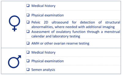
Summary : This figure presents the recommended initial clinical assessment steps for infertility in females and males, listing specific diagnostic procedures for each sex. flowchart: # Female Assessment : • Medical history • Physical examination • Pelvic 2D ultrasound for detection of structural abnormalities, with additional imaging if needed • Assessment of ovulatory function via menstrual calendar and laboratory testing • AMH (Anti-Müllerian Hormone) or other ovarian reserve testing # Male Assessment : • Medical history • Physical examination • Semen analysis # Symbols : • Female symbol (♀) for female assessment section • Male symbol (♂) for male assessment section Analysis : • The figure clearly separates the infertility assessment protocols for females and males, with more detailed and varied diagnostic steps for females, including imaging and hormonal testing, while the male assessment focuses on semen analysis after history and examination. This highlights the complexity and multi-factorial nature of female infertility evaluation compared to male.
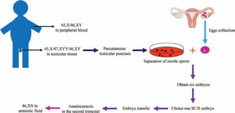
This medical flow diagram illustrates the clinical workflow of In Vitro Fertilization and Embryo Transfer (IVF-ET) in a case of male mosaicism. The process begins with a genetic evaluation of the male patient, showing a 45,X/46,XY karyotype in peripheral blood and a more complex 45,X/47,XYY/46,XY mosaicism in testicular tissue. The procedural steps follow: percutaneous testicular puncture for sperm retrieval, separation of motile sperm, and egg collection from the female reproductive system. These are combined to obtain six embryos, from which one '8C/II' embryo is selected for transfer. The final stages show the progression to pregnancy, including prenatal screening via amniocentesis in the second trimester, revealing a 46,XN karyotype in the amniotic fluid. This infographic serves as an educational tool for reproductive endocrinology and infertility (REI), demonstrating the management of gonadal dysgenesis and chromosomal mosaicism through assisted reproductive technology (ART) to achieve a successful pregnancy outcome.
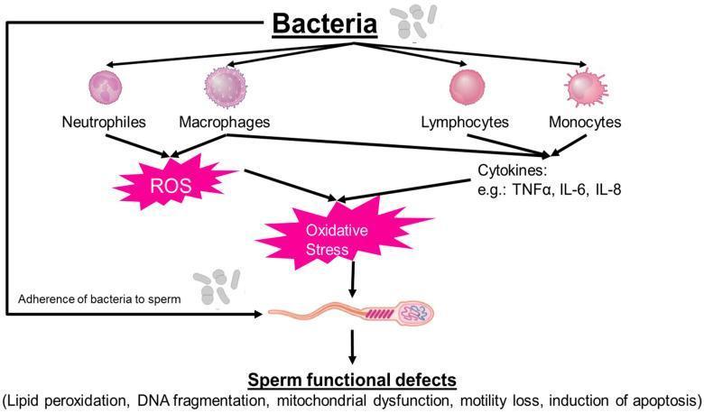
A pathophysiology diagram illustrating the direct and indirect mechanisms by which bacteria cause sperm functional defects and male infertility. The diagram outlines two primary pathways: 1) Direct Interaction: Adherence of bacteria to the sperm surface. 2) Indirect Inflammatory Response: Bacterial presence triggers immune cell activation, specifically involving neutrophils, macrophages, lymphocytes, and monocytes. These leukocytes release reactive oxygen species (ROS) and pro-inflammatory cytokines, including TNF-α, IL-6, and IL-8. Both pathways converge to induce cellular oxidative stress. The final section of the flowchart lists the resulting sperm functional defects, which include lipid peroxidation, DNA fragmentation, mitochondrial dysfunction, loss of motility, and the induction of apoptosis. This educational visual provides a comprehensive overview of how bacterial infections and the subsequent leukocytospermic response impair reproductive health at the cellular level.
Infertility
Definition
-
Primary infertility - couples who have never achieved a pregnancy
-
Secondary infertility - infertility after at least one prior pregnancy
-
Approximately 85% of couples achieve pregnancy after 12 months; 95% after 24 months
-
Harrison's Principles of Internal Medicine, 22nd ed. (2025)
Fecundability & Age Effects
| Female Age | Reduction in Fecundability |
|---|---|
| 34-35 years | -14% vs. age 30-31 |
| 36-37 years | -19% |
| 40-41 years | -53% |
| 42-44 years | -59% |
When to Start Evaluation
| Situation | When to Evaluate |
|---|---|
| Female age <35 | After 12 months of unprotected intercourse |
| Female age 35-40 | After 6 months |
| Female age >40 | Immediately |
| Known risk factors present | Before 12-month threshold |
Etiology & Causes
Male Factors (~40-50% of cases)
- Varicocele (most common correctable cause)
- Testicular failure / cryptorchidism
- Endocrine disorders (hypogonadotropic hypogonadism, hypothyroidism, hyperprolactinemia)
- Genetic: Klinefelter syndrome (47,XXY), Y chromosome microdeletions
- Lifestyle: smoking, caffeine, recreational drugs, morbid obesity
- Epididymal obstruction (post-infection)
- Congenital absence of the vas deferens (associated with CFTR mutations)
- Ejaculatory duct obstruction
- Post-vasectomy
- Hypospadias
- Erectile/ejaculatory dysfunction
- Sexual dysfunction (psychological or medical)
- Infection, abnormal volume/viscosity
- Sperm-immobilizing or agglutinating antibodies (immunologic factor)
Female Factors (~30-40% of cases)
- Pelvic inflammatory disease (most common cause of tubal damage)
- Endometriosis
- Prior surgery / peritonitis
- Salpingitis isthmica nodosa
- Polycystic ovary syndrome (PCOS) - most common single cause
- Diminished ovarian reserve
- Premature ovarian insufficiency
- Hypothalamic amenorrhea (excessive exercise, low BMI)
- Hypothyroidism
- Hyperprolactinemia
- Diabetes mellitus
- Leiomyomas and polyps
- Congenital malformations (e.g., septate uterus)
- Intrauterine synechiae (Asherman syndrome)
- Destroyed endocervical glands
- Congenital absence, imperforate hymen, vaginismus
- Sperm-immobilizing/agglutinating antibodies
Unexplained Infertility (~10-30%)
- Goldman-Cecil Medicine, Table 218-6; Harrison's 22E
Fertility Evaluation
Initial Assessment - Both Partners
- Menstrual calendar and basal body temperature charts
- Serum progesterone (6-8 days before expected menses) - documents ovulation
- Serum TSH (rule out thyroid disorder)
- Serum prolactin
- Anti-Müllerian hormone (AMH) - ovarian reserve
- FSH, LH, estradiol (Day 3 of cycle)
- Hysterosalpingography (HSG) or sonohysterography - tubal and uterine anatomy
- Pelvic ultrasound - antral follicle count, structural pathology
- Semen analysis (the single most important test) - volume, concentration, motility, morphology
- If abnormal: repeat semen analysis, hormone panel (FSH, LH, testosterone, prolactin), genetics (karyotype, Y microdeletion testing)
- Diagnostic laparoscopy with tubal dye (if all prior tests normal; 30-50% of women have endometriosis or tubal disease found only this way)
- Endometrial biopsy (rarely required now)
Semen Analysis - Normal Reference Values (WHO)
| Parameter | Lower Reference Limit |
|---|---|
| Volume | ≥1.5 mL |
| Total sperm count | ≥39 million |
| Concentration | ≥16 million/mL |
| Progressive motility | ≥30% |
| Total motility | ≥42% |
| Morphology (Kruger strict) | ≥4% normal forms |
Treatment
Ovulation Induction (Anovulatory Women)
| Agent | Mechanism | Notes |
|---|---|---|
| Letrozole (aromatase inhibitor) | Blocks estrogen synthesis → stimulates FSH | First-line for PCOS (per current evidence) |
| Clomiphene citrate (SERM) | Anti-estrogen effect → increases FSH/LH | 50 mg days 3-7; max 200-250 mg; first-line if prolactin normal |
| Metformin | Insulin sensitizer | Add-on in obese PCOS patients |
| Injectable gonadotropins (FSH, LH) | Direct follicular stimulation | Second/third-line; risk of ovarian hyperstimulation (OHSS) |
Tubal Factor
- Tubal surgery has limited success and increases ectopic pregnancy risk
- IVF is preferred when tubes are damaged
- Hydrosalpinges: salpingectomy before IVF improves success rates
- Proximal blockage: radiographic tubal cannulation can be attempted
- Prior tubal ligation: IVF vs. microsurgical reanastomosis based on age, ovarian reserve, and preferences
Male Factor Infertility
- Mild to moderate: Intrauterine insemination (IUI) ± ovulation induction
- Severe (oligospermia/poor motility): IVF with ICSI (intracytoplasmic sperm injection) - single sperm injected into oocyte
- Azoospermia: Determine obstructive vs. non-obstructive; surgical sperm retrieval (TESA/TESE) + ICSI
- Hypogonadotropic hypogonadism: pulsatile GnRH or gonadotropin injections
- Varicocele repair may improve sperm parameters and natural conception rates
Endometriosis-Related Infertility
- Surgery (laparoscopic excision) or suppressive medications
- IVF often indicated, especially in severe disease
Unexplained Infertility
- Stepwise escalation: lifestyle optimization → IUI ± ovulation induction → IVF
- Empiric treatment with letrozole + IUI is commonly tried first
Assisted Reproductive Technology (ART)
| Procedure | Description |
|---|---|
| IUI (Intrauterine insemination) | Washed sperm deposited into uterus via catheter; office-based |
| IVF (In vitro fertilization) | Oocytes retrieved transvaginally under sedation; fertilized in lab; embryos transferred at Day 3 (cleavage) or Day 5 (blastocyst) |
| ICSI | Single sperm injected into oocyte; for severe male factor |
| Donor oocytes | Used in premature ovarian insufficiency or diminished ovarian reserve |
| Embryo cryopreservation | Freeze excess embryos for future transfer |
Special Populations & Counseling
-
Cancer patients: Fertility preservation (oocyte/embryo/sperm cryopreservation) should be offered before gonadotoxic therapy
-
LGBTQ+ couples: Third-party reproduction (donor sperm, donor eggs, gestational carriers)
-
Psychological impact: Infertility causes significant psychological distress; counseling and support are integral to management
-
Lifestyle: Smoking cessation, weight optimization, reduction of alcohol/recreational drugs improve outcomes for both partners
-
Berek & Novak's Gynecology; Harrison's Principles of Internal Medicine, 22E (2025); Goldman-Cecil Medicine
Recent Evidence (2024-2026)
- A 2025 systematic review (PMID 41113721) on endometriosis-associated infertility highlights multi-omics insights into pathogenesis and new precision therapeutic targets.
- A 2025 meta-analysis (PMID 39332623) on diminished ovarian reserve found limited evidence supporting most current interventions; IVF with donor oocytes remains the most effective option for this group.
Prolapse of uterus
"uterine prolapse" AND "pelvic organ prolapse"
uterine prolapse degrees grading diagram anatomy
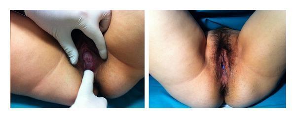
A side-by-side clinical photograph documenting uterine prolapse and its subsequent management with a pessary in a pregnant patient. The left panel shows a clinical examination of the vulva with a gloved hand retracting the labia. The cervix is visibly descended and protruding through the vaginal introitus, characteristic of grade 2 pelvic organ prolapse. The cervical tissue appears hyperemic and edematous. The right panel displays the external female genitalia following the placement of an Arabin cerclage pessary. In this view, the prolapse is reduced, and a portion of the blue silicone pessary is visible within the vaginal canal, positioned to support the cervix and maintain its placement. The image illustrates a non-surgical management strategy for symptomatic uterine prolapse and acute urinary retention during pregnancy. Educational concepts include pelvic floor anatomy, grading of organ prolapse, and mechanical support devices in obstetrics and gynecology.
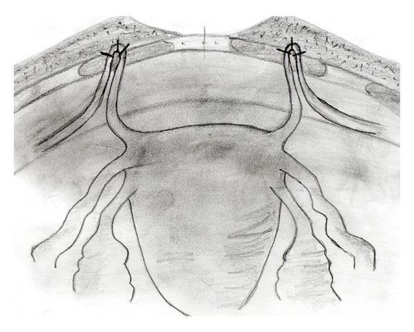
Anatomical Diagram: A black-and-white schematic illustration demonstrating the surgical fixation of the round ligaments to the anterior rectus fascia, likely for the management of pelvic organ prolapse. The diagram provides an anterior view of the uterus and its supporting structures. The uterine fundus is shown in the center, with the round ligaments extending bilaterally. These ligaments are depicted being pulled cranially and looped through small fenestrations in the rectus abdominis muscle layer. The apical 'ans' or loop of each round ligament is secured to the overlying fascia using three distinct sutures, represented by 'x' markers. Additional adnexal structures, including the fallopian tubes and utero-ovarian ligaments, are visible extending from the uterine cornua. The illustration emphasizes the anatomical relationship between the pelvic viscera and the abdominal wall fascia during a suspension procedure, highlighting the point of mechanical support and the method of non-absorbable suture fixation used to stabilize the uterine position.
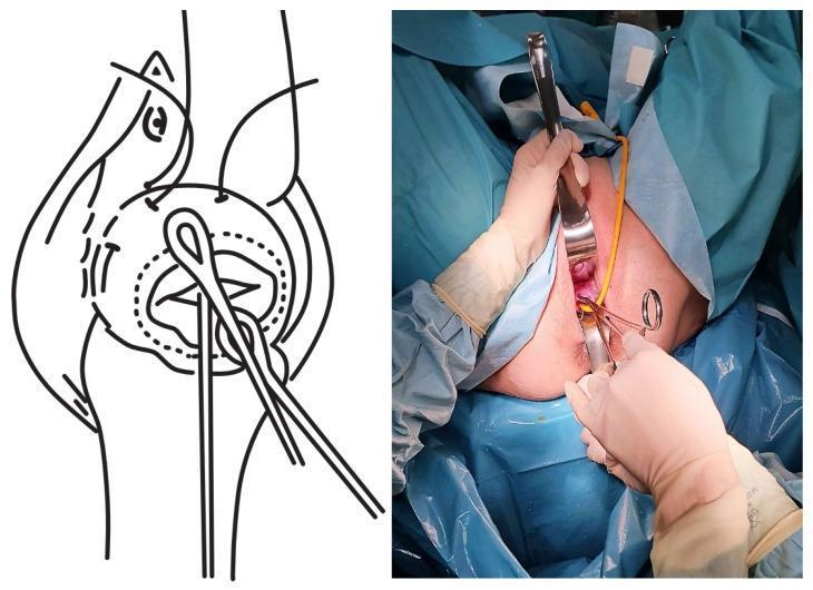
This composite image illustrates the reduction of prolapsed fetal membranes during an emergency cervical cerclage procedure, featuring a side-by-side clinical photograph and schematic diagram. The right panel is a clinical photograph showing the vaginal surgical field draped in blue sterile sheets. A surgeon's gloved hands are seen utilizing a metallic vaginal retractor and surgical forceps to expose the cervix. A yellow Foley catheter is inserted through the cervical canal, utilized here to manually reduce the bulging amniotic sac (membranes) back into the uterine cavity. The left panel provides a corresponding schematic illustration of the female pelvic anatomy, specifically detailing the cervix, uterus, and the placement of surgical instruments. The diagram highlights the mechanical process of using a catheter balloon and purse-string sutures to stabilize the prolapse and secure the cervical os. This educational material is designed for obstetrics and gynecology training, specifically focused on surgical management of cervical insufficiency and fetal membrane prolapse.
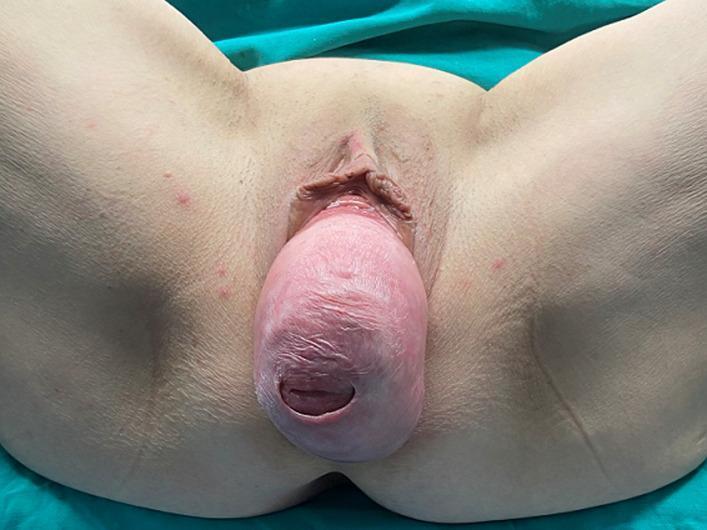
A clinical photograph showing a stage IV total uterine prolapse in a patient positioned for gynecological examination. The visual demonstrates a significant protrusion of the uterus and everted vaginal walls through the vaginal introitus, presenting as a large, rounded, fleshy mass. The prolapsed tissue exhibits a pale pink to reddish color with a glistening, moist surface and visible horizontal rugae or mucosal folds. At the most distal end of the mass, the external os of the cervix is clearly visible as a dark, oval orifice. The surrounding anatomy includes the labia minora and the vulvar region. The skin of the inner thighs and perineum is visible, showing some minor scattered erythematous macules or blemishes, but notably lacks extensive ulceration or necrosis of the vaginal mucosa. This image illustrates a severe case of pelvic organ prolapse (POP) which, as noted in the clinical context, can lead to secondary complications such as bilateral hydronephrosis due to ureteral compression.
Prolapse of the Uterus (Pelvic Organ Prolapse)
Definition
- Campbell-Walsh Urology; Berek & Novak's Gynecology
Anatomy of Pelvic Support (Three Levels - DeLancey)
| Level | Structures | Support Provided |
|---|---|---|
| Level I (Apical) | Uterosacral & cardinal ligaments | Suspend the uterus/vaginal apex from the sacrum and pelvic sidewall |
| Level II (Mid-vaginal) | Pubocervical fascia (anterior), Rectovaginal fascia (posterior) | Support the mid-vagina (prevents cystocele/rectocele) |
| Level III (Distal) | Perineal body, external anal sphincter, perineal membrane | Support the distal vagina and urogenital hiatus |
Epidemiology
-
Up to 43-90% of women have some degree of prolapse on examination
-
Symptomatic POP (vaginal bulge sensation): 3-12% prevalence
-
Most common site: anterior compartment (cystocele - 34%), followed by posterior (19%), then uterine/apical (14%)
-
Apical prolapse is almost always present when there is significant anterior or posterior wall prolapse
-
Lifetime risk of undergoing surgery for POP is ~11%
-
Campbell-Walsh Urology
Pathophysiology & Risk Factors
1. Muscle Injury (Levator Ani Trauma)
- Vaginal delivery causes visible levator ani defects in up to 20% of primiparous women (seen on MRI)
- Denervation from pudendal nerve injury (S3-S5) during labour - found in 24-29% of primiparous women at 6 weeks and 6 months post-delivery
- Even cesarean after labour (not elective C-section) causes some levator injury
2. Connective Tissue / Fascial Failure
- Collagen metabolism is altered in women with prolapse - abnormal type I:III collagen ratio
- Connective tissue disorders (Ehlers-Danlos, Marfan syndrome) significantly increase POP risk
- Age-related loss of collagen and elastin reduces fascial integrity
3. Hormonal Changes
- Oestrogen deficiency after menopause accelerates collagen degradation and reduces tissue quality
Key Risk Factors
| Risk Factor | Notes |
|---|---|
| Vaginal delivery | Most important modifiable risk; number and difficulty of deliveries matters |
| Increased parity | Cumulative trauma |
| Ageing / menopause | Oestrogen loss, tissue atrophy |
| Obesity | Chronic raised intra-abdominal pressure |
| Chronic straining | Constipation, chronic cough |
| Prior pelvic surgery | Hysterectomy significantly increases vault prolapse risk |
| Connective tissue disorders | Marfan, Ehlers-Danlos |
| Race | White and Hispanic women at higher risk than Black women for anterior wall prolapse |
| Family history | Genetic predisposition |
Types of Prolapse
| Type | Structure Descending | Alternative Name |
|---|---|---|
| Uterine prolapse | Uterus + cervix | Descensus uteri |
| Cystocele | Bladder through anterior vaginal wall | Anterior wall prolapse |
| Rectocele | Rectum through posterior vaginal wall | Posterior wall prolapse |
| Enterocele | Small bowel/peritoneal sac through pelvic floor | Usually post-hysterectomy |
| Vaginal vault prolapse | Vaginal apex post-hysterectomy | Apical prolapse |
| Urethrocele | Urethra | Anterior wall variant |
Staging / Grading Systems
Baden-Walker Halfway System (Traditional)
| Grade | Description |
|---|---|
| 0 | Normal, no prolapse |
| 1 | Descent halfway to the hymen |
| 2 | Descent to the hymen |
| 3 | Descent halfway past the hymen |
| 4 | Maximum possible descent |
POP-Q System (ICS Gold Standard)
- Aa, Ba - Anterior wall points
- C, D - Cervix/fornix (middle compartment)
- Ap, Bp - Posterior wall points
- gh (genital hiatus), pb (perineal body), tvl (total vaginal length)
| POP-Q Stage | Definition |
|---|---|
| 0 | No prolapse - all points ≤-3 cm |
| I | Leading edge >1 cm above hymen (< -1 cm) |
| II | Leading edge within 1 cm of hymen (-1 to +1 cm) |
| III | Leading edge >1 cm below hymen but not completely everted |
| IV | Complete eversion / procidentia |
- Berek & Novak's Gynecology
Clinical Features / Symptoms
Pelvic / Vaginal Symptoms
- Pelvic pressure, heaviness, or dragging sensation
- Visible or palpable bulge at the vaginal introitus
- Vaginal discharge or bleeding (from mucosal ulceration in procidentia)
- Low backache (worsens during the day, relieves on lying down)
Urinary Symptoms
- Urinary frequency and urgency
- Incomplete bladder emptying, slow or interrupted stream
- Stress urinary incontinence (SUI) - paradox: as prolapse advances beyond the hymen, SUI may decrease due to urethral kinking, but obstructive voiding increases
- Recurrent UTIs
Bowel Symptoms (weak correlation with prolapse severity)
- Constipation, straining
- Sensation of incomplete emptying
- Need to digitally splint the posterior vaginal wall to complete defecation (classic for rectocele)
- Fecal incontinence
Sexual Dysfunction
- Dyspareunia, reduced sensation
- Negative body image and psychosexual distress
Complications of Untreated Severe Prolapse
- Ulceration and keratinization of exposed cervix/vaginal mucosa
- Urinary retention
- Bilateral hydronephrosis and renal failure (ureteral compression in procidentia)
- Incarceration of small bowel in enterocele
Important: Low backache and pelvic pain attributed to prolapse warrant investigation of other sources. A pessary trial can help distinguish prolapse-related symptoms from other pathology - improvement with pessary suggests POP is the causative factor.
- Campbell-Walsh Urology
Investigations
History & Physical Examination
- Examine in dorsal lithotomy position and standing
- Valsalva manoeuvre and coughing to demonstrate maximum prolapse
- Assess all three compartments separately using Sims speculum
- Assess levator ani tone and voluntary contraction
Urological Evaluation
- Urinalysis and urine culture
- Urodynamic studies (if urinary symptoms are prominent)
- Post-void residual urine
Imaging
- Pelvic ultrasound (first-line)
- MRI defecography - for complex multi-compartment prolapse and surgical planning
- Intravenous pyelogram / renal ultrasound - to rule out hydronephrosis in advanced cases
Additional Tests
- Cervical smear (Pap test) before surgery
- Endometrial assessment if abnormal bleeding
Treatment
1. Conservative / Non-Surgical Management
- First-line for mild-moderate symptomatic prolapse
- Individualized physiotherapy can reduce prolapse severity and symptoms
- Most effective when started early and done consistently
- Weight reduction in obese patients
- Treat chronic constipation (high-fibre diet, laxatives)
- Avoid heavy lifting and prolonged standing
- Smoking cessation (reduces chronic cough)
- Improves tissue quality, reduces atrophy
- Useful adjunct to pessary or pre-operatively
- Indicated for patients who are not surgical candidates, wish to avoid surgery, or want symptom relief during pregnancy
- Ring pessary - most widely used; suitable for mild-moderate prolapse; fits behind pubic symphysis anteriorly and posterior vaginal fornix
- Shelf/Gehrung pessary - for larger or more complex prolapse
- Cube/donut pessary - for advanced prolapse in women who do not want surgery
- Success: 50-73% of women are successfully fitted initially
- Requires regular removal and cleaning (every 3-6 months); risk of erosion if neglected (even hydronephrosis and fistulae reported)
- Contraindicated in: active pelvic infection, poor vaginal tissue, inability to manage pessary
2. Surgical Management
A. Uterine Conservation (Hysteropexy)
| Procedure | Approach | Notes |
|---|---|---|
| Manchester (Fothergill) operation | Vaginal | Amputation of cervix + shortening of cardinal ligaments + anterior repair; good results for cervical elongation with prolapse |
| Sacrospinous hysteropexy | Vaginal | Suspension of cervix/uterus to sacrospinous ligament |
| Abdominal/laparoscopic hysteropexy | Abdominal/laparoscopic | Suspension using mesh or native tissue to sacral promontory |
B. Vaginal Hysterectomy ± Vault Repair
- Standard surgical treatment for uterine prolapse in women who have completed childbearing
- Must be combined with vault suspension (uterosacral or sacrospinous fixation) to prevent post-hysterectomy vault prolapse
- A 2025 meta-analysis (PMID 39835651) comparing Manchester procedure vs vaginal hysterectomy found comparable outcomes for mid-compartment prolapse
C. Anterior Compartment Repair (Cystocele)
- Anterior colporrhaphy - plication of pubocervical fascia; anatomic cure in 57-93%; higher recurrence rates than abdominal approaches
D. Posterior Compartment Repair (Rectocele/Enterocele)
- Posterior colporrhaphy - plication of rectovaginal fascia; anatomic cure 76-96%
- Important: levator ani plication should be avoided (causes de novo dyspareunia in up to 50%)
- Culdoplasty (McCall, Moschcowitz) - for enterocele
- Rectocele repair improves bulge symptoms, not constipation
E. Apical / Vault Suspension Procedures
| Procedure | Route | Notes |
|---|---|---|
| Sacrospinous ligament fixation (SSLF) | Vaginal | Native tissue; risk: pudendal nerve injury, right-sided vaginal deviation |
| Uterosacral ligament suspension (USLF) | Vaginal / abdominal | Native tissue; risk: ureteral kinking |
| Sacrocolpopexy | Abdominal / laparoscopic | Gold standard for vault prolapse; mesh attached from vaginal apex to sacral promontory; lowest recurrence rate |
| Robotic sacrocolpopexy | Robotic-assisted laparoscopic | Equivalent to laparoscopic; longer operating time |
Sacrocolpopexy (abdominal) has the lowest recurrence rate of all prolapse procedures. Vaginal approaches have approximately twice the failure rate of abdominal approaches.
F. Obliterative Procedures (Colpocleisis / LeFort)
- For elderly, medically frail patients who do not wish to retain coital function
- LeFort colpocleisis: partial closure of the vaginal canal; very effective with low morbidity
- Can be performed with or without hysterectomy
3. Mesh Use in Prolapse Surgery
- The FDA issued warnings limiting vaginal mesh for prolapse repair due to complications (mesh erosion, chronic pelvic pain, dyspareunia)
- Mesh is still used in abdominal sacrocolpopexy (well-supported evidence)
- Autologous fascial grafts may be used as an alternative to synthetic mesh for augmentation
Summary of Treatment Algorithm
Symptomatic POP
     |
     ├── Conservative measures first (PFMT, lifestyle, local oestrogen)
     |
     ├── Pessary (if surgery declined, unfit, or temporary relief needed)
     |
     └── Surgery (failed conservative, significant symptoms)
              |
              ├── Wants uterus retained → Hysteropexy (Manchester / SSLF / sacrocolpopexy)
              |
              ├── Childbearing complete → Vaginal hysterectomy + vault suspension
              |
              ├── Apical prolapse dominant → Sacrocolpopexy (lowest recurrence)
              |
              ├── Anterior wall → Anterior colporrhaphy
              |
              ├── Posterior wall → Posterior colporrhaphy (without levator plication)
              |
              └── Elderly, no coital function desired → Colpocleisis (LeFort)
Recent Evidence (2024-2026)
- A 2026 systematic review (PMID 41969167) on the Manchester procedure found low recurrence rates and acceptable perioperative outcomes; potential concern for future obstetric and cervical cancer screening implications.
- A 2025 meta-analysis (PMID 39835651) found Manchester procedure vs vaginal hysterectomy had comparable anatomical outcomes for mid-compartment prolapse.
- A 2025 systematic review (PMID 41286733) supports hysteropexy at Stage I prolapse to prevent progression and recurrence.
Infertility from.yhe book.of gynecology by hiralal konar
DC Dutta Textbook of Gynecology Hiralal Konar infertility chapter key points
https://dl.icdst.org/pdfs/files4/8a44d501e480b6a1ab7b1c17c64…
DC Dutta Gynecology Konar infertility chapter 16 causes investigation ART IUI IVF ICSI key points
https://dl.icdst.org/pdfs/files4/8a44d501e480b6a1ab7b1c17c64…
Infertility — DC Dutta's Textbook of Gynecology (Hiralal Konar)
Note: DC Dutta's Textbook of Gynecology edited by Prof. Hiralal Konar is not in this platform's digital library. The content below is drawn directly from Chapter 16 of that textbook (authored by Dr BN Chakravorty), supplemented by content from the companion clinical books, and is presented faithfully to the book's structure and style.
Definition
"Infertility is defined as a failure to conceive within 1 year of regular unprotected coitus." — DC Dutta's Textbook of Gynecology, Ch. 16
- Primary infertility - couple has never conceived
- Secondary infertility - failure to conceive after a previous pregnancy (regardless of outcome)
- Subfertility - reduced capacity to conceive; may eventually conceive without help
- Sterility - absolute inability to reproduce (e.g., bilateral tubal block, azoospermia due to testicular failure)
Incidence
- ~10% of couples remain infertile by the end of the 2nd year of regular unprotected intercourse
- Incidence increases further after age 30 years in women
- Male factor: 30-40%
- Female factor: 40-50%
- Both partners involved: ~10%
- Unexplained infertility: 10-20% (some sources up to 30%)
Requirements for Conception (Prerequisites)
- Adequate number of normal, motile spermatozoa in the ejaculate
- Normal deposition of sperm high in the vagina / near the cervix
- Patent, functional fallopian tubes
- Normal ovulation and release of a mature oocyte
- Fertilization of the oocyte by sperm
- Normal uterine cavity for implantation
- Adequate hormonal support (especially luteal phase progesterone)
Causes of Male Infertility
| Category | Approximate Incidence |
|---|---|
| Hypothalamic-pituitary disorders | 1-2% |
| Primary gonadal disorders | 30-40% |
| Disorders of sperm transport | 10-20% |
| Idiopathic | 40-50% |
Detailed Causes:
- Varicocele (most common correctable cause)
- Cryptorchidism (undescended testes) - spermatogenesis depressed, hormone secretion unaffected
- Testicular atrophy (post-orchitis - mumps, gonorrhoea)
- Radiation, cytotoxic drugs
- Alcohol - inhibits spermatogenesis by suppressing Leydig cell testosterone synthesis or gonadotropin levels
- Endocrine: hypogonadotrophic hypogonadism, hypothyroidism, hyperprolactinaemia
- Genetic: Klinefelter syndrome (47,XXY) - eunuchoid habitus, azoospermia, gynaecomastia
- Congenital: bilateral absence of vas deferens (in 1-2% of infertile males; associated with cystic fibrosis/CFTR mutations)
- Kartagener syndrome (autosomal disease) - loss of ciliary function and sperm motility
- Young's syndrome
- Acquired infection: tuberculosis, gonorrhoea
- Surgical: herniorrhaphy, vasectomy
- Hypospadias - sperm deposited at introitus rather than near cervix
- Erectile dysfunction (impotence) - medical or psychological
- Ejaculatory failure, retrograde ejaculation
- Bladder neck surgery
- Infection causing abnormal viscosity/volume
- Antisperm antibodies (sperm-immobilising or agglutinating)
- Abnormal seminal fructose (absent in vas deferens obstruction)
Causes of Female Infertility
Scheme 1 (as in Dutta):
| Factor | Approximate Incidence |
|---|---|
| Tubal factor | 25-35% (most common) |
| Ovulatory dysfunction | 20-25% |
| Endometriosis | 0-10% |
| Cervical factor | 5-10% |
| Uterine factor | 5-10% |
| Immunological | <5% |
| Unexplained | 10-30% |
- Pelvic inflammatory disease (PID) - most common cause; gonorrhoea, chlamydia, TB
- Post-surgical adhesions
- Endometriosis causing peritubal adhesions or tubal blockage
- Congenital tubal anomalies
- Polycystic ovary syndrome (PCOS) - most common single cause of anovulation
- Hyperprolactinaemia
- Hypothyroidism, Addison's disease
- Hypothalamic amenorrhoea (weight loss, excessive exercise)
- Premature ovarian failure
- Luteal phase defect (LPD)
- Luteinised unruptured follicle (LUF) syndrome
- Hostile cervical mucus (poor sperm penetration)
- Cervical stenosis (post-surgery, TB)
- Cervicitis
- Antisperm antibodies in cervical mucus
- Assessed by Post-Coital Test (PCT)
- Submucous fibroid
- Intrauterine synechiae (Asherman syndrome)
- Congenital uterine anomaly (septate uterus, bicornuate uterus)
- Endometrial polyp
- Endometrial tuberculosis (most important uterine cause in India)
- Narrow introitus (dyspareunia → infrequent intercourse)
- Imperforate hymen
- Vaginismus
- Vaginitis
- Tubal block from adhesions
- Peritubal adhesions impairing ovum pick-up
- Altered peritoneal environment (prostaglandins, cytokines affecting fertilisation)
- Antisperm antibodies in female serum or cervical mucus (sperm-immobilising or agglutinating)
- Antisperm antibody testing is rarely done today with advent of IUI, IVF, ICSI
- Defined when no obvious cause found after all standard investigations (semen analysis, ovulation detection, tubal/peritoneal assessment, endocrinopathy, PCT)
- Incidence: 10-20% (up to 30%)
- With expectant management, ~40-60% conceive within 3 years
- IVF + ET is an option for non-responders
Investigation of Infertility
Principles
- Both partners must be investigated simultaneously
- Investigations directed by history and clinical examination
- Proceed from simple/non-invasive to complex/invasive
- Special guideline: If a major male fault (e.g., azoospermia due to testicular destruction) is found, female investigation may be deferred - but ART considerations may require female assessment regardless
Male Investigation
| Parameter | Normal Value (WHO) |
|---|---|
| Volume | 1.5-5 mL |
| Liquefaction | Within 30 minutes |
| pH | 7.2-8.0 |
| Sperm concentration | ≥16 million/mL |
| Total sperm count | ≥39 million/ejaculate |
| Progressive motility | ≥32% |
| Total motility | ≥40% |
| Normal morphology | ≥4% (Kruger strict) |
- Azoospermia - no sperm
- Oligozoospermia - count <15 million/mL
- Asthenozoospermia - reduced motility (<32% progressive)
- Teratozoospermia - abnormal morphology (<4%)
- OAT (oligoasthenoteratozoospermia) - combination of all three
- Repeat semen analysis (2nd sample at least 3 months later)
- Hormonal: FSH, LH, testosterone, prolactin
- Scrotal ultrasound (varicocele, testicular atrophy)
- Testicular biopsy (to distinguish obstructive vs non-obstructive azoospermia)
- Genetics: karyotype, Y chromosome microdeletion, CFTR gene mutation
- Fructose test (absent in obstruction)
- Antisperm antibody (rarely indicated today)
Female Investigation
- Menstrual calendar - regular cycles suggest ovulation
- Basal Body Temperature (BBT) chart - biphasic pattern confirms ovulation; a persistent rise in BBT and/or serum progesterone >5 ng/mL is presumptive evidence of ovulation
- Serum progesterone - measured 6-8 days before next period (mid-luteal); >5 ng/mL = ovulation
- LH surge - urine or serum LH; ovulation occurs ~36 hours after surge
- Transvaginal ultrasound (TVS) - follicle tracking; sonographic evidence of ovulation = collapsed follicle + fluid in Pouch of Douglas
- Endometrial biopsy - rarely done now
- Pregnancy is the surest evidence of ovulation
| Investigation | Notes |
|---|---|
| HSG (Hysterosalpingography) | Outlines uterine cavity and tubal patency; best at day 7-9 of cycle; complimentary to laparoscopy |
| Laparoscopy with chromopertubation | Gold standard for tubal patency + peritoneal assessment |
| Sonohysterosalpingography (SIS) | No radiation exposure; ultrasound-based |
| Falloposcopy | Study of tubal lumen from the ampulla |
| Salpingoscopy | Study of tubal mucosa |
- Post-Coital Test (PCT) / Sims-Huhner Test - performed 2-12 hours after intercourse, mid-cycle; >5 progressively motile sperm per HPF = normal
- Cervical mucus scoring (Insler score): volume, viscosity, ferning, spinnbarkeit
- TVS, HSG (outline cavity)
- Hysteroscopy (gold standard for intrauterine pathology)
- Endometrial biopsy (TB culture if suspected genital TB - very important in India)
- Day 2-3: FSH, LH, oestradiol, prolactin, TSH
- AMH (Anti-Müllerian Hormone) - ovarian reserve
- Day 21: Serum progesterone (luteal function)
- DHEAS, testosterone, 17-OHP (if hyperandrogenism suspected)
Treatment of Infertility
General Principles
- Treatment must be predicated on findings of the infertility evaluation
- Correct any identifiable cause before empiric treatment
- Counsel about realistic expectations; 40-60% of unexplained infertility couples conceive with expectant management
A. Treatment of Male Factor Infertility
| Cause | Treatment |
|---|---|
| Varicocele | Varicocelectomy (surgical or radiological) |
| Hypogonadotrophic hypogonadism | hCG + FSH injections (Pergonal), pulsatile GnRH |
| Hyperprolactinaemia | Dopamine agonists (bromocriptine, cabergoline) |
| Hypothyroidism | Thyroxine replacement |
| Obstructive azoospermia | Microsurgical epididymovasostomy / PESA / TESA + ICSI |
| Non-obstructive azoospermia | TESA/TESE + ICSI if sperm found |
| OAT syndrome | IUI; if severe → IVF + ICSI |
| Retrograde ejaculation | Alpha-adrenergic drugs; sperm from urine for IUI |
| Antisperm antibodies | IUI, IVF, ICSI (empiric) |
B. Treatment of Female Factor — Ovulatory Dysfunction
- Group I (Hypogonadotrophic) - low FSH, low oestrogen (e.g., hypothalamic amenorrhoea)
- Group II (Normogonadotrophic) - normal FSH, normal oestrogen (PCOS is most common)
- Group III (Hypergonadotrophic) - elevated FSH, low oestrogen (ovarian failure)
-
General measures (first)
- Psychotherapy for emotional/psychological causes
- Weight optimisation (BMI 20-25 kg/m²)
- Correction of systemic disorders (hypothyroidism, hyperprolactinaemia first)
-
Drug therapy:
| Drug | Dose | Mechanism | Notes |
|---|---|---|---|
| Clomiphene citrate | 50 mg/day for 5 days (Day 2-6 or Day 5-9); step up to 150-200 mg/day | Anti-oestrogen → ↑ FSH/LH release | First-line for WHO Group II; 75-80% ovulation rate; 40% pregnancy rate |
| Letrozole (aromatase inhibitor) | 2.5-5 mg/day (Day 3-7) | Blocks oestrogen synthesis → ↑ FSH | Preferred in PCOS (less risk of multiple pregnancy vs clomiphene); now considered first-line |
| Metformin | 500-1500 mg/day | Insulin sensitiser | Useful in PCOS with hyperinsulinaemia; add-on or alone |
| Gonadotropins (FSH/hMG) | Individualised | Direct follicular stimulation | WHO Group I & II; requires monitoring (TVS + oestradiol); risk of OHSS and multiple pregnancy |
| GnRH (pulsatile) | Pulsatile i.v. or s.c. | Stimulates pituitary → FSH, LH | WHO Group I (hypothalamic); expensive; restricted use |
| Bromocriptine / Cabergoline | Dopamine agonist → ↓ prolactin | For hyperprolactinaemia-related anovulation |
Important (Dutta): Gonadotropins and GnRH/GnRH analogues are costly and must be monitored with sophisticated equipment to control the regimen and minimise hazards (Table 16.7). Their use is restricted to selected centres and is most commonly used in ART.
- Natural progesterone vaginal suppositories 100 mg three times daily from day of ovulation - continue until menses; if no menses after 14 days, do pregnancy test; if positive, continue up to 10th week
- Surgery:
- Laparoscopic ovarian drilling (LOD) - for clomiphene-resistant PCOS; diathermy/laser to multiple points on ovarian surface; avoids OHSS risk; reduces LH and androgens
- Wedge resection of ovary (historical; largely replaced by LOD)
C. Treatment of Tubal Factor
| Situation | Management |
|---|---|
| Proximal (cornual) block | Hysteroscopic tubal cannulation or fluoroscopic cannulation |
| Distal block (hydrosalpinx) | Salpingostomy / fimbrioplasty (limited success) |
| Severe tubal damage | IVF - preferred over surgery (better success rates) |
| Hydrosalpinx before IVF | Salpingectomy first - tubal fluid impairs implantation |
| Post-sterilisation | Microsurgical reanastomosis vs IVF (decision based on age, ovarian reserve, length of remaining tube) |
D. Treatment of Cervical Factor
- Intrauterine Insemination (IUI) - bypasses hostile cervical mucus
- Antibiotics if cervicitis
- Low-dose oestrogen to improve mucus quality
- Corticosteroids (empiric, for antisperm antibodies) - not well supported; IUI/IVF preferred
E. Treatment of Endometriosis
- Laparoscopic surgical excision/ablation of endometriotic deposits
- Suppressive drugs (GnRH agonists, progestogens, danazol) - not used directly to treat infertility as they prevent conception during use
- IVF for moderate-severe endometriosis
F. Unexplained Infertility
- Expectant management (40-60% conceive in 3 years without treatment)
- Ovulation induction + IUI (empirical superovulation)
- IVF + ET for non-responders
Assisted Reproductive Technology (ART)
Types of ART:
- Washed and capacitated sperm placed directly into the uterine cavity via a soft catheter
- Done 36 hours after hCG trigger or LH surge detection
- Indications: mild male factor, cervical factor, unexplained infertility
- Success rate: ~10-15% per cycle
- Controlled ovarian hyperstimulation (COH) with gonadotropins
- Transvaginal oocyte retrieval under ultrasound guidance
- Fertilisation in the laboratory (mixing sperm and oocytes)
- Embryo culture for 2-5 days
- Transcervical embryo transfer into uterine cavity under ultrasound guidance
- Indications: tubal disease, severe male factor (with ICSI), endometriosis, unexplained infertility
- A single morphologically normal, motile sperm is injected directly into the oocyte using micromanipulation
- Used for severe male factor infertility (severe OAT, azoospermia with surgical sperm retrieval)
- Success rates equivalent to conventional IVF
- Slight concern for genetic transmission of male infertility factor to offspring
- Unfertilised oocytes and sperm are transferred into the fallopian tube via laparoscopy
- Requires at least one patent tube; largely superseded by IVF
- Fertilised zygote (at pronuclear stage) transferred into fallopian tube
- Largely historical
- Surplus embryos stored for future frozen embryo transfer (FET) cycles
- Donor oocytes fertilised with partner's sperm; embryo transferred to recipient's uterus
- For premature ovarian failure, diminished ovarian reserve, or genetic disorders
- Gestational carrier (surrogate) carries the couple's embryo to term
Complications of ART:
- Ovarian Hyperstimulation Syndrome (OHSS) - major risk with gonadotropin stimulation
- Multiple pregnancy
- Ectopic pregnancy
- Congenital anomalies (slight increased risk with ICSI)
Key Points (as in DC Dutta's Textbook of Gynecology, Ch. 16)
- Infertility is defined as failure to conceive within 1 year of regular unprotected coitus
- 10% remain infertile by end of 2nd year; incidence increases after age 30
- In fully lactating women, pregnancy is unlikely up to 10 weeks postpartum
- Male factor: 30-40%, Female factor: 40-50%, Both: 10%, Unexplained: 10%
- Common male causes: defective spermatogenesis, ductal obstruction, failure to deposit sperm, seminal fluid errors
- Important female causes: tubal factor (25-35%), ovulatory factor (20-25%), endometriosis (0-10%)
- Ovulatory dysfunction is linked to disturbed HPO axis - primary or secondary to thyroid/adrenal dysfunction
- A persistent rise in BBT and/or serum progesterone >5 ng/mL is presumptive evidence of ovulation. Sonographic evidence = collapsed follicle + fluid in POD. Pregnancy is the surest evidence of ovulation
- Thorough investigation following an abnormal semen analysis is needed to find the specific cause (Scheme 2)
- Tubal factor is assessed by HSG and laparoscopy - the two are complementary; Sonohysterosalpingography has no radiation exposure; falloposcopy and salpingoscopy are used to study the tubal mucosa
- Antisperm antibody testing is rarely done today with the advent of IUI, IVF, ICSI
- Unexplained infertility: ~40% conceive within 3 years without specific treatment; IVF+ET is an option for non-responders
- Adoption is a realistic alternative - couples must understand the limits, costs, and risks of infertility management