Complete radius bone
Reading File
Finding Sources
Finding Sources
Searching Images
radius bone anatomy
Reading File
Finding Sources
Searching Images
radius bone labeled anatomy forearm ulna
Reading File
Reading File
Reading File
Loading Image
Loading Image
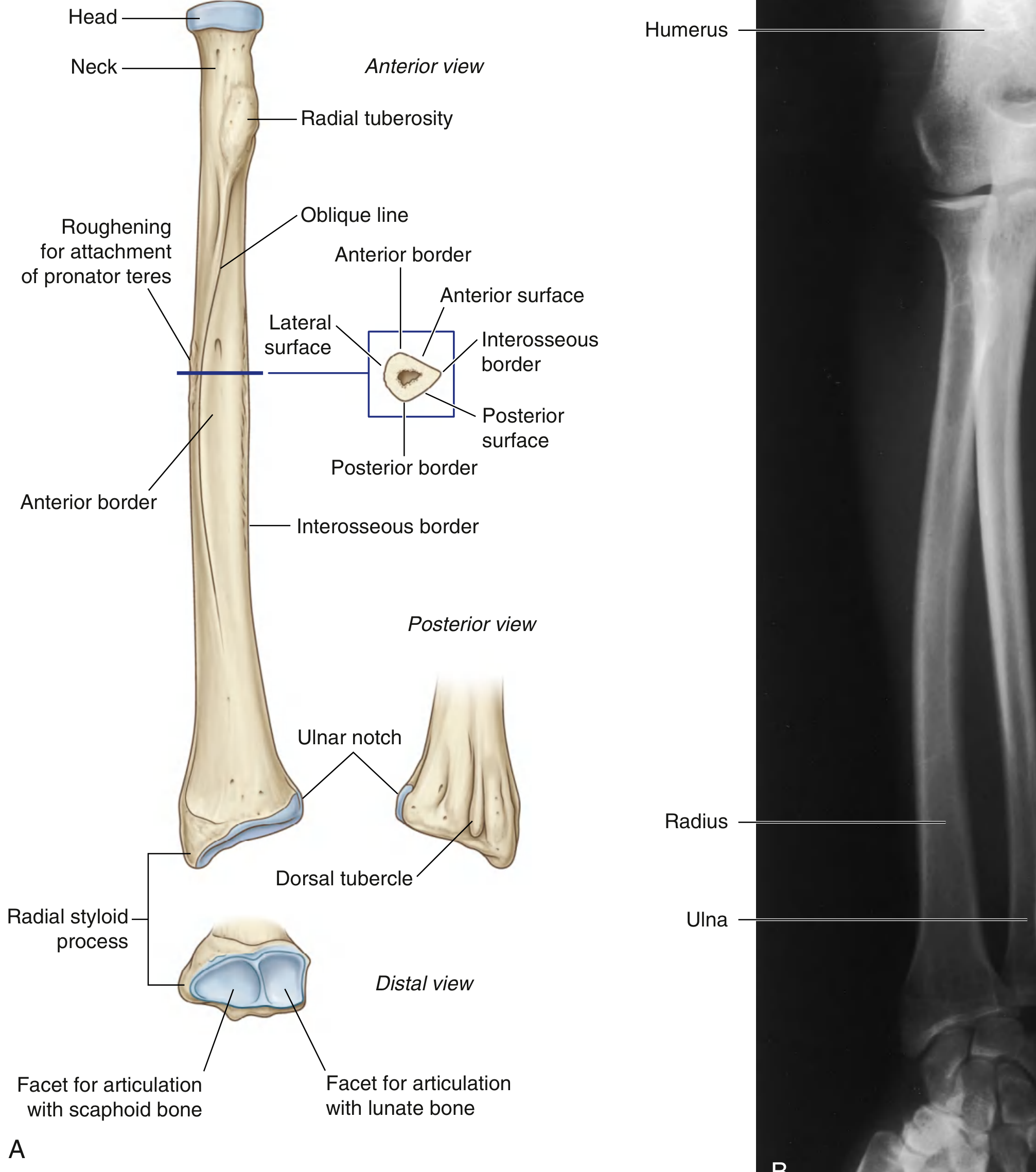
The radius is one of the two forearm bones, located on the thumb (lateral) side.
Complete anatomy of the radius (quick full summary)
- Type: Long bone
- Position: Lateral forearm (anatomical position)
- Shape pattern:
- Small proximally
- Broad distally (forms most of wrist articulation)
Parts
-
Proximal end
- Head of radius: disc-shaped; superior fovea articulates with capitulum of humerus
- Neck of radius
- Radial tuberosity: insertion of biceps brachii tendon
-
Shaft (body)
- Triangular cross-section
- 3 borders: anterior, posterior, interosseous
- 3 surfaces: anterior, posterior, lateral
- Interosseous border gives attachment to interosseous membrane (between radius and ulna)
-
Distal end
- Radial styloid process (lateral)
- Dorsal tubercle (Lister tubercle) posteriorly
- Ulnar notch medially (for distal radioulnar joint)
- Inferior articular facets for scaphoid and lunate (radiocarpal joint)
Major articulations
- Proximal radioulnar joint (with ulna)
- Humeroradial joint (with capitulum)
- Distal radioulnar joint
- Radiocarpal joint (with scaphoid and lunate)
Main functional points
- Essential for pronation-supination of forearm
- Transmits load from hand to elbow
- Distal radius is a common fracture site (for example, Colles fracture)
Source: Imaging anatomy text and atlas Volume 3, Bones, joints, Vessels and Nerves, p. 93-95; Gray's Anatomy for Students, p. 892-894.
This is a shared conversation. Sign in to Orris to start your own chat.