
Make a powepoint presentation of 10 to 15 slides on the topic highlighting introduction to usg guided regional blocks, advantages and disadvantages of usg guided regional blocks
ultrasound guided nerve block regional anesthesia probe needle
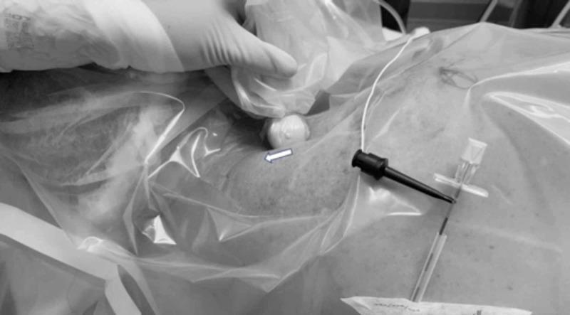
This clinical photograph captures a regional anesthesia procedure, specifically an ultrasound-guided interscalene brachial plexus block at the level of the upper trunk. The image displays a sterilely prepared field covered by a transparent plastic drape. A clinician's gloved hand is visible on the left, manipulating an ultrasound probe that is sheathed in a sterile cover. An arrow indicates the cephalad rotation of the probe's lateral edge. To the right, a specialized regional anesthesia needle is inserted through the drape and into the skin in an in-plane orientation relative to the ultrasound beam. Visible components of the needle assembly include a clear hub with a white cap and a black stimulator connector, indicating the use of neurostimulation for nerve localization. The procedure demonstrates the lateral-to-medial advancement of the needle through the middle scalene muscle toward the upper trunk of the brachial plexus. This visual serves as an educational guide for probe orientation and needle-probe alignment during catheter placement for continuous peripheral nerve blocks.
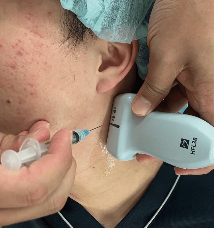
This clinical procedural photograph demonstrates an ultrasound-guided selective glossopharyngeal nerve block (UGSGNB). The image shows a high-frequency linear ultrasound probe (model HFL38, labeled 13-6 MHz) positioned in a transverse orientation on the upper lateral neck, just inferior to the earlobe and posterior to the mandible. A thin needle, attached to a transparent syringe with a blue hub, is being inserted into the submandibular/upper cervical region using an in-plane technique. The needle's trajectory is directed toward the deep cervical fascia beneath the probe's footprint, where clear ultrasound gel has been applied for acoustic coupling. The procedure is aimed at delivering local anesthetic (e.g., ropivacaine) near the styloid process and stylohyoid muscle to achieve neural blockade. This educational visual is relevant for anesthesiology and pain management training, highlighting probe placement and needle approach for regional anesthesia of the head and neck.
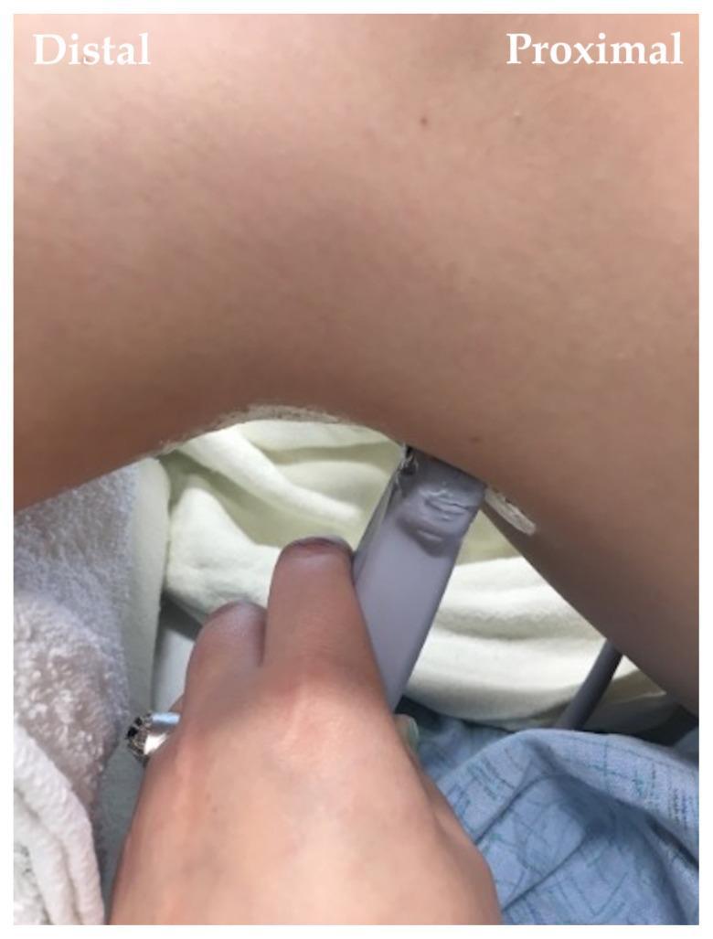
A clinical photograph demonstrating the procedural setup for a regional anesthesia technique, specifically a popliteal fossa block. The image shows a practitioner's hand holding a linear ultrasound probe against the posterior aspect of a patient's leg at the level of the popliteal crease. The probe is positioned transversely to visualize the sciatic nerve and its bifurcation into the tibial and common peroneal nerves. Text overlays indicate the 'Distal' (lower leg) and 'Proximal' (thigh) orientations. Acoustic coupling gel is visible at the interface between the transducer and the skin. This visual serves as a guide for needle-probe coordination and anatomical landmark identification during ultrasound-guided nerve blocks. The patient is in a prone or lateral position, resting on clinical linens, typical of an anesthetic workshop or operative environment.
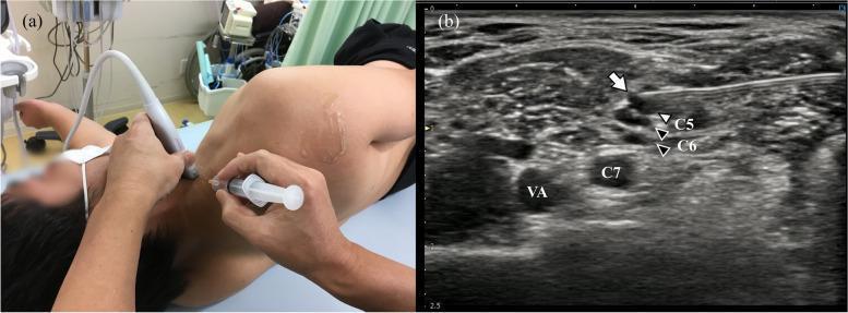
This composite image illustrates the clinical application of ultrasound-guided regional anesthesia. Image (a) is a clinical photograph showing a patient in the lateral recumbent position. A practitioner is performing an ultrasound-guided cervical nerve block, with a linear probe placed on the lateral neck and a syringe/needle assembly inserted via an in-plane approach. Image (b) is the corresponding transverse sonogram of the interscalene brachial plexus region. It highlights anatomical landmarks including the C5 and C6 nerve roots (marked with white and black arrowheads), the C7 nerve root located more deeply and medially, and the pulsatile vertebral artery (VA). A hyperechoic needle (white arrow) is visualized with the tip positioned precisely near the C5 and C6 nerve roots, which are merging to form the superior trunk. This educational material demonstrates the correct needle-to-nerve relationship and anatomical localization required for safe and effective interscalene or cervical nerve blocks in pain management and orthopedic surgery.
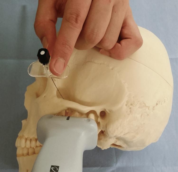
A clinical photograph demonstrating the procedural setup for an ultrasound-guided maxillary nerve block (UGMNB) using a supra-zygomatic approach on a skull model. A high-frequency linear ultrasound probe is positioned horizontally over the zygomatic arch and infratemporal fossa region to visualize deep anatomical landmarks, specifically the lateral pterygoid plate and the pterygopalatine fossa. A needle is shown entering through a clear, winged needle guide held by a hand. The needle trajectory is directed inferiorly and medially toward the target area posterior to the maxilla. This educational visual illustrates the spatial relationship between the ultrasound transducer, the needle insertion point, and the underlying osseous anatomy of the skull. This setup is used in regional anesthesia and pain management to provide sensory blockade for maxillofacial surgeries. The use of the needle guide ensures precise angle control and alignment with the ultrasound beam for real-time visualization of the needle tip.
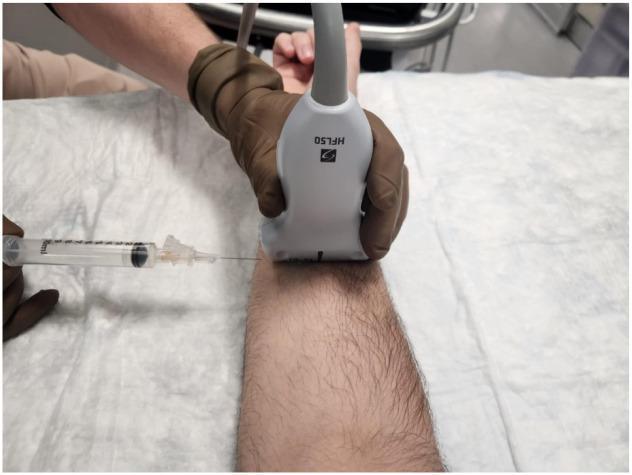
Clinical procedural photograph demonstrating the setup for an ultrasound-guided median nerve block in the mid-forearm. A clinician, wearing brown sterile gloves, holds a high-frequency linear ultrasound probe (labeled HFL50) in a transverse orientation on the volar aspect of a patient's forearm. Simultaneously, a clear syringe with a fine-gauge needle is shown approaching the skin from the lateral side in an in-plane technique. The in-plane approach is characterized by the needle being parallel to the long axis of the probe, allowing for continuous visualization of the needle shaft and tip as it traverses the soft tissue towards the target nerve. This image illustrates the ergonomic positioning and spatial relationship between the ultrasound transducer and the needle used for hydrodissection or regional anesthesia in the management of conditions such as Carpal Tunnel Syndrome (CTS).
ultrasound image brachial plexus interscalene block sonogram
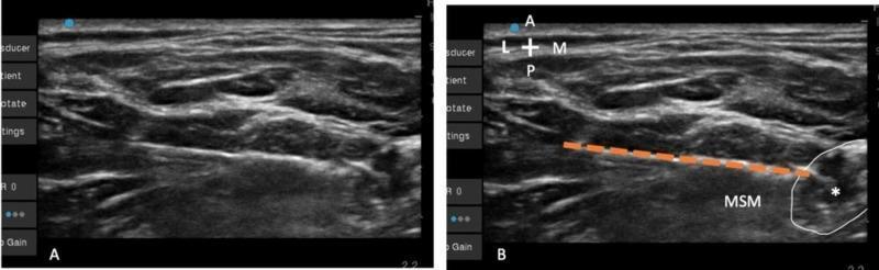
This diagnostic ultrasound image demonstrates an interscalene brachial plexus block procedure. The visual is divided into two panels: (A) an unlabeled sonogram and (B) an annotated version showing a lateral-to-medial in-plane needle approach. Anatomical landmarks include the middle scalene muscle (MSM), which appears as a relatively hypoechoic structure with a striated fibrillar texture, and the upper trunk of the brachial plexus, marked by an asterisk (*) and outlined by a white circle. A Tuohy needle path is represented by an interrupted orange line, illustrating its trajectory through the MSM to reach the lateral aspect of the upper trunk. The imaging displays the relationship between the deep cervical fascia and the neural structures, highlighting the needle's positioning for regional anesthesia. This clinical image is intended for medical education regarding ultrasound-guided nerve blocks and regional anesthetic techniques, specifically focusing on needle visualization and anatomical target identification in the cervical region.
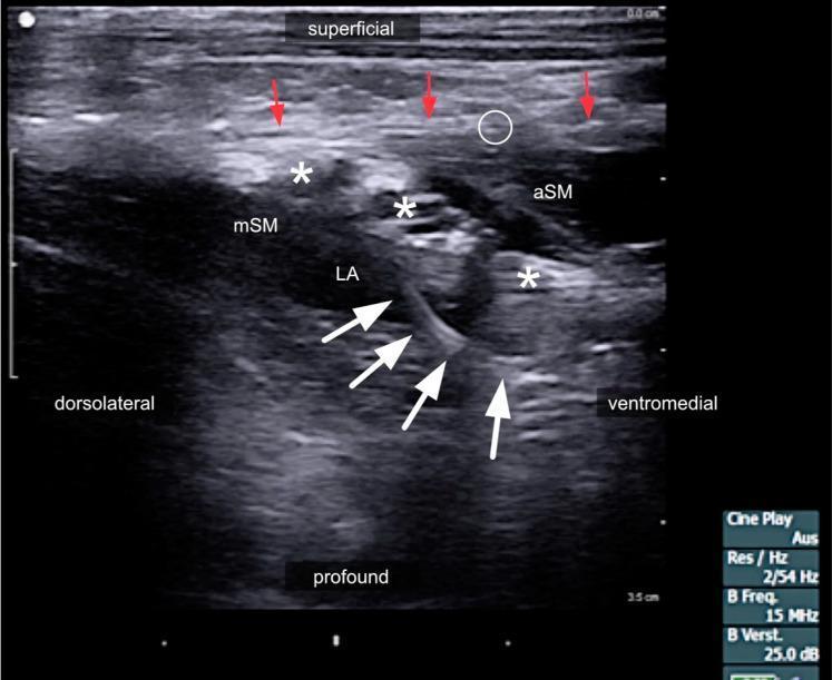
This diagnostic ultrasound image demonstrates the left lateral neck region during a continuous interscalene brachial plexus block. The sonogram identifies the anterior scalene muscle (aSM) and middle scalene muscle (mSM) with the interscalene groove located between them. The ventral rami of the brachial plexus are highlighted by white asterisks (*) appearing as hypoechoic oval structures. A white circle indicates the phrenic nerve situated on the anterior surface of the aSM. Red arrows delineate the hyperechoic prevertebral fascia, which acts as a structural boundary for local anesthetic (LA) spread. The LA is visible as a dark, hypoechoic fluid collection surrounding the nerve roots. A medical catheter is identifiable in situ, marked by white arrowheads, positioned deep to the sternocleidomastoid muscle (SCM) and targeting the interscalene space. This image serves as a clinical example of regional anesthesia placement, illustrating the anatomical relationships between cervical musculature, fascia, and neural structures necessary for safe and effective anesthetic delivery.
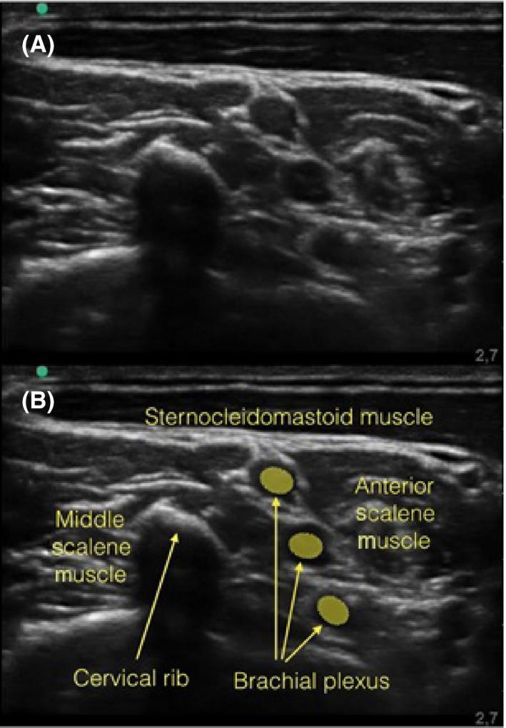
This diagnostic image displays two panels (A and B) of a musculoskeletal ultrasound scan in the cervical region, specifically showing an interscalene approach to the brachial plexus. Image A is the original sonogram, while image B provides anatomical labels. Key structures include the hypoechoic, rounded nerve roots of the brachial plexus located in the interscalene groove between the anterior scalene muscle and the middle scalene muscle. Superficial to these is the sternocleidomastoid muscle. A significant clinical finding is the presence of a cervical rib, which appears as a prominent, irregular hyperechoic structure. This osseous variation is seen casting a dense acoustic shadow that obscures the underlying neural anatomy, potentially complicating regional anesthesia procedures such as a brachial plexus block. The educational focus is on the ultrasonographic identification of anatomical variations and their impact on clinical interventions like nerve blocks.
femoral nerve block ultrasound guided lower limb
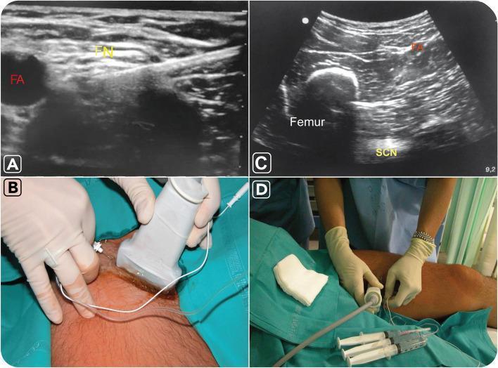
This composite figure illustrates ultrasound-guided nerve block procedures of the lower limb. Panels A and B focus on the femoral nerve block. Image A is a transverse ultrasound view of the femoral region showing the femoral nerve (FN) as a cluster of intermediate echogenicity situated lateral to the anechoic (black) femoral artery (FA). Panel B shows the corresponding clinical setup with a linear transducer placed at the inguinal crease and a needle inserted in-plane. Panels C and D demonstrate the sciatic nerve block via an anterior approach. Ultrasound image C reveals the sciatic nerve (SCN) as a hyperechoic, oval structure located deep to the femur and femoral artery, positioned between the adductor magnus and hamstring muscles at a depth of approximately 6–8 cm. Clinical photograph D displays the positioning for the anterior sciatic block with the patient in a supine position, showing the needle insertion at the medial aspect of the thigh. This visual guide demonstrates key anatomical landmarks and probe positioning for regional anesthesia of the femoral and sciatic nerves.
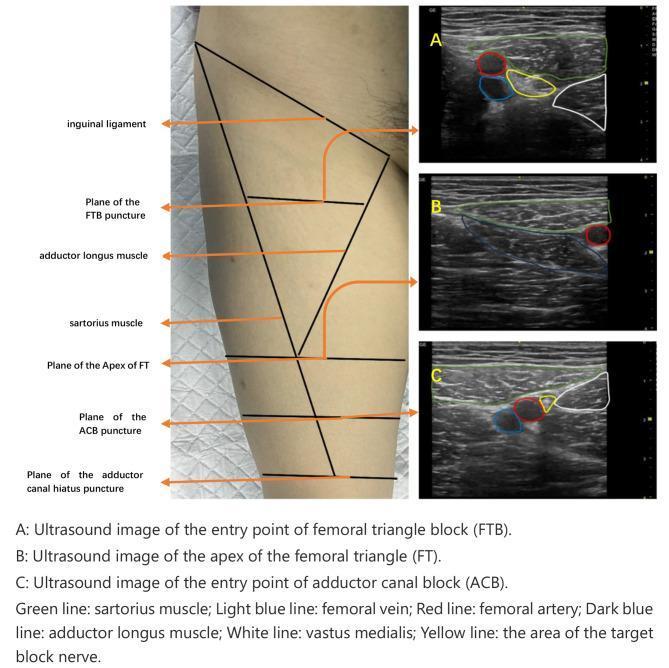
This composite educational graphic details the ultrasound-guided regional anesthesia of the lower limb, specifically comparing the femoral triangle block (FTB) and adductor canal block (ACB). A clinical photograph of a patient's medial thigh demonstrates the surface anatomy and puncture planes relative to the inguinal ligament, sartorius muscle, and adductor longus. Three corresponding transverse ultrasound images (A, B, C) illustrate key cross-sectional anatomy. Image A shows the FTB entry point, identifying the femoral artery (red), femoral vein (light blue), and target nerve area (yellow) situated between the sartorius (green), adductor longus (dark blue), and vastus medialis (white). Image B displays the apex of the femoral triangle, where the sartorius muscle begins to cross over the femoral vessels. Image C identifies the ACB entry point, showing the further medial migration of the sartorius muscle over the vessels. These images are essential for anesthesiologists to distinguish between the proximal femoral triangle and the more distal adductor canal for effective saphenous nerve blockade while minimizing motor weakness.
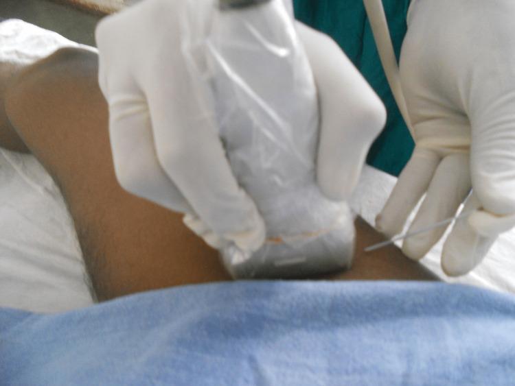
A clinical photograph illustrating the setup for an ultrasound-guided femoral nerve block. The image shows a practitioner's hands, donned in sterile white gloves, performing a procedure on the anterior thigh of a patient. One hand holds a high-frequency linear ultrasound transducer, which is encased in a sterile transparent plastic sleeve to maintain aseptic conditions. The probe is positioned transversely on the skin near the inguinal crease. The other hand is seen introducing a long, thin metallic needle—likely an 18G Quincke spinal needle—using an in-plane or out-of-plane approach relative to the transducer. The patient's limb is partially covered with a blue surgical drape, indicating a sterile field. This visual demonstrates the real-time needle guidance technique used in regional anesthesia to ensure accurate local anesthetic delivery near the femoral nerve while avoiding vascular structures.
mkdir -p /tmp/workspace/usg-regional-blocks && ls /tmp/workspace/
~/usg-regional-blocks/build_pptx.py
import json, subprocess, base64
from io import BytesIO
from pptx import Presentation
from pptx.util import Inches, Pt, Emu
from pptx.dml.color import RGBColor
from pptx.enum.text import PP_ALIGN, MSO_ANCHOR
from pptx.oxml.ns import qn
from lxml import etree

# ── colour palette ──────────────────────────────────────────────────────────
DARK_BLUE   = RGBColor(0x0D, 0x2E, 0x5A)   # deep navy
MID_BLUE    = RGBColor(0x1A, 0x5C, 0x9E)   # medium blue
ACCENT_TEAL = RGBColor(0x00, 0xB2, 0xB9)   # teal accent
LIGHT_BG    = RGBColor(0xF0, 0xF5, 0xFF)   # very light blue-white
WHITE       = RGBColor(0xFF, 0xFF, 0xFF)
DARK_TEXT   = RGBColor(0x1A, 0x1A, 0x2E)
GRAY_TEXT   = RGBColor(0x55, 0x55, 0x66)
GREEN       = RGBColor(0x07, 0x7E, 0x4A)
RED_DARK    = RGBColor(0xB0, 0x1C, 0x2E)
LIGHT_GREEN = RGBColor(0xE8, 0xF8, 0xF0)
LIGHT_RED   = RGBColor(0xFD, 0xEC, 0xEE)
GOLD        = RGBColor(0xF5, 0xC5, 0x18)

W = Inches(13.333)
H = Inches(7.5)

prs = Presentation()
prs.slide_width  = W
prs.slide_height = H
blank = prs.slide_layouts[6]  # fully blank

# ── helper: solid fill on a shape ───────────────────────────────────────────
def solid_fill(shape, rgb):
    fill = shape.fill
    fill.solid()
    fill.fore_color.rgb = rgb

# ── helper: add a rounded-rectangle shape ───────────────────────────────────
def add_rect(slide, l, t, w, h, rgb, radius_emu=0):
    shape = slide.shapes.add_shape(1, Inches(l), Inches(t), Inches(w), Inches(h))
    solid_fill(shape, rgb)
    shape.line.fill.background()  # no border
    if radius_emu:
        sp = shape._element
        prstGeom = sp.find(qn('p:spPr')).find(qn('a:prstGeom'))
        if prstGeom is None:
            spPr = sp.find(qn('p:spPr'))
            prstGeom = etree.SubElement(spPr, qn('a:prstGeom'))
        prstGeom.set('prst', 'roundRect')
        avLst = prstGeom.find(qn('a:avLst'))
        if avLst is None:
            avLst = etree.SubElement(prstGeom, qn('a:avLst'))
        for gd in avLst.findall(qn('a:gd')):
            avLst.remove(gd)
        gd = etree.SubElement(avLst, qn('a:gd'))
        gd.set('name', 'adj')
        gd.set('fmla', f'val {radius_emu}')
    return shape

# ── helper: add textbox ──────────────────────────────────────────────────────
def add_tb(slide, l, t, w, h, text, sz, bold=False, color=DARK_TEXT,
           align=PP_ALIGN.LEFT, wrap=True, italic=False, font="Calibri",
           va=None):
    tb = slide.shapes.add_textbox(Inches(l), Inches(t), Inches(w), Inches(h))
    tf = tb.text_frame
    tf.word_wrap = wrap
    tf.margin_left = 0; tf.margin_right = 0
    tf.margin_top = 0;  tf.margin_bottom = 0
    if va:
        tf.vertical_anchor = va
    p = tf.paragraphs[0]
    p.alignment = align
    run = p.add_run()
    run.text = text
    run.font.name = font
    run.font.size = Pt(sz)
    run.font.bold = bold
    run.font.italic = italic
    run.font.color.rgb = color
    return tf

# ── helper: multi-bullet textbox ─────────────────────────────────────────────
def add_bullets(slide, l, t, w, h, bullets, sz=17, color=DARK_TEXT,
                bullet_color=ACCENT_TEAL, font="Calibri", line_spacing_pt=None):
    tb = slide.shapes.add_textbox(Inches(l), Inches(t), Inches(w), Inches(h))
    tf = tb.text_frame
    tf.word_wrap = True
    tf.margin_left = Pt(4); tf.margin_right = 0
    tf.margin_top = 0; tf.margin_bottom = 0
    for i, btext in enumerate(bullets):
        p = tf.paragraphs[0] if i == 0 else tf.add_paragraph()
        p.alignment = PP_ALIGN.LEFT
        if line_spacing_pt:
            p.line_spacing = Pt(line_spacing_pt)
        # bullet character
        run0 = p.add_run()
        run0.text = "● "
        run0.font.name = font
        run0.font.size = Pt(sz)
        run0.font.color.rgb = bullet_color
        run0.font.bold = True
        # text
        run1 = p.add_run()
        run1.text = btext
        run1.font.name = font
        run1.font.size = Pt(sz)
        run1.font.color.rgb = color
    return tf

# ── helper: download image ───────────────────────────────────────────────────
def fetch_img(url):
    result = json.loads(subprocess.check_output(
        ["python", "/tmp/skills/shared/scripts/fetch_images.py", url]
    ))
    item = result[0]
    if item.get("base64"):
        parts = item["base64"].split(",", 1)
        b64 = parts[1] if len(parts) == 2 else parts[0]
        return BytesIO(base64.b64decode(b64))
    return None

# ── helper: add image to slide ───────────────────────────────────────────────
def add_img(slide, url, l, t, w, h=None):
    stream = fetch_img(url)
    if stream:
        if h is None:
            slide.shapes.add_picture(stream, Inches(l), Inches(t), width=Inches(w))
        else:
            slide.shapes.add_picture(stream, Inches(l), Inches(t), Inches(w), Inches(h))

# ── helper: header bar ───────────────────────────────────────────────────────
def header_bar(slide, title, subtitle=None):
    add_rect(slide, 0, 0, 13.333, 1.35, DARK_BLUE)
    add_rect(slide, 0, 1.35, 13.333, 0.07, ACCENT_TEAL)  # accent line
    add_tb(slide, 0.4, 0.18, 12.5, 0.85, title,
           sz=32, bold=True, color=WHITE, align=PP_ALIGN.LEFT)
    if subtitle:
        add_tb(slide, 0.4, 0.88, 12.5, 0.45, subtitle,
               sz=16, bold=False, color=ACCENT_TEAL, align=PP_ALIGN.LEFT)

# ── helper: footer ───────────────────────────────────────────────────────────
def footer(slide, text="USG-Guided Regional Blocks"):
    add_rect(slide, 0, 7.2, 13.333, 0.3, DARK_BLUE)
    add_tb(slide, 0.4, 7.21, 12.5, 0.28, text,
           sz=11, color=RGBColor(0xAA, 0xCC, 0xFF), align=PP_ALIGN.LEFT)

# ═══════════════════════════════════════════════════════════════════════════
#  SLIDE 1 – Title Slide
# ═══════════════════════════════════════════════════════════════════════════
s = prs.slides.add_slide(blank)
# full gradient-like background
add_rect(s, 0, 0, 13.333, 7.5, DARK_BLUE)
add_rect(s, 0, 0, 13.333, 7.5, RGBColor(0x0A, 0x28, 0x50))  # slight overlay

# teal accent bar (vertical left)
add_rect(s, 0, 0, 0.25, 7.5, ACCENT_TEAL)

# decorative circle (large, translucent-like)
add_rect(s, 8.5, 0.5, 4.5, 6.0, RGBColor(0x13, 0x40, 0x78))

# Title
add_tb(s, 0.7, 1.5, 8.0, 1.4,
       "Ultrasound-Guided",
       sz=44, bold=True, color=WHITE)
add_tb(s, 0.7, 2.85, 8.0, 1.2,
       "Regional Nerve Blocks",
       sz=44, bold=True, color=ACCENT_TEAL)

add_tb(s, 0.7, 4.15, 8.0, 0.7,
       "Introduction · Advantages · Disadvantages",
       sz=20, bold=False, color=RGBColor(0xCC, 0xDD, 0xFF), italic=True)

add_tb(s, 0.7, 5.2, 8.0, 0.55,
       "Department of Anaesthesiology",
       sz=16, bold=False, color=RGBColor(0xAA, 0xBB, 0xDD))

add_tb(s, 0.7, 5.75, 8.0, 0.45,
       "2026",
       sz=15, bold=False, color=RGBColor(0x88, 0xAA, 0xCC))

# image on right panel
add_img(s,
        "https://cdn.orris.care/cdss_images/pmc_clinical_VQA_ee17de4366a26dde78616bfd7fbfd039c0b53053f7361138be778c6bb5c7d8f7.jpg",
        8.8, 0.9, 4.1, 5.7)

# ═══════════════════════════════════════════════════════════════════════════
#  SLIDE 2 – Overview / Contents
# ═══════════════════════════════════════════════════════════════════════════
s = prs.slides.add_slide(blank)
add_rect(s, 0, 0, 13.333, 7.5, LIGHT_BG)
header_bar(s, "Contents", "What this presentation covers")
footer(s)

topics = [
    "01   Historical Background & Evolution",
    "02   What is Ultrasound-Guided Regional Anesthesia?",
    "03   Ultrasound Physics & Equipment",
    "04   Needle Techniques: In-Plane & Out-of-Plane",
    "05   Common Block Types",
    "06   Advantages – Safety & Efficacy",
    "07   Advantages – Patient & System Benefits",
    "08   Disadvantages – Technical Challenges",
    "09   Disadvantages – Clinical Limitations",
    "10   Comparison: USG vs. Nerve Stimulator",
    "11   Applications in Special Populations",
    "12   Summary & Key Takeaways",
]

col1 = topics[:6]
col2 = topics[6:]

for i, item in enumerate(col1):
    add_rect(s, 0.4, 1.6 + i*0.88, 5.8, 0.75, WHITE, radius_emu=40000)
    add_tb(s, 0.6, 1.68 + i*0.88, 5.5, 0.58, item,
           sz=14, color=DARK_BLUE, bold=False)

for i, item in enumerate(col2):
    add_rect(s, 6.9, 1.6 + i*0.88, 6.0, 0.75, WHITE, radius_emu=40000)
    add_tb(s, 7.1, 1.68 + i*0.88, 5.7, 0.58, item,
           sz=14, color=DARK_BLUE, bold=False)

# ═══════════════════════════════════════════════════════════════════════════
#  SLIDE 3 – Historical Background
# ═══════════════════════════════════════════════════════════════════════════
s = prs.slides.add_slide(blank)
add_rect(s, 0, 0, 13.333, 7.5, LIGHT_BG)
header_bar(s, "Historical Background & Evolution")
footer(s)

milestones = [
    ("1950s–1980s", "Landmark / paresthesia techniques: purely anatomical landmark guidance with risk of nerve injury."),
    ("1980s–1990s", "Nerve stimulators introduced: electrical stimulation to identify motor responses, reducing but not eliminating blind needle placement."),
    ("1994",        "La Grange et al. first report ultrasound used to assist supraclavicular brachial plexus block."),
    ("Late 1990s",  "Miniaturized high-frequency probes (10–15 MHz) made peripheral nerve imaging practical in everyday clinical settings."),
    ("2000s",       "Exponential adoption: ultrasound replaces nerve stimulators as primary guidance method in many centres worldwide."),
    ("2010s–now",   "ASRA & ESRA endorse USG guidance; interfascial plane blocks (PECS, TAP, QL, ESP) become possible only because of ultrasound."),
]

for i, (yr, txt) in enumerate(milestones):
    y = 1.55 + i*0.9
    add_rect(s, 0.4, y, 2.1, 0.72, MID_BLUE, radius_emu=30000)
    add_tb(s, 0.42, y+0.08, 2.05, 0.55, yr,
           sz=13, bold=True, color=WHITE, align=PP_ALIGN.CENTER)
    add_tb(s, 2.65, y+0.04, 10.0, 0.65, txt,
           sz=13.5, color=DARK_TEXT, wrap=True)

# ═══════════════════════════════════════════════════════════════════════════
#  SLIDE 4 – What is USG-Guided Regional Anesthesia?
# ═══════════════════════════════════════════════════════════════════════════
s = prs.slides.add_slide(blank)
add_rect(s, 0, 0, 13.333, 7.5, LIGHT_BG)
header_bar(s, "Ultrasound-Guided Regional Anesthesia",
           "Definition & Core Concept")
footer(s)

add_rect(s, 0.4, 1.55, 8.1, 1.25, WHITE, radius_emu=40000)
add_tb(s, 0.6, 1.62, 7.8, 1.1,
       "USG-guided regional anesthesia uses real-time high-frequency ultrasound imaging "
       "to directly visualize target nerves, surrounding anatomic structures, and the advancing "
       "needle, enabling precise local anesthetic deposition under direct vision.",
       sz=15, color=DARK_TEXT, wrap=True)

bullets_def = [
    "Replaces purely anatomical landmark or nerve stimulator approaches",
    "Allows real-time visualization of nerves, fascia, vessels and the needle",
    "Local anesthetic 'spread' around the nerve can be confirmed in real time",
    "Applicable to single-injection and continuous catheter techniques",
    "Used in upper limb, lower limb, truncal, and neuraxial procedures",
]
add_bullets(s, 0.4, 2.95, 7.9, 3.9, bullets_def, sz=15.5)

# image on right
add_img(s,
        "https://cdn.orris.care/cdss_images/pmc_clinical_VQA_27b5fa0beb45ba207b83dd4b4284bf3990b00636bba7f76cc3fab91ba12e45bc.jpg",
        9.05, 1.55, 3.9, 5.3)

# ═══════════════════════════════════════════════════════════════════════════
#  SLIDE 5 – Ultrasound Physics & Equipment
# ═══════════════════════════════════════════════════════════════════════════
s = prs.slides.add_slide(blank)
add_rect(s, 0, 0, 13.333, 7.5, LIGHT_BG)
header_bar(s, "Ultrasound Physics & Equipment")
footer(s)

cards = [
    ("Transducer Frequency",
     "High-frequency linear probes (10–15 MHz) for superficial nerves; "
     "low-frequency curved probes (2–5 MHz) for deep structures (e.g., sciatic, lumbar plexus)."),
    ("Nerve Appearance",
     "Nerves appear as hyperechoic (bright) round or oval honeycomb-like structures "
     "(fascicular pattern) in short-axis view."),
    ("Key Sonographic Terms",
     "Hyperechoic = bright (bone, needle, fascia). "
     "Hypoechoic = dark (nerves, vessels in some views). "
     "Anechoic = fluid (arteries, veins)."),
    ("Echogenic Needles",
     "Textured/echogenic needles improve needle tip visualisation, especially with steep "
     "insertion angles or deeper targets."),
    ("Scanning Planes",
     "Short-axis (transverse): nerve seen as circle/oval — most common. "
     "Long-axis (longitudinal): nerve seen as striped tube — confirms needle along nerve."),
    ("Machine Settings",
     "Gain, depth, and focus are adjusted to optimise visualisation. "
     "Colour Doppler avoids inadvertent vascular injection."),
]

for i, (title, body) in enumerate(cards):
    col = i % 3
    row = i // 3
    x = 0.35 + col * 4.28
    y = 1.6  + row * 2.65
    add_rect(s, x, y, 4.0, 2.45, WHITE, radius_emu=45000)
    add_rect(s, x, y, 4.0, 0.42, MID_BLUE, radius_emu=45000)
    add_tb(s, x+0.15, y+0.05, 3.7, 0.35, title,
           sz=13.5, bold=True, color=WHITE)
    add_tb(s, x+0.12, y+0.5, 3.75, 1.85, body,
           sz=12.5, color=DARK_TEXT, wrap=True)

# ═══════════════════════════════════════════════════════════════════════════
#  SLIDE 6 – In-Plane & Out-of-Plane Techniques
# ═══════════════════════════════════════════════════════════════════════════
s = prs.slides.add_slide(blank)
add_rect(s, 0, 0, 13.333, 7.5, LIGHT_BG)
header_bar(s, "Needle Guidance Techniques",
           "In-Plane vs. Out-of-Plane Approaches")
footer(s)

# Left panel – In-Plane
add_rect(s, 0.35, 1.55, 5.95, 5.35, WHITE, radius_emu=40000)
add_rect(s, 0.35, 1.55, 5.95, 0.5, MID_BLUE, radius_emu=40000)
add_tb(s, 0.55, 1.6, 5.6, 0.4, "IN-PLANE TECHNIQUE",
       sz=16, bold=True, color=WHITE, align=PP_ALIGN.CENTER)

ip_bullets = [
    "Needle inserted from side of probe, parallel to ultrasound beam",
    "Entire needle shaft AND tip visualised throughout procedure",
    "Preferred technique — provides complete needle tracking",
    "Easier to confirm tip position before injection",
    "Longer skin-to-nerve path but greater safety margin",
    "Used for most brachial plexus, femoral, sciatic blocks",
]
add_bullets(s, 0.5, 2.2, 5.65, 4.5, ip_bullets, sz=14.5, line_spacing_pt=18)

# Right panel – Out-of-Plane
add_rect(s, 7.05, 1.55, 5.95, 5.35, WHITE, radius_emu=40000)
add_rect(s, 7.05, 1.55, 5.95, 0.5, RGBColor(0x5A, 0x2E, 0x7A), radius_emu=40000)
add_tb(s, 7.25, 1.6, 5.6, 0.4, "OUT-OF-PLANE TECHNIQUE",
       sz=16, bold=True, color=WHITE, align=PP_ALIGN.CENTER)

oop_bullets = [
    "Needle crosses ultrasound beam — only tip seen as bright dot",
    "Shorter path to nerve; useful for narrow access areas",
    "Challenging — difficult to confirm tip vs. shaft",
    "Requires experienced operator to avoid tissue spread errors",
    "Risk of passing tip beyond target unseen",
    "Used for intercostal, ophthalmic, and some truncal blocks",
]
add_bullets(s, 7.2, 2.2, 5.65, 4.5, oop_bullets, sz=14.5,
            bullet_color=RGBColor(0xAA, 0x55, 0xFF), line_spacing_pt=18)

# divider
add_rect(s, 6.5, 1.55, 0.12, 5.35, ACCENT_TEAL)

# image
add_img(s,
        "https://cdn.orris.care/cdss_images/pmc_clinical_VQA_fa1600b0ce8a8c62eb6ff9d2549f1a1273615309d6fa54afd0539d4c1576411c.jpg",
        6.55, 3.4, 0.3, 2.0)  # tiny divider image not needed

# ═══════════════════════════════════════════════════════════════════════════
#  SLIDE 7 – Common Block Types
# ═══════════════════════════════════════════════════════════════════════════
s = prs.slides.add_slide(blank)
add_rect(s, 0, 0, 13.333, 7.5, LIGHT_BG)
header_bar(s, "Common USG-Guided Block Types",
           "Upper Limb · Lower Limb · Truncal · Neuraxial")
footer(s)

regions = [
    ("UPPER LIMB", MID_BLUE, [
        "Interscalene brachial plexus",
        "Supraclavicular brachial plexus",
        "Infraclavicular brachial plexus",
        "Axillary brachial plexus",
        "Forearm / terminal nerve blocks",
    ]),
    ("LOWER LIMB", RGBColor(0x1A, 0x7A, 0x5E), [
        "Femoral nerve block",
        "Adductor canal block (saphenous)",
        "Sciatic nerve block",
        "Popliteal sciatic block",
        "Ankle block",
    ]),
    ("TRUNCAL", RGBColor(0x7A, 0x3E, 0x00), [
        "Transversus Abdominis Plane (TAP)",
        "PECS I & II (pectoral blocks)",
        "Erector Spinae Plane (ESP)",
        "Quadratus Lumborum (QL) block",
        "Rectus sheath block",
    ]),
    ("NEURAXIAL\nADJUNCTS", RGBColor(0x55, 0x22, 0x88), [
        "Ultrasound for epidural depth",
        "Paravertebral block",
        "Caudal block (esp. paediatric)",
        "Cervical plexus block",
        "Fascial plane blocks",
    ]),
]

for col, (title, color, items) in enumerate(regions):
    x = 0.3 + col * 3.22
    add_rect(s, x, 1.55, 3.05, 5.55, WHITE, radius_emu=40000)
    add_rect(s, x, 1.55, 3.05, 0.52, color, radius_emu=40000)
    add_tb(s, x+0.1, 1.59, 2.85, 0.44, title,
           sz=13, bold=True, color=WHITE, align=PP_ALIGN.CENTER)
    for j, item in enumerate(items):
        add_tb(s, x+0.18, 2.2+j*0.95, 2.75, 0.85, f"› {item}",
               sz=13, color=DARK_TEXT, wrap=True)

# ═══════════════════════════════════════════════════════════════════════════
#  SLIDE 8 – Advantages: Safety & Efficacy
# ═══════════════════════════════════════════════════════════════════════════
s = prs.slides.add_slide(blank)
add_rect(s, 0, 0, 13.333, 7.5, LIGHT_BG)
header_bar(s, "Advantages: Safety & Efficacy",
           "Why ultrasound guidance has transformed regional anesthesia")
footer(s)

adv_safety = [
    ("Real-Time Visualisation", "Nerves, vessels, fascia, and adjacent structures are directly seen, eliminating 'blind' needle advancement."),
    ("Reduced Vascular Puncture", "Colour Doppler identifies arteries and veins, dramatically cutting inadvertent intravascular injection risk."),
    ("Lower Local Anaesthetic Dose", "Cochrane review confirms USG reduces LA volume needed, cutting systemic toxicity risk (LAST)."),
    ("Fewer Needle Passes", "Reduced number of needle insertions shortens procedure time and decreases tissue trauma."),
    ("Detect Intraneural Injection", "Distension of nerve fascicles can be seen in real time, allowing immediate stop — reducing permanent nerve injury."),
    ("Confirms LA Spread", "Operator watches local anaesthetic spread circumferentially around the nerve, ensuring an effective block before the patient enters theatre."),
]

for i, (title, body) in enumerate(adv_safety):
    row = i % 3
    col = i // 3
    x = 0.35 + col * 6.45
    y = 1.6  + row * 1.78
    add_rect(s, x, y, 6.15, 1.6, WHITE, radius_emu=40000)
    add_rect(s, x, y, 0.18, 1.6, GREEN, radius_emu=15000)
    add_tb(s, x+0.3, y+0.1, 5.75, 0.45, title,
           sz=14.5, bold=True, color=GREEN)
    add_tb(s, x+0.3, y+0.52, 5.75, 1.0, body,
           sz=13, color=DARK_TEXT, wrap=True)

# ═══════════════════════════════════════════════════════════════════════════
#  SLIDE 9 – Advantages: Patient & System Benefits
# ═══════════════════════════════════════════════════════════════════════════
s = prs.slides.add_slide(blank)
add_rect(s, 0, 0, 13.333, 7.5, LIGHT_BG)
header_bar(s, "Advantages: Patient & System Benefits")
footer(s)

# left content
pat_adv = [
    "Superior analgesia: Longer duration and improved quality vs. nerve stimulator techniques (Miller's Anesthesia 10e).",
    "Faster onset: Precise deposition around nerve speeds uptake of local anaesthetic.",
    "Reduced opioid consumption: Better blocks mean less post-op morphine — fewer opioid side effects (PONV, sedation, respiratory depression).",
    "Applicability in obesity & anatomical variants: Direct visualisation overrides surface landmarks distorted by body habitus.",
    "Use in anticoagulated patients: Compressible sites can be assessed for haematoma risk with Doppler.",
    "ERAS integration: USG blocks are central to enhanced recovery protocols — earlier ambulation, shorter hospital stay.",
    "Paediatric safety: Large paediatric databases confirm high safety profile; ASRA/ESRA recommend USG for all paediatric regional techniques.",
    "Novel blocks enabled: Interfascial plane blocks (TAP, PECS, ESP, QL) were not possible without USG — opened entirely new analgesic options.",
]
add_bullets(s, 0.35, 1.55, 8.1, 5.5, pat_adv, sz=14.5, line_spacing_pt=20)

# right image
add_img(s,
        "https://cdn.orris.care/cdss_images/pmc_clinical_VQA_10dadaf20fbc0acd7a38eedbd70a15b63f39cdcac9b0e4c1d6de04a3bc7d1a5d.jpg",
        8.65, 1.55, 4.3, 5.5)

# ═══════════════════════════════════════════════════════════════════════════
#  SLIDE 10 – Disadvantages: Technical Challenges
# ═══════════════════════════════════════════════════════════════════════════
s = prs.slides.add_slide(blank)
add_rect(s, 0, 0, 13.333, 7.5, LIGHT_BG)
header_bar(s, "Disadvantages: Technical Challenges",
           "Barriers and limitations of ultrasound guidance")
footer(s)

disadv = [
    ("Steep Learning Curve",
     "Requires mastery of sonoanatomy, probe manipulation, and simultaneous needle handling. "
     "Dedicated simulation training is mandatory."),
    ("Hand-Eye Coordination",
     "Operator must coordinate probe position, needle direction, and image interpretation in real time — "
     "different skill set from traditional techniques."),
    ("Needle Tip Visualisation",
     "The needle tip (not just shaft) may be lost in deeper structures or at steep angles — "
     "a cause of inadvertent vascular or neural puncture even with USG."),
    ("Image Quality Limitations",
     "Deep nerves, obese patients, oedematous tissue, and scarred areas degrade image quality. "
     "Gas, bone, and calcified tissue cause acoustic shadowing."),
    ("Equipment Cost & Maintenance",
     "High-frequency portable ultrasound machines are expensive; probe covers, gel, and servicing add "
     "ongoing costs. Availability in resource-limited settings remains challenging."),
    ("Sterility Requirements",
     "Sterile probe covers and gel are mandatory for invasive procedures, adding preparation time and cost."),
]

for i, (title, body) in enumerate(disadv):
    row = i % 3
    col = i // 3
    x = 0.35 + col * 6.45
    y = 1.6  + row * 1.78
    add_rect(s, x, y, 6.15, 1.6, WHITE, radius_emu=40000)
    add_rect(s, x, y, 0.18, 1.6, RED_DARK, radius_emu=15000)
    add_tb(s, x+0.3, y+0.1, 5.75, 0.45, title,
           sz=14.5, bold=True, color=RED_DARK)
    add_tb(s, x+0.3, y+0.52, 5.75, 1.0, body,
           sz=13, color=DARK_TEXT, wrap=True)

# ═══════════════════════════════════════════════════════════════════════════
#  SLIDE 11 – Disadvantages: Clinical Limitations
# ═══════════════════════════════════════════════════════════════════════════
s = prs.slides.add_slide(blank)
add_rect(s, 0, 0, 13.333, 7.5, LIGHT_BG)
header_bar(s, "Disadvantages: Clinical Limitations")
footer(s)

clin_lim = [
    "USG does NOT eliminate all complications — pneumothorax, vascular puncture, and nerve injury still occur.",
    "Block failure: Even with correct LA spread visualised, nerve stimulation or patient response may still be absent.",
    "Time-consuming setup: Probe preparation, sterile draping, and machine setup lengthen pre-procedure time vs. landmark technique.",
    "Operator fatigue: Prolonged procedures in difficult patients can cause significant physical and mental fatigue.",
    "Not universally available: Emergency and rural settings often lack USG-capable machines or trained operators.",
    "Interference from previous surgery or pathology: Scar tissue, metallic implants, and prior nerve damage alter sonoanatomy.",
    "Dynamic anatomy: Patient movement, breathing, and volume changes shift nerve/vessel positions during the procedure.",
    "Over-reliance risk: Sole reliance on ultrasound without knowledge of traditional anatomy is dangerous — knowledge of landmark technique remains essential.",
]

# two columns
for i, txt in enumerate(clin_lim[:4]):
    add_rect(s, 0.35, 1.6+i*1.35, 5.9, 1.2, WHITE, radius_emu=40000)
    add_rect(s, 0.35, 1.6+i*1.35, 0.18, 1.2, RED_DARK, radius_emu=15000)
    add_tb(s, 0.62, 1.66+i*1.35, 5.5, 1.0, txt,
           sz=13.5, color=DARK_TEXT, wrap=True)

for i, txt in enumerate(clin_lim[4:]):
    add_rect(s, 7.0, 1.6+i*1.35, 5.9, 1.2, WHITE, radius_emu=40000)
    add_rect(s, 7.0, 1.6+i*1.35, 0.18, 1.2, RGBColor(0xDD, 0x55, 0x00), radius_emu=15000)
    add_tb(s, 7.28, 1.66+i*1.35, 5.5, 1.0, txt,
           sz=13.5, color=DARK_TEXT, wrap=True)

# divider
add_rect(s, 6.55, 1.55, 0.12, 5.35, ACCENT_TEAL)

# ═══════════════════════════════════════════════════════════════════════════
#  SLIDE 12 – USG vs Nerve Stimulator Comparison
# ═══════════════════════════════════════════════════════════════════════════
s = prs.slides.add_slide(blank)
add_rect(s, 0, 0, 13.333, 7.5, LIGHT_BG)
header_bar(s, "USG vs. Nerve Stimulator: Comparison",
           "Evidence-based head-to-head analysis")
footer(s)

headers = ["Parameter", "Ultrasound Guidance", "Nerve Stimulator"]
hcolors = [DARK_BLUE, GREEN, MID_BLUE]
rows_data = [
    ["Success Rate",     "Higher (90–98%)",              "Moderate (80–90%)"],
    ["Onset Time",       "Faster",                       "Moderate"],
    ["LA Dose Required", "Lower",                        "Higher"],
    ["Needle Passes",    "Fewer",                        "More"],
    ["Vascular Puncture","Greatly reduced (Doppler)",    "Blind — not excluded"],
    ["Intraneural Inj.", "Detectable in real time",      "Not directly detectable"],
    ["Learning Curve",   "Steep (anatomy + US skills)",  "Moderate"],
    ["Equipment Cost",   "Higher (machine + probes)",    "Lower (stimulator only)"],
    ["In Uncooperative  Pts", "Feasible (visual)",       "Difficult (no motor response)"],
    ["Novel Blocks",     "Interfascial plane blocks possible", "Not feasible"],
]

col_widths = [3.1, 4.55, 4.55]
col_starts = [0.35, 3.55, 8.2]
row_h = 0.51

# Header row
for c, (hdr, hcol, wid, xs) in enumerate(zip(headers, hcolors, col_widths, col_starts)):
    add_rect(s, xs, 1.55, wid, 0.55, hcol, radius_emu=25000)
    add_tb(s, xs+0.1, 1.6, wid-0.15, 0.45, hdr,
           sz=14.5, bold=True, color=WHITE, align=PP_ALIGN.CENTER)

for r, row in enumerate(rows_data):
    bg = LIGHT_BG if r % 2 == 0 else WHITE
    for c, (cell, xs, wid) in enumerate(zip(row, col_starts, col_widths)):
        add_rect(s, xs, 2.18+r*row_h, wid, row_h-0.03, bg)
        clr = DARK_BLUE if c == 0 else (GREEN if c == 1 else MID_BLUE)
        add_tb(s, xs+0.1, 2.2+r*row_h, wid-0.15, row_h-0.08, cell,
               sz=12, color=clr, bold=(c == 0))

# ═══════════════════════════════════════════════════════════════════════════
#  SLIDE 13 – Special Populations & Applications
# ═══════════════════════════════════════════════════════════════════════════
s = prs.slides.add_slide(blank)
add_rect(s, 0, 0, 13.333, 7.5, LIGHT_BG)
header_bar(s, "Applications in Special Populations")
footer(s)

pop_data = [
    ("Paediatrics", MID_BLUE,
     ["Smaller nerves require high-frequency probes",
      "ASRA/ESRA: USG recommended for all paediatric blocks",
      "Performed under general anaesthesia — USG ensures safety",
      "Reduced LA doses prevent toxicity in low body weight"]),
    ("Obstetrics", RGBColor(0x8E, 0x22, 0x7A),
     ["Epidural depth estimation pre-procedure",
      "TAP blocks for post-caesarean analgesia",
      "Avoids systemic opioids — safer for neonate",
      "Useful in morbidly obese parturients"]),
    ("Geriatrics", RGBColor(0x5A, 0x40, 0x00),
     ["Calcified/fibrotic anatomy makes landmark technique unreliable",
      "USG navigates altered sonoanatomy",
      "Lower LA doses safe in reduced hepatic clearance",
      "Reduces opioid-related confusion and falls"]),
    ("Anticoagulated", RGBColor(0x7A, 0x00, 0x1A),
     ["Compressibility test with Doppler confirms patent vessels",
      "Avoids neuraxial techniques when coagulopathic",
      "Peripheral USG blocks preferred over central neuraxial",
      "Real-time haematoma detection"]),
]

for col, (title, color, pts) in enumerate(pop_data):
    x = 0.3 + col * 3.22
    add_rect(s, x, 1.55, 3.0, 5.55, WHITE, radius_emu=40000)
    add_rect(s, x, 1.55, 3.0, 0.5, color, radius_emu=40000)
    add_tb(s, x+0.08, 1.59, 2.84, 0.42, title,
           sz=14, bold=True, color=WHITE, align=PP_ALIGN.CENTER)
    for j, pt in enumerate(pts):
        add_tb(s, x+0.18, 2.2+j*1.18, 2.7, 1.1, f"› {pt}",
               sz=12.5, color=DARK_TEXT, wrap=True)

# ═══════════════════════════════════════════════════════════════════════════
#  SLIDE 14 – Summary & Key Takeaways
# ═══════════════════════════════════════════════════════════════════════════
s = prs.slides.add_slide(blank)
add_rect(s, 0, 0, 13.333, 7.5, DARK_BLUE)
add_rect(s, 0, 0, 0.25, 7.5, ACCENT_TEAL)

add_tb(s, 0.55, 0.25, 12.0, 0.85, "Summary & Key Takeaways",
       sz=34, bold=True, color=WHITE)
add_rect(s, 0.55, 1.1, 12.5, 0.06, ACCENT_TEAL)

key_pts = [
    ("✔", GREEN, "USG guidance provides real-time visualisation of nerves, needles and LA spread — the most significant advance in regional anaesthesia in decades."),
    ("✔", GREEN, "Advantages: higher success rates, reduced LA dose, fewer vascular punctures, detectable intraneural injection, enabled novel interfascial blocks."),
    ("✔", GREEN, "Patient benefits: better analgesia, less opioids, faster recovery, applicable in obese/paediatric/geriatric patients."),
    ("✘", RGBColor(0xFF, 0x66, 0x66), "Disadvantages: steep learning curve, equipment cost, image quality limitations, and does not eliminate complications."),
    ("✘", RGBColor(0xFF, 0x66, 0x66), "Operator must combine USG skills WITH a thorough understanding of traditional anatomy — USG is a tool, not a substitute for knowledge."),
    ("★", GOLD, "ASRA & ESRA endorse USG as the preferred guidance modality; it is now standard of care in most academic and regional anaesthesia centres."),
]

for i, (sym, clr, txt) in enumerate(key_pts):
    y = 1.35 + i * 0.97
    add_rect(s, 0.5, y, 0.55, 0.75, RGBColor(0x13, 0x40, 0x78), radius_emu=30000)
    add_tb(s, 0.52, y+0.1, 0.5, 0.55, sym,
           sz=20, bold=True, color=clr, align=PP_ALIGN.CENTER)
    add_tb(s, 1.2, y+0.05, 11.6, 0.75, txt,
           sz=14.5, color=WHITE, wrap=True)

# ═══════════════════════════════════════════════════════════════════════════
#  SLIDE 15 – Thank You / References
# ═══════════════════════════════════════════════════════════════════════════
s = prs.slides.add_slide(blank)
add_rect(s, 0, 0, 13.333, 7.5, LIGHT_BG)
add_rect(s, 0, 0, 13.333, 1.5, DARK_BLUE)
add_rect(s, 0, 1.5, 13.333, 0.07, ACCENT_TEAL)

add_tb(s, 0.5, 0.3, 12.0, 1.1, "Thank You",
       sz=48, bold=True, color=WHITE, align=PP_ALIGN.CENTER)

add_tb(s, 1.0, 1.75, 11.5, 0.5,
       "References & Further Reading",
       sz=18, bold=True, color=DARK_BLUE)

refs = [
    "1. Miller RD et al. Miller's Anesthesia, 10th ed. Elsevier, 2023. Chapter 42 (Regional Anaesthesia) & Chapter 53 (Paediatric Regional Anaesthesia).",
    "2. Barash PG et al. Barash, Cullen & Stoelting's Clinical Anesthesia, 9th ed. Wolters Kluwer, 2023. Chapter 36.",
    "3. Roberts JR, Custalow CB. Roberts and Hedges' Clinical Procedures in Emergency Medicine, 7th ed. Elsevier, 2019. Chapter 31.",
    "4. Schwartz SI et al. Schwartz's Principles of Surgery, 11th ed. McGraw-Hill, 2019. Chapter on Regional Anaesthesia.",
    "5. Cochrane Review: Confidence JK et al. Ultrasound guidance for peripheral nerve blockade. Cochrane Database Syst Rev. (confirmed reduced LA volume with USG).",
    "6. ASRA & ESRA Joint Practice Advisory on Ultrasound-Guided Regional Anesthesia. Reg Anesth Pain Med. 2019.",
]

for i, ref in enumerate(refs):
    add_tb(s, 1.0, 2.35+i*0.77, 11.5, 0.7, ref,
           sz=12.5, color=DARK_TEXT, wrap=True)

# ── save ────────────────────────────────────────────────────────────────────
out = "/tmp/workspace/usg-regional-blocks/USG_Guided_Regional_Blocks.pptx"
buf = BytesIO()
prs.save(buf)
with open(out, "wb") as f:
    f.write(buf.getvalue())
print(f"Saved: {out}")
cd /tmp/workspace/usg-regional-blocks && python build_pptx.py 2>&1
**/fetch_images.py in /tmp/skills
python /tmp/skills/shared/scripts/fetch_images.py "https://cdn.orris.care/cdss_images/pmc_clinical_VQA_ee17de4366a26dde78616bfd7fbfd039c0b53053f7361138be778c6bb5c7d8f7.jpg" 2>&1 | head -5
cd /tmp/workspace/usg-regional-blocks && python build_pptx.py 2>&1
ls -lh /tmp/workspace/usg-regional-blocks/USG_Guided_Regional_Blocks.pptx && python -c " from pptx import Presentation prs = Presentation('/tmp/workspace/usg-regional-blocks/USG_Guided_Regional_Blocks.pptx') print(f'Total slides: {len(prs.slides)}') for i, slide in enumerate(prs.slides, 1): title = '' for shape in slide.shapes: if shape.has_text_frame and shape.text_frame.text.strip(): title = shape.text_frame.text.strip()[:60] break print(f' Slide {i:2d}: {title}') "
USG Guided Regional Blocks
Presentation · PPTX
| # | Slide | Content |
|---|---|---|
| 1 | Title Slide | Full title with clinical image of interscalene block |
| 2 | Contents | 12-topic overview in two columns |
| 3 | Historical Background | Timeline from 1950s landmark technique to 2020s interfascial blocks |
| 4 | What is USG Regional Anesthesia? | Definition, core concept, 5 key points + clinical image |
| 5 | Ultrasound Physics & Equipment | 6-card grid: frequency, nerve appearance, echogenicity terms, echogenic needles, scanning planes, machine settings |
| 6 | In-Plane vs. Out-of-Plane Technique | Two-column comparison with bullet points for each needle approach |
| 7 | Common Block Types | 4-column layout: Upper limb, Lower limb, Truncal, Neuraxial adjuncts |
| 8 | Advantages - Safety & Efficacy | 6 cards (green accent): real-time visualisation, vascular safety, reduced LA dose, fewer passes, intraneural detection, LA spread confirmation |
| 9 | Advantages - Patient & System Benefits | 8 bullets + femoral block clinical image: ERAS, paediatric safety, novel interfascial blocks |
| 10 | Disadvantages - Technical Challenges | 6 cards (red accent): learning curve, hand-eye coordination, needle tip loss, image quality, equipment cost, sterility |
| 11 | Disadvantages - Clinical Limitations | Two-column layout of 8 real-world limitations |
| 12 | USG vs. Nerve Stimulator Comparison | Colour-coded table comparing 10 parameters |
| 13 | Special Populations | 4-column layout: Paediatrics, Obstetrics, Geriatrics, Anticoagulated patients |
| 14 | Summary & Key Takeaways | Dark slide with 6 key points (green ticks, red crosses, gold star) |
| 15 | Thank You / References | 6 textbook and guideline references (Barash, Miller, Roberts & Hedges, Schwartz, Cochrane, ASRA/ESRA) |