
Gynecology 1.Endometriosis 2.Prolapse 3.PID 4.Ectopic pregnancy 5.Carcinoma Cervix Obstetrics 1.Mechanism of labour 2.Twins 3.Breech 4.Placenta 5.APH Detailed answers from OBG DC DUTTA textbook
Gynecology
- Endometriosis - pathogenesis, sites, symptoms, infertility, diagnosis, medical and surgical treatment.
- Genital prolapse - supports of uterus, degrees/types, clinical assessment, pessary and operative management.
- Pelvic inflammatory disease (PID) - etiology, clinical diagnosis, complications, antibiotic regimens, prevention.
- Ectopic pregnancy - risk factors, diagnosis including beta-hCG/TVS, expectant, medical and surgical management.
- Carcinoma cervix - screening, FIGO staging, clinical features, investigations, treatment by stage, prevention.
Obstetrics
- Mechanism of normal labour - cardinal movements, fetal head diameters, mechanism in occipitoanterior position.
- Twin pregnancy - diagnosis, chorionicity, antenatal surveillance, complications, delivery planning.
- Breech presentation - types, diagnosis, external cephalic version, selection for vaginal breech birth, assisted breech delivery.
- Placenta - formation, structure, circulation, functions, placental hormones, abnormalities.
- Antepartum hemorrhage (APH) - definition, causes, evaluation, placenta previa, placental abruption, resuscitation and delivery decisions.
Explain mechanism of labour with diagrams
diagram cardinal movements mechanism of normal labour occipitoanterior fetal head engagement internal rotation extension restitution external rotation
"labor, obstetric" AND mechanism
mechanism of normal labour cardinal movements occipitoanterior authoritative obstetrics
https://geekymedics.com/mechanism-of-labour
Educational medical diagram of the cardinal movements in normal vaginal labour in left occipitoanterior vertex presentation. Show eight clearly labelled sequential panels from maternal pelvic view and sagittal view: engagement, descent, flexion, internal rotation to occipitoanterior, extension and crowning beneath pubic symphysis, restitution, external rotation with delivery of shoulders, and expulsion. Use accurate maternal pelvis and fetal head orientation, clean textbook line-art.
Mechanism of normal labour
The fetal head adapts its smallest possible diameters to the most favourable diameters of the maternal pelvis.
Cardinal movements
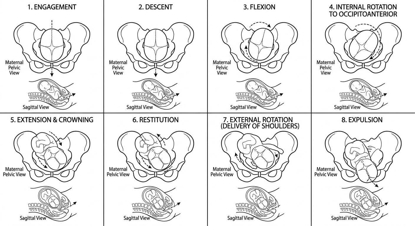
Prerequisites for normal mechanism
- Adequate uterine contractions
- Normal maternal bony pelvis and soft tissues
- Fetus of average size
- Vertex presentation with good flexion
- The fetal head and shoulders must adapt to the changing shape of the pelvic inlet, cavity, and outlet.
Important diameters
| Structure | Important diameter |
|---|---|
| Engaging transverse diameter of fetal head | Biparietal diameter: 9.5 cm |
| Diameter of a well-flexed vertex | Suboccipitobregmatic: 9.5 cm |
| AP diameter of outlet | Approximately 11.5 cm |
| Transverse diameter of outlet, intertuberous | Approximately 11 cm |
Mechanism in left occipitoanterior presentation
1. Engagement
- Clinically, it corresponds approximately to the presenting part at the level of the ischial spines, station 0.
- In primigravidae, engagement often occurs before labour or early in labour.
- In multiparae, it may occur later, sometimes during labour.
Diagram: engagement at the pelvic inlet
Maternal pelvic inlet, viewed from above

          Sacrum
            ↑
      ┌───────────┐
      │  •─────•  │  ← Biparietal diameter has entered
      │   Fetal   │
      │   head    │
      └───────────┘
            ↓
         Symphysis pubis
2. Descent
Forces responsible
- Uterine contractions
- Maternal bearing-down efforts in the second stage
- Straightening of the fetal body
- Extension of the fetal trunk after delivery of the shoulders
Diagram: stations during descent
         Pelvic inlet
              |
          -3  |  Head high
          -2  |
          -1  |
--------------|--------------  Ischial spines = 0 station
          +1  |
          +2  |
          +3  |  Head visible at introitus
              |
         Pelvic outlet
3. Flexion
Why is flexion important?
Deflexed head                Well-flexed head

Occipitofrontal              Suboccipitobregmatic
diameter: 11.5 cm            diameter: 9.5 cm
       ↓                            ↓
Less favourable              Most favourable
4. Internal rotation of the head
- In LOA, the occiput generally rotates 45 degrees anteriorly to reach direct OA.
- If the head began in left occipitotransverse, it rotates approximately 90 degrees anteriorly.
- This occurs because the pelvic floor directs the presenting part forward.
Diagram: internal rotation
View from below: maternal pelvic outlet

A. Before rotation             B. After rotation

        Sacrum                       Sacrum
          ↑                            ↑
   O  ←  Head  →                    Head
                                     O
   Left OT position             Direct OA position
          ↓                            ↓
      Symphysis pubis             Symphysis pubis
5. Extension
- Occiput
- Bregma
- Forehead
- Face
- Chin
Diagram: extension around the pubic symphysis
                 Symphysis pubis
                       ↓
                  [ Occiput ]
                       \
                        \  ← Head extends
                         \
                    forehead
                       nose
                      mouth
                       chin
                     Perineum
Crowning
6. Restitution
- The head had rotated independently during internal rotation.
- Restitution corrects this twist in the fetal neck.
- In LOA, the head generally turns toward the maternal left thigh after delivery.
Head delivered in OA        Restitution

        O                        ↖ O
        |                          |
  Symphysis pubis            Turns toward fetal back
7. External rotation
Shoulders at outlet

       Sacrum
         ↑
     Posterior shoulder

       Fetal trunk
         |
  Anterior shoulder
         ↓
   Symphysis pubis
8. Expulsion
- The anterior shoulder is delivered first from beneath the symphysis pubis.
- The posterior shoulder then sweeps over the perineum.
- The rest of the trunk and lower limbs are delivered by lateral flexion.
1. Anterior shoulder: beneath symphysis
2. Posterior shoulder: over perineum
3. Trunk and limbs: follow rapidly
Sequence in one diagram
Engagement
    ↓
Descent with flexion
    ↓
Internal rotation of occiput anteriorly
    ↓
Extension: head born
    ↓
Restitution: head untwists
    ↓
External rotation: shoulders rotate into AP diameter
    ↓
Anterior shoulder, posterior shoulder, trunk delivered
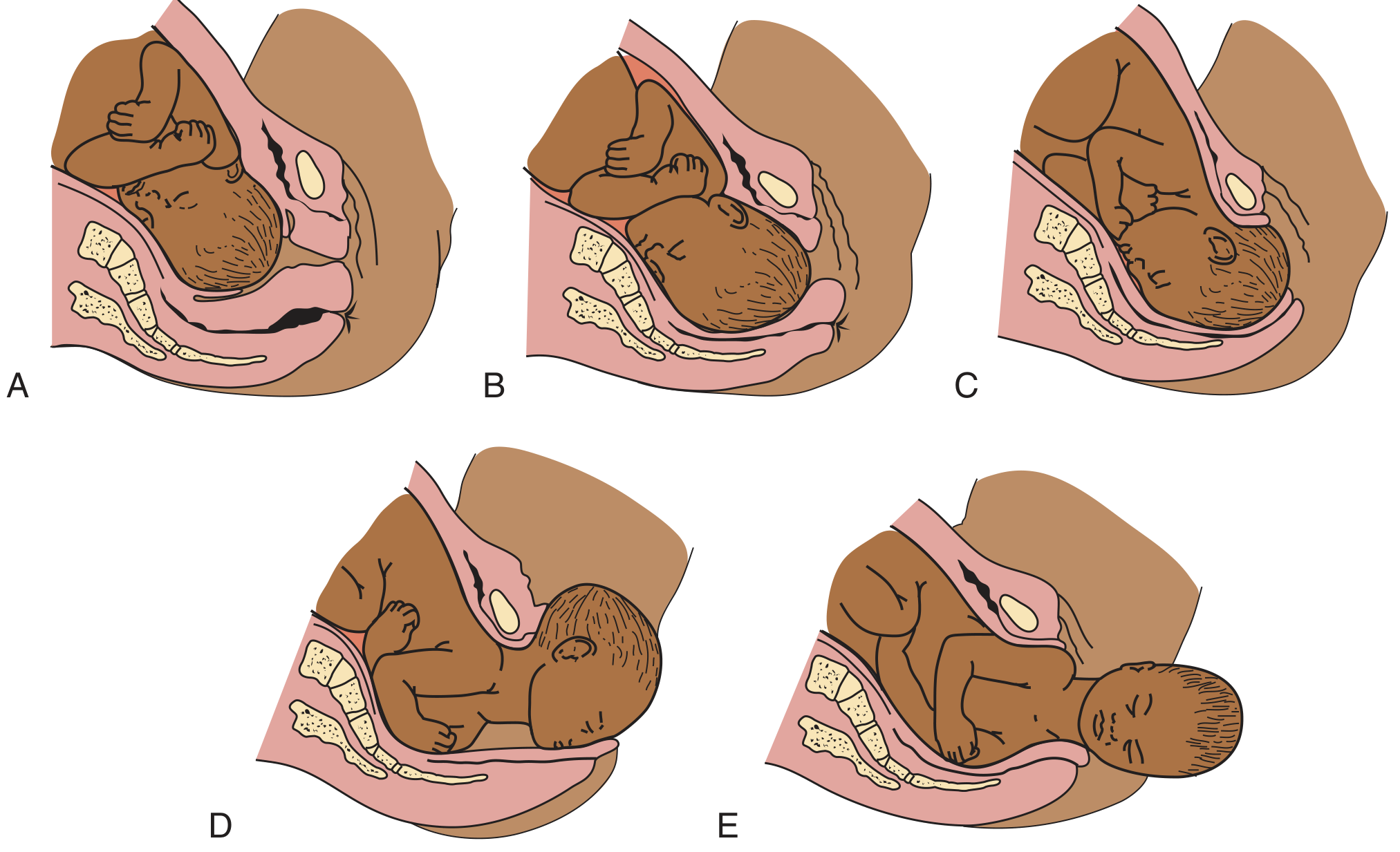
Short exam answer
"PROLAPSE" Give answer of this topic in very detailed from DC DUTTA textbook Of obg+ marrow obg notes edition 8
medical diagram pelvic organ prolapse cystocele rectocele uterine prolapse compartments POP-Q
"Pelvic Organ Prolapse"[MeSH Terms] AND treatment
ACOG pelvic organ prolapse evaluation management practice guidance
- Definition and terminology
- Pelvic supports of uterus and vagina
- Etiology and risk factors
- Pathogenesis and types of prolapse
- Cystocele, urethrocele, rectocele, enterocele, uterine prolapse, vault prolapse
- Clinical features and complications
- Clinical examination and grading, including POP-Q
- Investigations
- Conservative treatment and pessaries
- Surgical management, with uterus-preserving versus hysterectomy-based procedures
- Special situations: young women, women desiring fertility, elderly or medically unfit patients
- Recurrence, prevention, and viva points
Explain genital prolapse in full detail, section by section
labelled medical diagram female pelvic organ prolapse anterior apical posterior compartments cystocele uterine prolapse rectocele
NICE guideline pelvic organ prolapse assessment conservative surgical management pessary pelvic floor training
A labelled medical textbook diagram of female genital prolapse in a sagittal pelvic section. Compare normal anatomy with anterior compartment cystocele, apical uterine prolapse, posterior compartment rectocele, and enterocele. Clearly label bladder, uterus, cervix, vagina, rectum, pubic symphysis, sacrum and pelvic floor.
"Pelvic Organ Prolapse"[MeSH Terms] AND treatment
Genital Prolapse
Note: I do not have the text of the requested D.C. Dutta or Marrow OBG Notes edition available for verbatim reproduction. The following is an original, detailed examination note organized in the traditional D.C. Dutta style and cross-checked against standard urogynecology teaching. Pelvic organ prolapse definitions, compartmental approach, and POP-Q details are consistent with Berek & Novak's Gynecology, pp. 1592, 1610-1611.
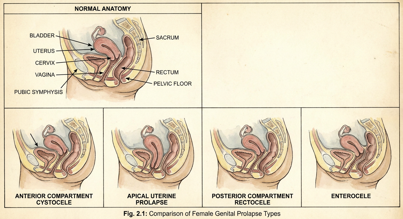
1. Definition
- Anterior vaginal wall
- Posterior vaginal wall
- Uterus and cervix
- Vaginal vault after hysterectomy
2. Normal supports of uterus and vagina
A. Primary supports
1. Pelvic diaphragm
- Puborectalis
- Pubococcygeus
- Iliococcygeus
Functions
- Supports the pelvic viscera from below.
- Maintains the levator hiatus.
- Resists rise in intra-abdominal pressure during coughing, straining, lifting, and defecation.
- Maintains the normal vaginal axis.
- Keeps the upper vagina directed posteriorly toward the sacrum rather than downward toward the vaginal opening.
2. Urogenital diaphragm and perineal membrane
- Perineal membrane
- Deep transverse perineal muscles
- Sphincter urethrae complex
- Associated fascia
3. Perineal body
- Bulbospongiosus
- Superficial transverse perineal muscles
- External anal sphincter
- Fibers of levator ani
B. Ligamentous and fascial supports
1. Uterosacral ligaments
- Extend from cervix and upper vagina backward to sacrum, usually around S2-S4.
- They support the cervix and upper vagina posteriorly.
- They are important for apical support.
2. Cardinal ligaments, also called transverse cervical or Mackenrodt ligaments
- Extend from cervix and upper vagina laterally toward the pelvic sidewall.
- Provide lateral support to cervix and upper vagina.
- They contain uterine vessels.
3. Pubocervical fascia
- Lies between bladder and anterior vaginal wall.
- Supports bladder base and proximal urethra.
- Defect or attenuation leads to anterior vaginal wall prolapse, especially cystocele.
4. Rectovaginal fascia
- Lies between rectum and posterior vaginal wall.
- Supports posterior vaginal wall.
- Damage leads to rectocele.
5. Endopelvic fascia
C. Traditional classification of uterine supports
True supports
- Pelvic diaphragm
- Urogenital diaphragm
- Perineal body
- Uterosacral ligaments
- Cardinal ligaments
- Pubocervical and rectovaginal fascia
False supports
- Round ligaments
- Broad ligaments
- Uterovesical fold of peritoneum
- Position of uterus, normally anteverted and anteflexed
3. Etiology and risk factors
A. Congenital and constitutional factors
- Congenital weakness of connective tissue
- Familial predisposition
- Connective-tissue disorders, for example Ehlers-Danlos syndrome
- Naturally wide pelvic outlet
- Poorly developed pelvic floor
- Spina bifida or neuropathic pelvic floor disorders
B. Obstetric trauma
- Multiple vaginal births
- Prolonged second stage of labour
- Obstructed labour
- Large baby
- Instrumental vaginal delivery
- Malposition, especially persistent occipitoposterior position
- Inadequately repaired perineal tear
- Third- or fourth-degree perineal tear
- Repeated childbirth at short intervals
- Difficult or traumatic delivery
Mechanism of obstetric injury
- Stretching or avulsion of levator ani muscles
- Pudendal nerve injury
- Stretching or tears in endopelvic fascia
- Damage to perineal body
- Injury to uterosacral-cardinal ligament support
C. Increased intra-abdominal pressure
- Chronic cough, such as COPD, chronic bronchitis, asthma, smoking-related cough
- Chronic constipation and habitual straining
- Heavy manual work or repeated heavy lifting
- Obesity
- Ascites
- Pelvic or abdominal mass
D. Menopause and aging
- Estrogen deficiency causes vaginal atrophy.
- Connective tissue becomes weaker.
- Muscle mass and pelvic-floor tone decline.
- Tissue healing capacity is reduced.
E. Previous pelvic surgery
- Hysterectomy without adequate vault suspension can lead to post-hysterectomy vaginal vault prolapse.
- Previous prolapse surgery may fail or recur.
- Radical pelvic surgery can damage nerve and fascial support.
4. Pathogenesis
Key pathological events
- Weakness or injury of levator ani widens the levator hiatus.
- Vaginal walls become less firmly attached to the pelvic sidewalls.
- Fascial supports stretch or tear.
- Uterosacral-cardinal ligament complex fails.
- Uterus or vaginal vault descends.
- The vaginal axis becomes more vertical.
- Repeated gravitational force and straining increase the prolapse.
Important concept
- Uterine prolapse may coexist with cystocele and rectocele.
- Repairing only the visible anterior wall defect without correcting apical support may lead to recurrence.
5. Types of genital prolapse
| Compartment | Main structure involved | Common clinical name |
|---|---|---|
| Anterior | Bladder, urethra | Cystocele, urethrocele |
| Apical or middle | Uterus, cervix, vaginal vault, small bowel | Uterine prolapse, vault prolapse, enterocele |
| Posterior | Rectum, small bowel | Rectocele, enterocele |
A. Anterior compartment prolapse
1. Cystocele
Features
- Anterior vaginal wall bulge
- Frequency of micturition
- Urgency
- Stress urinary incontinence
- Difficulty in emptying bladder
- Feeling of incomplete evacuation
- Recurrent UTI
- Need to reduce the bulge manually before voiding in advanced cases
Types of cystocele
- Central defect: weakness of central pubocervical fascia.
- Lateral defect or paravaginal defect: detachment of fascia from arcus tendineus fascia pelvis.
- Apical cystocele: associated with loss of upper vaginal or apical support.
2. Urethrocele
B. Apical or middle compartment prolapse
1. Uterine prolapse
- Cystocele
- Rectocele
- Enterocele
- Elongation of cervix
2. Vaginal vault prolapse
3. Enterocele
- With uterine prolapse
- After hysterectomy
- As a posterior vaginal wall bulge
- In association with vault prolapse
C. Posterior compartment prolapse
Rectocele
Symptoms
- Difficulty in defecation
- Sensation of incomplete evacuation
- Need for repeated straining
- Need to press the posterior vaginal wall or perineum to pass stool, called digital splinting
- Constipation
- Feeling of a posterior vaginal bulge
6. Degrees of uterine prolapse
First-degree uterine prolapse
- Cervix descends into vagina but does not reach the introitus.
- Usually mild.
- May be asymptomatic or associated with pelvic heaviness and backache.
Second-degree uterine prolapse
- Cervix descends to or through the vaginal introitus.
- The uterus is partly within the vagina.
- Usually associated with cystocele and rectocele.
Third-degree uterine prolapse, procidentia
- Entire uterus lies outside the vaginal introitus.
- Vaginal walls are everted.
- Often associated with cystocele, rectocele, enterocele, and decubitus ulcer.
Complete procidentia
7. Cervical elongation
A. Supravaginal elongation of cervix
- Cervix is elongated.
- Fundus may remain relatively high.
- Uterosacral and cardinal supports may be relatively preserved.
- Commonly associated with uterine prolapse.
- Measured by a long cervical length relative to uterine corpus.
B. Infravaginal elongation
- Elongation of ectocervix below vaginal attachment.
- May be congenital or acquired.
- Vaginal fornices remain at normal level.
- May mimic prolapse.
8. Clinical features
A. Local symptoms
- Feeling of something coming down per vaginam
- Vaginal bulge or mass
- Pelvic dragging sensation
- Pelvic heaviness
- Low backache
- Symptoms increase with standing, exertion, coughing, and lifting
- Symptoms improve on lying down
- Vaginal discharge due to congestion or ulceration
- Contact bleeding from decubitus ulcer
- Difficulty walking in severe procidentia
B. Urinary symptoms
- Frequency
- Urgency
- Nocturia
- Stress urinary incontinence
- Urge urinary incontinence
- Hesitancy
- Poor urinary stream
- Incomplete bladder emptying
- Retention
- Recurrent UTI
- Overflow incontinence in severe obstruction
Occult stress urinary incontinence
C. Bowel symptoms
- Constipation
- Straining
- Incomplete evacuation
- Obstructed defecation
- Digital splinting
- Fecal urgency or incontinence in some patients
D. Sexual symptoms
- Dyspareunia
- Reduced sexual satisfaction
- Vaginal dryness due to atrophy
- Fear or embarrassment due to visible bulge
9. Complications
Local complications
- Congestion and edema of prolapsed tissues
- Keratinization of exposed vaginal epithelium
- Excoriation
- Infection
- Decubitus ulcer
- Bleeding
- Offensive vaginal discharge
- Rarely, malignant change must be excluded in a chronic nonhealing ulcer
Urinary complications
- Recurrent urinary tract infection
- Acute urinary retention
- Chronic retention
- Hydroureter
- Hydronephrosis
- Renal impairment in neglected severe cases
Bowel complications
- Chronic constipation
- Obstructed defecation
- Fecal impaction
10. Clinical examination
A. General examination
- Age and parity
- Nutritional state
- BMI
- Chronic cough
- Signs of COPD
- Abdominal mass or ascites
- Previous scars
- Mobility and fitness for surgery
B. Abdominal examination
- Mass
- Ascites
- Distended bladder
- Previous abdominal scars
- Hernia
C. Local examination
Inspection
- Degree of uterine descent
- Anterior vaginal wall prolapse
- Posterior vaginal wall prolapse
- Eversion of vaginal walls
- Decubitus ulcer
- Cervical elongation
- Vaginal atrophy
- Perineal body deficiency
- Widened genital hiatus
- Previous episiotomy scar
- Urinary leakage on coughing
Speculum examination
- Retract posterior vaginal wall to assess anterior prolapse.
- Retract anterior wall to assess posterior prolapse.
- Support apex when evaluating isolated wall defects.
- Inspect cervix and vagina for ulceration, discharge, growth, and atrophy.
Bimanual examination
- Uterine size
- Uterine mobility
- Cervical length
- Adnexal mass
- Associated pelvic pathology
Rectovaginal examination
- Rectocele
- Enterocele
- Integrity of rectovaginal septum
- Perineal body
- Anal sphincter tone
- Rectal pathology
11. POP-Q classification
- Measurements above hymen are negative.
- Measurements beyond hymen are positive.
POP-Q points
| Point | Site |
|---|---|
| Aa | Fixed point on anterior wall, 3 cm proximal to urethral meatus |
| Ba | Most distal point of upper anterior vaginal wall |
| C | Cervix or vaginal cuff scar |
| D | Posterior fornix, used only when uterus is present |
| Ap | Fixed point on posterior wall, 3 cm proximal to hymen |
| Bp | Most distal point of upper posterior vaginal wall |
| gh | Genital hiatus |
| pb | Perineal body |
| tvl | Total vaginal length |
POP-Q stages
| Stage | Definition |
|---|---|
| Stage 0 | No prolapse |
| Stage I | Leading edge remains more than 1 cm above hymen |
| Stage II | Leading edge lies between 1 cm above and 1 cm below hymen |
| Stage III | Leading edge is more than 1 cm below hymen but not complete eversion |
| Stage IV | Complete or near-complete vaginal eversion |
12. Investigations
Basic investigations
- Urinalysis
- Urine culture if urinary symptoms
- Complete blood count if ulcer bleeding or preoperative assessment
- Blood glucose, renal function, and preoperative tests as required
- Cervical cytology according to screening eligibility
- Biopsy of suspicious, bleeding, indurated, or nonhealing ulcer
Bladder assessment
- Postvoid residual urine
- Cough stress test with prolapse reduced
- Uroflowmetry in voiding difficulty
- Urodynamic study in selected patients, especially if:
- Mixed urinary symptoms
- Voiding dysfunction
- Prior incontinence surgery
- Suspected occult SUI
- Unclear diagnosis
Imaging
- Pelvic ultrasound for uterine or adnexal pathology
- Renal ultrasound in severe procidentia or suspected urinary obstruction
- MRI or defecography in complex posterior-compartment and defecatory disorders
13. Management principles
- Symptoms and their effect on quality of life
- Compartment(s) involved
- Severity of prolapse
- Age
- Desire for future fertility and preservation of uterus
- Sexual activity
- General health and anesthetic fitness
- Associated urinary and bowel symptoms
- Previous surgery
- Surgeon expertise and patient preference
A. Conservative treatment
1. Lifestyle modification
- Weight reduction where appropriate
- Treat chronic cough
- Smoking cessation
- Treat constipation
- Avoid repeated heavy lifting
- Optimize diabetes and general health
2. Pelvic floor muscle training
- Improve pelvic-floor strength
- Reduce symptoms
- Improve support
- Help coexisting urinary incontinence
3. Local vaginal estrogen
- Before pessary insertion
- Before surgery
- For vaginal dryness and irritation
- To improve comfort and epithelial health
B. Pessary treatment
Indications
- Patient does not want surgery
- Pregnancy or desire to defer surgery
- Elderly or medically unfit patient
- Temporary management before surgery
- Patient awaiting optimization of comorbidities
- Recurrent prolapse
- Woman wishing to retain fertility
Common types
| Pessary | Usual use |
|---|---|
| Ring pessary with or without support | Mild to moderate cystocele or uterine prolapse |
| Hodge pessary | Cystocele, uterine displacement |
| Shelf pessary | More advanced uterovaginal prolapse |
| Gellhorn pessary | Advanced prolapse, especially if ring fails |
| Donut pessary | Severe prolapse or wide introitus |
| Cube pessary | Advanced prolapse, but needs frequent removal |
Principles of fitting
- Empty bladder.
- Select the largest comfortable pessary that remains retained.
- It should support prolapse without causing pain or pressure.
- Patient should be able to void normally.
- Reassess for discomfort, erosion, discharge, bleeding, and retention.
Complications of pessary
- Vaginal discharge
- Foul odor
- Vaginal ulceration or erosion
- Bleeding
- Infection
- Embedded or incarcerated pessary if neglected
- Rarely, fistula formation
14. Surgical management
General surgical principles
- Correct all significant compartment defects.
- Ensure apical support.
- Treat associated urinary or bowel dysfunction appropriately.
- Decide whether uterus should be preserved or removed.
- Choose reconstructive versus obliterative surgery.
- Counsel regarding recurrence, sexual function, operative risks, and mesh-related issues.
A. Vaginal hysterectomy with pelvic floor repair
Components
- Vaginal hysterectomy
- Apical suspension, often uterosacral ligament suspension or McCall culdoplasty
- Anterior colporrhaphy if cystocele is significant
- Posterior colporrhaphy and perineorrhaphy if rectocele/perineal deficiency is present
- Enterocele repair if present
Important point
B. Anterior colporrhaphy
Indication
Principle
- Vaginal epithelium is separated from the underlying pubocervical fascia.
- Defective fascia is plicated in the midline.
- Excess vaginal epithelium is trimmed if needed.
- Vaginal wall is closed.
Limitation
C. Paravaginal repair
Indication
Principle
D. Posterior colporrhaphy
Indication
Principle
- Dissect posterior vaginal epithelium off rectovaginal fascia.
- Plicate the fascial defect in the midline.
- Repair deficient perineal body by perineorrhaphy where required.
Caution
E. McCall culdoplasty
Purpose
- Obliterates or reduces the cul-de-sac.
- Supports vaginal vault.
- Helps prevent enterocele and vault prolapse.
F. Sacrospinous ligament fixation
Indications
- Vaginal vault prolapse
- Uterine prolapse, often after vaginal hysterectomy
- Recurrent apical prolapse
Principle
Advantages
- Vaginal approach
- Avoids abdominal incision
- Useful in older or comorbid women
Possible complications
- Buttock pain
- Hemorrhage
- Nerve injury
- Vaginal axis deviation
- Dyspareunia
- Recurrence, especially anterior compartment recurrence
G. Uterosacral ligament suspension
Advantages
- Restores relatively central vaginal axis
- Can be done at vaginal hysterectomy or for vault prolapse
Important risk
H. Sacrocolpopexy
Indications
- Post-hysterectomy vaginal vault prolapse
- Advanced apical prolapse
- Recurrent prolapse
- Sexually active woman desiring maximal vaginal length and support
Principle
Advantages
- Strong and durable apical support
- Maintains vaginal length
- Lower recurrence for apical prolapse in many settings
Risks
- Bowel, bladder, ureteric, or vascular injury
- Ileus or bowel obstruction
- Mesh exposure or erosion
- Sacral osteomyelitis, rare
- Thromboembolism
- Pain and dyspareunia
I. Uterus-preserving procedures, hysteropexy
- Sacrospinous hysteropexy
- Uterosacral hysteropexy
- Sacrohysteropexy
- Laparoscopic hysteropexy
- Manchester-Fothergill operation in selected cases
J. Manchester-Fothergill operation
Traditional indication
- Uterine prolapse
- Cervical elongation, especially supravaginal elongation
- Desire to preserve uterus and menstrual function
- No desire for immediate fertility, although fertility may remain possible
Main steps
- Amputation of elongated cervix.
- Plication and shortening of cardinal ligaments in front of cervical stump.
- Anterior colporrhaphy if required.
- Posterior colpoperineorrhaphy if required.
Drawbacks and concerns
- Cervical stenosis
- Dysmenorrhea
- Infertility or subfertility
- Cervical incompetence or obstetric complications in future pregnancy
- Persistent or recurrent prolapse
K. Le Fort partial colpocleisis
Indications
- Elderly woman
- Severe prolapse
- No desire for future vaginal intercourse
- High anesthetic or surgical risk where a shorter obliterative procedure is preferred
Principle
Advantages
- Short operation
- Effective for advanced prolapse
- Less invasive than reconstructive procedures
Contraindication
15. Mesh in prolapse surgery
- Transvaginal mesh for prolapse has been associated with exposure, erosion, pain, dyspareunia, infection, bleeding, and need for repeat surgery.
- In several jurisdictions, transvaginal mesh for POP has been restricted or withdrawn from routine use.
- Abdominal or laparoscopic mesh sacrocolpopexy is different from transvaginal mesh placement and remains an option in selected patients.
- Mesh should be used only after informed consent and by an appropriately trained surgeon.
16. Management according to patient profile
A. Young woman desiring future childbearing
- Prefer conservative management when possible.
- Pelvic floor physiotherapy.
- Pessary where appropriate.
- Correct chronic cough, constipation, obesity, and heavy lifting.
- Defer definitive surgery until family is complete if symptoms permit.
- If surgery is unavoidable, consider uterus-preserving options after specialist counselling.
B. Completed family, symptomatic uterovaginal prolapse
- Vaginal hysterectomy with apical suspension
- Anterior and/or posterior repair as required
- Laparoscopic or abdominal sacrocolpopexy in selected cases
- Uterus-preserving hysteropexy if preferred and appropriate
C. Elderly woman who is not sexually active
- Pessary if suitable
- Le Fort colpocleisis for advanced symptomatic prolapse if no future vaginal intercourse is desired
- Vaginal reconstructive surgery if medically fit and desired
D. Medically unfit patient
- Pessary
- Vaginal estrogen if atrophic
- Treatment of cough and constipation
- Pelvic floor exercises
- Regular surveillance for ulceration and urinary complications
17. Prevention
- Good antenatal and intrapartum care
- Avoid unnecessary traumatic vaginal delivery
- Proper repair of episiotomy and perineal tears
- Timely diagnosis and repair of obstetric anal sphincter injury
- Pelvic floor muscle exercises after childbirth
- Weight optimization
- Treatment of chronic cough and constipation
- Avoid repeated heavy lifting
- Smoking cessation
- Apical support at the time of hysterectomy when indicated