
Pituitary gland histology 5m anatomy
Reading File
This is a visual/anatomy/histology query. Searching the medical library with parallel queries now.
Searching sources...
Finding Sources
Finding Sources
Reading File
Reading File
Finding Sources
Reading File
Reading File
Finding Sources
Excellent - Junqueira's Basic Histology has detailed content. Let me read the histology sections from Junqueira's and also fetch the diagrams.
Reading File
Reading File
Reading File
I now have comprehensive content from Junqueira's Basic Histology. Let me fetch the key histology images to include in the answer.
Loading Image
Loading Image
Loading Image
Loading Image
Reading File
Loading Image
I now have everything needed for a thorough 5-mark anatomy/histology answer. Here is the complete response:
Pituitary Gland - Histology & Anatomy (5-mark)
Gross Anatomy
The pituitary gland (hypophysis cerebri) is an ovoid body measuring approximately 8 mm AP x 12 mm transverse x 4 mm high, weighing ~100-500 mg. It sits in the sella turcica of the sphenoid bone, connected to the hypothalamus via the infundibular stalk.
It has two major divisions:
| Division | Parts |
|---|---|
| Adenohypophysis | Pars distalis (anterior lobe), pars intermedia, pars tuberalis |
| Neurohypophysis | Pars nervosa (posterior lobe), infundibular stem, median eminence |
The adenohypophysis constitutes two-thirds of the total volume. It is derived from Rathke's pouch (oral ectoderm); the neurohypophysis is a downgrowth of the diencephalon.
Low-Power Histology: Overall Organization
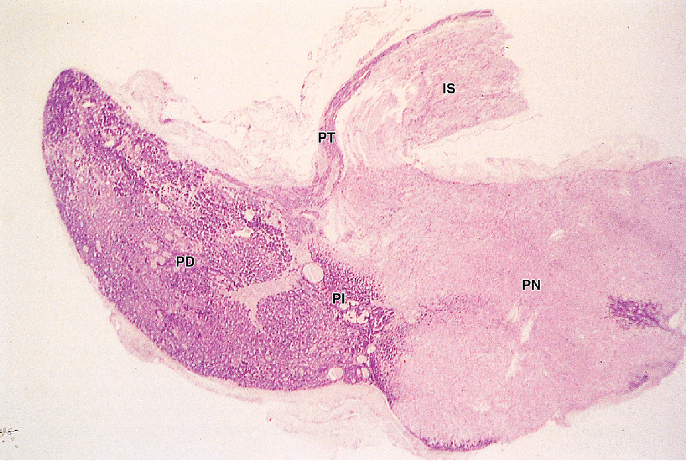
Low-power H&E of entire pituitary gland. The adenohypophysis (PD, PI, PT) is deeply basophilic/stained, contrasting with the pale, neural pars nervosa (PN). IS = infundibular stalk. (x30 H&E) - Junqueira's Basic Histology, 17e
Adenohypophysis (Anterior Pituitary)
Pars Distalis (accounts for 75% of the adenohypophysis)
Composed of cords and nests of epithelial cells supported by a delicate reticular connective tissue skeleton, interspersed with wide-lumen fenestrated sinusoidal capillaries (the secondary portal plexus). A thin fibrous capsule encloses it.
Classic staining (H&E / Azan) divides cells into two broad categories:
1. Chromophils - secretory cells with hormone-containing cytoplasmic granules:
- Acidophils (α-cells) - ~40% of cells; stain orange/pink with eosin or Orange G
- Basophils (β-cells) - ~10% of cells; stain blue/purple; PAS-positive (glycoproteins)
2. Chromophobes (γ-cells) - ~50% of cells; sparse cytoplasm, poorly staining; represent either stem cells (precursors) or degranulated secretory cells of any type.
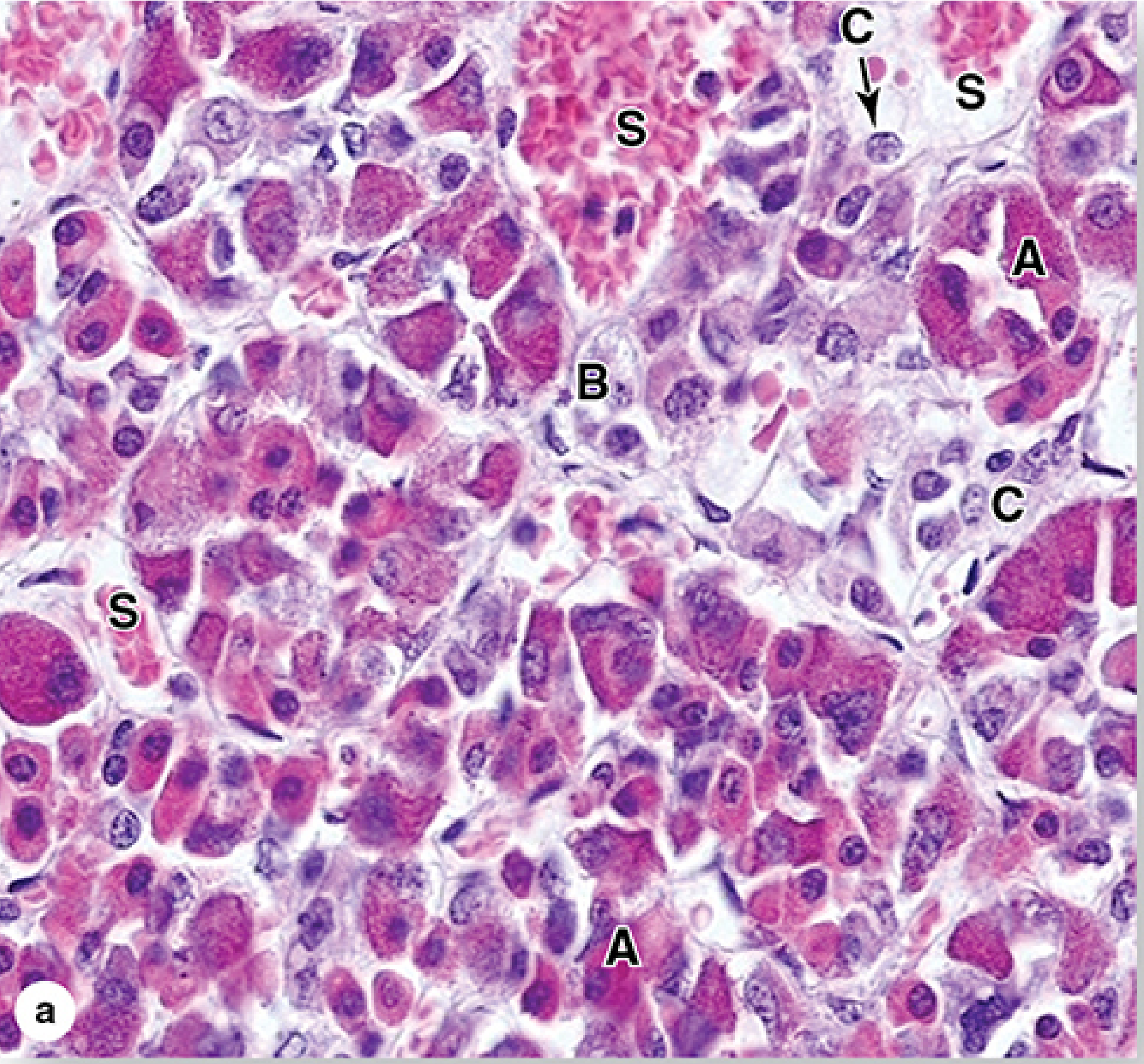
High-power H&E of pars distalis showing acidophils (A), basophils (B), chromophobes (C), and sinusoids (S). - Junqueira's Basic Histology, 17e
Immunohistochemical Cell Classification (modern)
| Cell Type | Staining | Hormone(s) | Location in Pars Distalis |
|---|---|---|---|
| Somatotrophs | Acidophilic | GH (somatotropin) | Lateral parts |
| Mammotrophs (Lactotrophs) | Acidophilic | Prolactin (PRL) | Dominant in pregnancy |
| Mammosomatotrophs | Acidophilic | GH + PRL | Variable |
| Corticotrophs | Basophilic | ACTH, β-LPH, β-endorphin (from POMC) | Central/anterior |
| Thyrotrophs | Basophilic | TSH | Anterior, central |
| Gonadotrophs | Basophilic | FSH + LH | Pars tuberalis-rich |
| Folliculostellate cells | Supporting cells | Growth factors, cytokines | Throughout |
Electron microscopy: cells are identified by membrane-enclosed secretory granules (60-900 nm diameter), the size varies by hormone type; secretion is by exocytosis.
Pars Tuberalis
A funnel-shaped extension surrounding the infundibulum. Rich in blood vessels and gonadotrophs (FSH/LH). Contains melatonin receptors in some species.
Pars Intermedia
A narrow zone between pars distalis and pars nervosa. Contains corticotrophs, chromophobes, and characteristically colloid-filled cysts (remnants of Rathke's pouch lumen). Corticotrophs here cleave POMC differently to produce primarily MSH, γ-LPH, and β-endorphin.
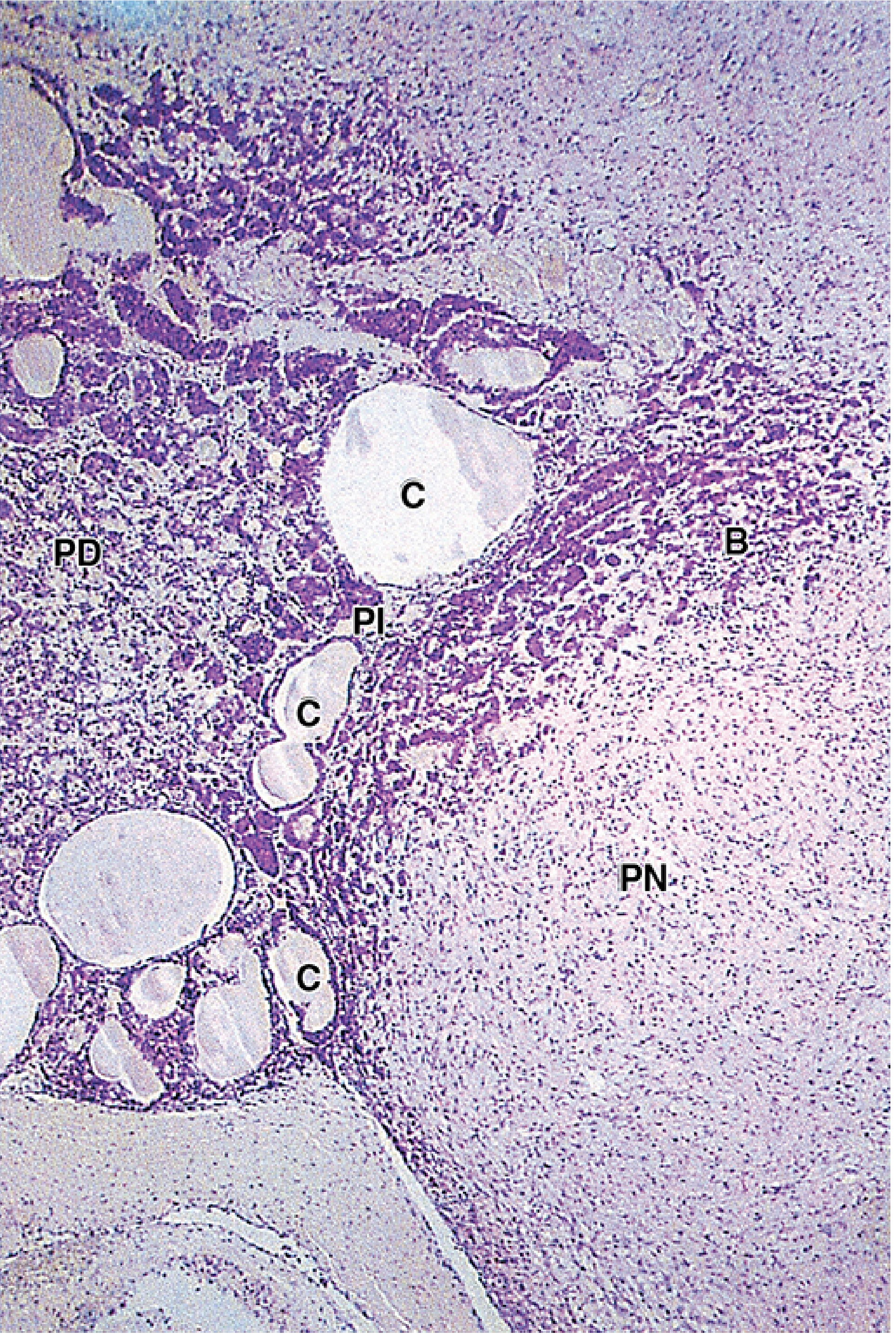
Pars intermedia (PI) between pars distalis (PD) and pars nervosa (PN), with colloid-filled cysts (C) and invading basophils (B). (H&E) - Junqueira's Basic Histology, 17e
Neurohypophysis (Posterior Pituitary - Pars Nervosa)
Unlike the adenohypophysis, the posterior pituitary does not synthesize hormones itself. It consists of:
- ~100,000 unmyelinated axons from hypothalamic supraoptic and paraventricular nuclei
- Pituicytes - modified glial cells (astrocyte-like), the most abundant cell type
- Fenestrated capillaries for hormone uptake
Key histological feature: Neurosecretory (Herring) bodies - faintly eosinophilic swellings/dilatations along axons, visible by LM, containing membrane-bound granules of ADH or oxytocin bound to carrier proteins (neurophysin I and II).
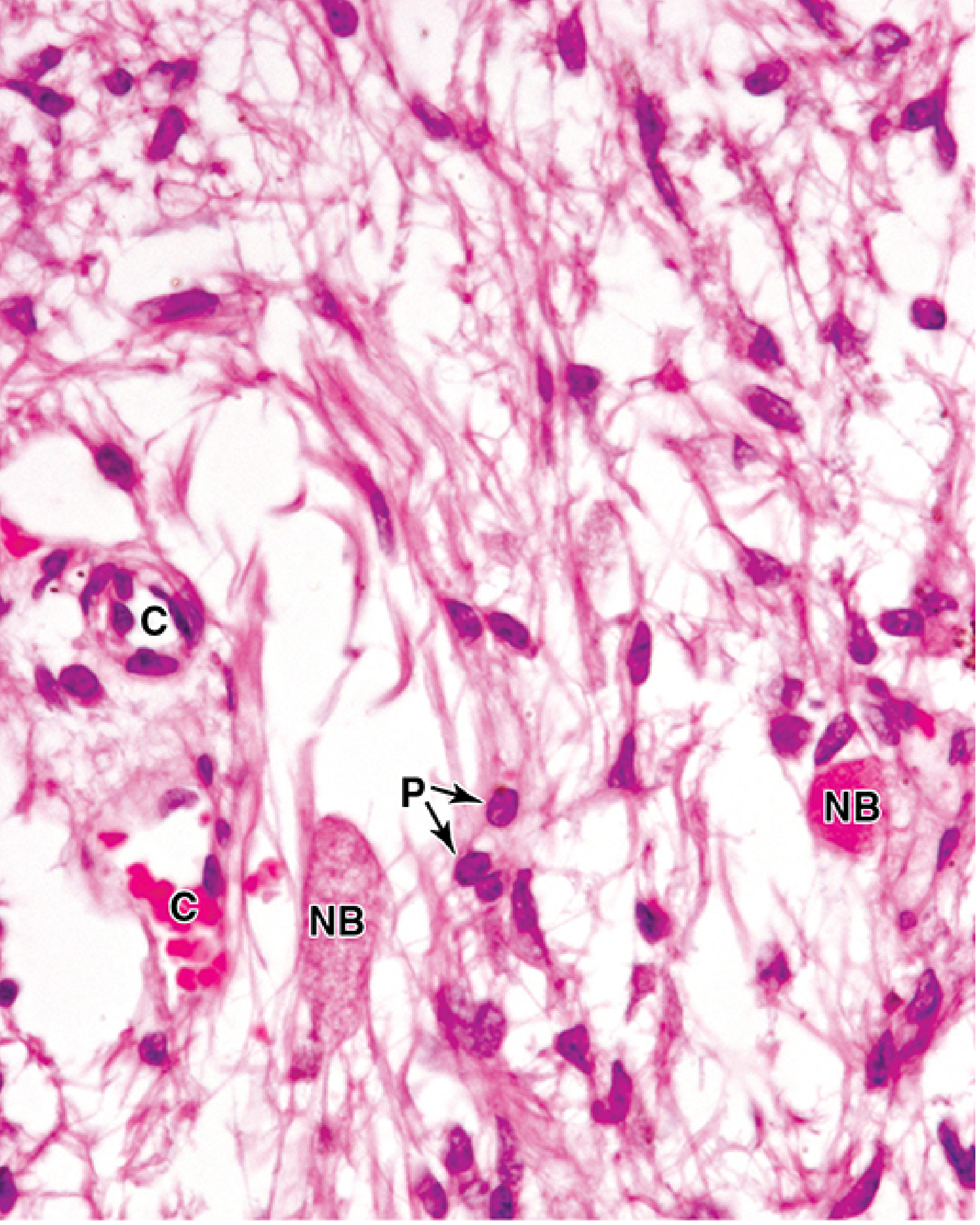
Pars nervosa showing pituicytes (P), Herring bodies / neurosecretory bodies (NB), and capillaries (C). (x400 H&E) - Junqueira's Basic Histology, 17e
| Hormone | Source Nucleus | Carrier Protein | Main Function |
|---|---|---|---|
| ADH (Vasopressin) | Supraoptic | Neurophysin II | Increases renal collecting duct water permeability |
| Oxytocin | Paraventricular | Neurophysin I | Uterine contraction; milk ejection |
Blood Supply
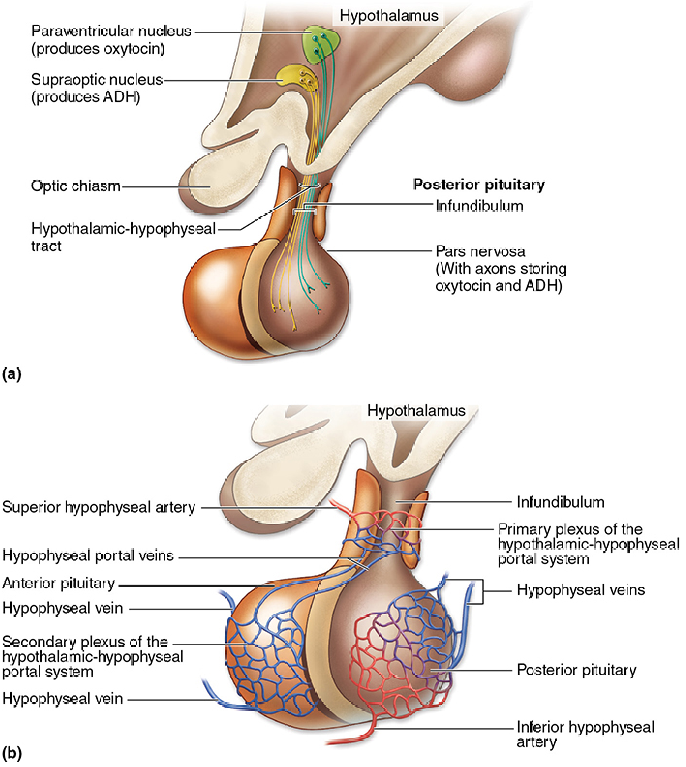
Hypothalamic-hypophyseal tract (a) and portal vascular system (b). - Junqueira's Basic Histology, 17e
- Superior hypophyseal arteries (branches of internal carotid) supply the median eminence and infundibular stalk, forming a primary capillary plexus
- Portal veins carry hypothalamic releasing/inhibiting hormones to the adenohypophysis via a secondary capillary plexus (the portal system)
- Inferior hypophyseal arteries supply the neurohypophysis directly
- Drained by hypophyseal veins
The adenohypophysis has no direct arterial supply - it depends entirely on the hypothalamic-hypophyseal portal system.
Quick Summary for 5 Marks
| Point | Key Fact |
|---|---|
| 1. Two parts | Adenohypophysis (Rathke's pouch) + neurohypophysis (diencephalon) |
| 2. Adenohypophysis cells | Acidophils (GH, PRL), basophils (ACTH, TSH, FSH, LH), chromophobes (~50%) |
| 3. Pars intermedia | Colloid cysts + MSH-producing corticotrophs |
| 4. Neurohypophysis | Pituicytes + Herring bodies; stores/releases ADH + oxytocin |
| 5. Blood supply | Portal system (no direct artery to adenohypophysis); inferior arteries to neurohypophysis |
Sources: Junqueira's Basic Histology 17e, p. 1002-1017 | Color Atlas of Human Anatomy Vol. 2 (Thieme), p. 566-568 | Scott-Brown's Otorhinolaryngology Head & Neck Surgery Vol. 1, p. 660-661
This is a shared conversation. Sign in to Orris to start your own chat.