
Frality score index from standard anaesthesia textbook . Long answer for MD anaesthesia exam
Reading File
Now let me search the library for frailty content in anaesthesia textbooks, running parallel searches:
Finding Sources
Finding Sources
Miller's Anesthesia has multiple frailty sections. Let me read them all in parallel:
Reading File
Reading File
Finding Sources
Excellent content. Now let me get more detail on the Clinical Frailty Scale, the modified Frailty Index, and anaesthetic implications:
Reading File
Finding Sources
Finding Sources
Finding Sources
Reading File
Finding Sources
Barash has a dedicated frailty section and a frailty screening tools table. Let me read that:
Reading File
Reading File
Searching PubMed
frailty index perioperative anaesthesia outcomes
I now have very rich content from Miller's Anesthesia (10e), Barash's Clinical Anesthesia (9e), and Current Surgical Therapy (14e). Let me also get the full Barash table and fetch the Edmonton Frail Scale image:
Reading File
Loading Image
I now have comprehensive content from three major anaesthesia textbooks. Let me compose the full long-answer response for the MD Anaesthesia exam.
FRAILTY SCORE INDEX IN ANAESTHESIA
Long Answer - MD Anaesthesia Examination
INTRODUCTION AND DEFINITION
Frailty is a complex, multidimensional syndrome of physiologic decline characterised by increased vulnerability to physiologic stressors, resulting in diminished capacity to maintain homeostasis after a perturbation. It represents a state of decreased physiologic reserve across multiple organ systems that renders an individual susceptible to adverse health outcomes following medical or surgical interventions.
The concept of frailty is distinct from - but overlapping with - disability and comorbidity (Fig. 34-2, Barash). A patient may be comorbid without being frail, disabled without being frail, or frail without having established disease. All three conditions, however, frequently coexist in elderly surgical patients.
Frailty phenotypes can present even in patients below 65 years of age, making chronologic age an imprecise surrogate for physiologic reserve.
Sources: Miller's Anesthesia 10e, Barash's Clinical Anesthesia 9e, Current Surgical Therapy 14e
PATHOPHYSIOLOGY AND MODELS
There are two dominant theoretical frameworks:
1. The Frailty Phenotype (Fried Model)
Described by Linda Fried and colleagues (2001) from a large prospective cohort study (Cardiovascular Health Study), this model defines frailty as a clinical syndrome based on five physical domains:
| Domain | Criterion |
|---|---|
| Unintentional weight loss | >10 lb (4.5 kg) in the past year |
| Self-reported exhaustion | Difficulty initiating movements / fatigue |
| Reduced grip strength | Below gender/BMI-adjusted threshold |
| Slow walking speed | Slowed gait speed |
| Low physical activity | Reduced energy expenditure per week |
Scoring:
- 0 criteria: Robust / Non-frail
- 1-2 criteria: Pre-frail
- ≥3 criteria: Frail
The Fried Phenotype is the most widely studied frailty instrument. It is strongly associated with postoperative complications including postoperative delirium, and predicts hospitalization, falls, disability, and death. It requires patient activity and special equipment and takes 5-20 minutes to administer.
2. The Deficit Accumulation Model (Rockwood Model)
Described by Rockwood and colleagues, this model conceptualises frailty as the accumulation of deficits across 92 items including signs, symptoms, functional impairments, and laboratory abnormalities. Each positive deficit is proportionally weighted to yield a Frailty Index (FI):
Frailty Index = Number of positive deficits / Total deficits assessed
Example: 10 positives out of 40 assessed = FI of 0.25
This model was used to develop the Clinical Frailty Scale (CFS), the most clinically feasible distillation of the deficit accumulation framework.
VALIDATED FRAILTY ASSESSMENT INSTRUMENTS
A. Clinical Frailty Scale (CFS)
Developed by Rockwood (Canadian Study of Health and Aging, 2005), the CFS is a nine-level observational scale that summarises information from a clinical encounter. It is not a questionnaire but a clinical judgement tool.
| CFS Score | Description |
|---|---|
| 1 | Very Fit - robust, active, energetic |
| 2 | Well - no active disease symptoms |
| 3 | Managing Well - medical problems well-controlled |
| 4 | Vulnerable - not dependent, but slowed; symptoms limit activity |
| 5 | Mildly Frail - limited dependence in instrumental ADLs |
| 6 | Moderately Frail - help needed with IADLs and some ADLs |
| 7 | Severely Frail - completely dependent on others for ADLs |
| 8 | Very Severely Frail - completely dependent and approaching end of life |
| 9 | Terminally Ill - life expectancy <6 months |
Frailty is defined by CFS score ≥ 4
Clinical advantages:
- Takes <2 minutes to administer
- No special equipment or extra space required
- Web-based training modules available for standardised application
- In direct comparison with the modified Fried Index, clinicians found it easier to use, more feasible in clinical settings, and quicker (<1 min vs 5 min)
- Predicts postoperative mortality and non-home discharge
- In systematic review and meta-analysis, the CFS had the strongest accuracy and feasibility among available scales
B. Fried Phenotype (Modified Frailty Phenotype)
As detailed above. Time to administer: 5-20 minutes. Requires patient activity and measurement of grip strength. Most strongly associated with postoperative delirium.
C. Edmonton Frail Scale (EFS)
An 11-item scale that encompasses:
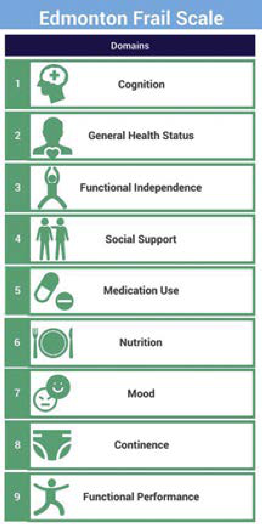
Edmonton Frail Scale - 9 domains (Current Surgical Therapy 14e)
The 11 items include:
- Clock drawing test (cognition)
- Timed Up-And-Go (TUG) test (functional performance)
- General health status (two questions)
- Functional independence (ADLs)
- Social support
- Medication use (polypharmacy; ≥5 medications)
- Nutrition (weight loss)
- Mood (depression screening)
- Continence
- Functional performance (TUG)
- Nine standardized questions
Scoring:
- 0-5: Not frail
- 6-7: Vulnerable
- 8-9: Mild frailty
- 10-11: Moderate frailty
- 12-17: Severe frailty
Advantages: Can be administered by non-medical personnel; validated against comprehensive geriatric assessment (gold standard); best predictor of postoperative complications. Takes <5 minutes. A software application is available. Requires space for TUG test.
D. FRAIL Scale
A simple 5-item questionnaire categorising patients as non-frail, pre-frail, and frail. The acronym stands for:
| Letter | Domain | Question |
|---|---|---|
| F | Fatigue | Are you fatigued? |
| R | Resistance | Can you climb one flight of stairs? |
| A | Ambulation | Can you walk one block? |
| I | Illnesses | Do you have >5 illnesses? |
| L | Loss of weight | Have you lost >5% body weight in the past year? |
Scoring:
- 0: Robust
- 1-2: Pre-frail
- 3-5: Frail
Advantages: Simple, quick (5-10 min), patient-reported, no equipment needed. Not as widely studied as CFS or Fried phenotype.
E. Modified Frailty Index (mFI / mFI-5)
Derived from the NSQIP (National Surgical Quality Improvement Program) database, the original mFI uses 11 variables from the Canadian Study of Health and Aging (CSHA) frailty index mapped against NSQIP variables:
| Variable | Mapped NSQIP Item |
|---|---|
| Functional status (dependent vs independent) | |
| Diabetes mellitus | |
| Chronic obstructive pulmonary disease | |
| Congestive heart failure | |
| Hypertension requiring medication | |
| History of TIA or CVA | |
| Impaired sensorium | |
| History of myocardial infarction | |
| Peripheral vascular disease / rest pain / gangrene | |
| Wound infection history | |
| History of angina |
The simplified mFI-5 uses 5 of these variables. Each positive variable scores 1 point.
In a study of 52,671 spine surgery patients (Flexman et al.), the mFI independently predicted:
- Major postoperative complications (OR 1.58)
- Prolonged length of stay (OR 1.89)
- Discharge to higher level of care (OR 2.29)
- 30-day mortality (OR 2.05 per 0.1 increase in frailty score)
F. Robinson's Surgical Frailty Definitions
Robinson and colleagues developed two efficient preoperative frailty assessment definitions:
Definition 1:
- Mini-Cog score ≤3
- Serum albumin ≤30 g/L
- One or more falls in the prior 6 months
- Haematocrit <35%
Definition 2:
- Timed Up-And-Go (TUG) ≥15 seconds
- Activity of daily living (ADL) dependence
- Charlson Comorbidity Index score ≥3
G. Risk Analysis Index (RAI)
A 5-10 minute patient-reported questionnaire that includes items typically part of standard nursing interviews. Useful for perioperative risk stratification.
COMPARISON TABLE OF FRAILTY TOOLS (Barash 9e, Table 34-1)
| Frailty Tool | Time | Type | Perioperative Significance |
|---|---|---|---|
| Clinical Frailty Scale (CFS) | <2 min | Observational | Mortality, non-home discharge |
| Edmonton Frailty Scale (EFS) | <5 min | Observational/patient-reported | Best predictor of postop complications |
| FRAIL Scale | 5-10 min | Patient-reported | Simple; not as widely studied |
| ACS NSQIP / mFI | 5-10 min | Chart review | Limited to 30-day outcomes |
| Risk Analysis Index | 5-10 min | Patient-reported | Includes standard nursing questions |
| Fried Phenotype | 5-20 min | Observational | Postop delirium; gold standard research |
| Frailty Index (Rockwood) | 10-13 min | Patient-reported | Postop complications; no equipment needed |
PERIOPERATIVE SIGNIFICANCE OF FRAILTY
Association with Adverse Outcomes
- Surgical complications: Frailty is independently associated with higher postoperative complication rates across general surgery, orthopaedic surgery, cardiac surgery, and spine surgery.
- Postoperative delirium: Frailty (especially assessed by Fried Phenotype) is the strongest predictor; incidence of delirium 5-50% in older surgical patients.
- Prolonged hospital stay: Frail patients have significantly longer length of stay.
- Non-home discharge: CFS ≥4 independently predicts discharge to skilled nursing/assisted-living facilities.
- Mortality: 7.7% one-year mortality in frail patients undergoing elective orthopaedic surgery. Degree of frailty independently and dose-dependently predicts readmission or death.
- Frailty augments other risk scores: Adds prognostic value beyond ASA score, RCRI, and Eagle's Cardiac Risk Index.
Current Practice Gaps
Despite evidence supporting frailty assessment, routine implementation is limited. A survey of 251 surgical oncologists showed only 6% currently perform geriatric assessments in older patients. Barriers identified include:
- Focus on cardiorespiratory evaluation
- Need for more training
- Absence of formal guidelines for evidence-based frailty assessment
- Time constraints in busy clinical settings
FRAILTY IN ANAESTHETIC PRACTICE
Preoperative Assessment
- Frailty screening should be incorporated into the preoperative assessment of all patients aged ≥65 years, and younger patients with clinical suspicion of reduced physiologic reserve.
- The CFS is recommended as the most feasible clinical approach: it takes <1 minute, requires no equipment, and has comparable accuracy to longer tools.
- Comprehensive Geriatric Assessment (CGA) - the gold standard - examines: burden of comorbidity, polypharmacy, physical function, psychological status, nutrition, delirium risk, and social support.
- CGA is more powerful than ASA score alone for predicting perioperative risk in older patients (mean age 78 years).
Anaesthetic Implications of Frailty
- Pharmacokinetics/Pharmacodynamics: Frail patients have reduced muscle mass (sarcopenia), altered body composition, decreased hepatic and renal blood flow - leading to unpredictable drug distribution and prolonged drug effect.
- Airway management: Reduced physiologic reserve limits apnoea tolerance; careful preoxygenation is essential.
- Haemodynamic instability: Reduced cardiovascular reserve increases sensitivity to anaesthetic agents and volume changes.
- Postoperative delirium prevention: Frail patients at high risk; prefer shorter-acting agents, minimise benzodiazepines, use multimodal analgesia.
- Hypothermia: Reduced muscle mass and subcutaneous fat increase risk.
- Pain management: Altered pharmacodynamics; opioids require dose reduction.
- Regional vs general anaesthesia: No definitive evidence that one technique is superior in frail patients, but regional techniques may reduce physiologic stress.
- Informed consent and goals of care: Frailty identified preoperatively allows for informed shared decision-making and realistic outcome counselling.
Prehabilitation
Frailty is a potentially modifiable risk factor. Preoperative rehabilitation (prehabilitation) includes:
- Nutritional assessment and optimisation (increase protein intake)
- Physical exercise and optimisation of functional status
- Lifestyle modification (smoking cessation, alcohol reduction)
- Psychological preparation and stress management
Evidence suggests at least 2-4 weeks of prehabilitation is necessary, and frail patients may have the greatest benefit. The PREPARE Trial (NCT04221295) is an RCT examining prehabilitation in frail patients across 11 Canadian hospitals.
OBJECTIVE/RADIOGRAPHIC MARKERS OF FRAILTY
An emerging area seeks to correlate objective radiographic markers with clinical frailty assessments:
- Psoas muscle area on CT (marker of sarcopenia)
- Preoperative ultrasound measurement of quadriceps depth - a simple, non-invasive bedside tool for identifying high-risk elderly patients
- These opportunistic imaging-based assessments could help operationalise frailty screening using imaging already obtained for surgical planning.
SUMMARY
Frailty assessment is an integral part of perioperative risk stratification in elderly patients. The two primary conceptual models are the Fried Phenotype (5 physical domains, frail if ≥3 present) and the Rockwood Deficit Accumulation Model (Frailty Index and Clinical Frailty Scale). For routine clinical anaesthesia practice, the Clinical Frailty Scale offers the best balance of accuracy, feasibility, and speed (<2 minutes). The Edmonton Frail Scale is preferred when predicting postoperative complications is the primary goal. Identifying frailty preoperatively enables risk communication, targeted optimisation through prehabilitation, and tailored perioperative management to minimise adverse outcomes.
References: Miller's Anesthesia 10e (pp. 3858-3859, p. 9153-9155); Barash's Clinical Anesthesia 9e (pp. 2715-2718, Table 34-1); Current Surgical Therapy 14e (Edmonton Frail Scale section)
Cite from clinical frailty index score from only anaesthesia book
Let me search specifically for the Clinical Frailty Scale content in anaesthesia textbooks.
Searching sources...
Finding Sources
Finding Sources
Good - only two pure anaesthesia textbooks: Miller's and Barash. Let me pull all CFS-specific content from both simultaneously.
Finding Sources
Miller's has a dedicated Box 68.1 for the CFS. Let me read that section and the other key passages simultaneously.
Reading File
Reading File
Reading File
Miller's Box 68.1 was referenced. Let me find and read it directly.
Finding Sources
Finding Sources
Found it - Box 68.1 is at line 3368. Let me read it now.
Reading File
I now have everything needed. Here is the complete, textbook-cited answer drawing exclusively from Miller's Anesthesia 10e and Barash's Clinical Anesthesia 9e.
CLINICAL FRAILTY SCALE (CFS)
Cited exclusively from Anaesthesia Textbooks
DEFINITION AND CONCEPTUAL BASIS
"Frailty, which is defined as a state of increased vulnerability to physiologic stressors, is associated with adverse health outcomes after medical and surgical interventions and limited life expectancy."- Miller's Anesthesia, 10e, p. 3857
There are two primary conceptual models from which frailty scoring tools derive:
- The Frailty Phenotype (Fried et al.) - based on five physical traits
- The Deficit Accumulation Model (Rockwood et al.) - based on 92 signs, symptoms, functional impairments, and laboratory abnormalities proportionally weighted into a Frailty Index
"The accumulating deficit model was used to develop the nine-level Clinical Frailty Scale (CFS), where frailty is defined by a CFS score ≥4."- Miller's Anesthesia, 10e, p. 3858
THE CLINICAL FRAILTY SCALE (CFS)
Developed by Rockwood K, Song X, MacKnight C, et al. - "A global clinical measure of fitness and frailty in elderly people." CMAJ. 2005;173(5):489-495 - cited in Miller's Anesthesia, 10e, Box 68.1, p. 9958-9959.
The CFS is a nine-level observational scale (the textbook version below presents 7 levels; the full Rockwood 2005 scale has 9):
Box 68.1 - Clinical Frailty Scale
(Miller's Anesthesia, 10e, p. 9958)
| Score | Category | Description |
|---|---|---|
| 1 | Very Fit | Robust, active, energetic, and motivated. Commonly exercise regularly. |
| 2 | Well | No active disease symptoms but less fit than category 1. |
| 3 | Well, with treated comorbid disease | Disease symptoms are well controlled. |
| 4 | Apparently Vulnerable | Not dependent on others for daily help, but symptoms often limit activities. |
| 5 | Mildly Frail | Demonstrate need for help in some activities of daily living. |
| 6 | Moderately Frail | Help is needed with all activities of daily living. |
| 7 | Severely Frail | Completely dependent on others for activities of daily living. |
Frailty is defined by CFS score ≥ 4
(From Rockwood K et al., CMAJ. 2005, as cited in Miller's Anesthesia, 10e, Box 68.1)
CFS vs. OTHER FRAILTY TOOLS - ANAESTHESIA TEXTBOOK DATA
From Barash's Clinical Anesthesia, 9e (Table 34-1, p. 2717):
| Frailty Tool | Time | Type | Perioperative Significance |
|---|---|---|---|
| Clinical Frailty Scale | <2 min | Observational | Associated with non-home discharge and mortality. No extra space or equipment needed. |
| Edmonton Frailty Scale | <5 min | Observational/patient-reported | Predicts postop complications. Needs space. |
| FRAIL Scale | 5-10 min | Patient-reported | Simple; not as widely studied. |
| ACS NSQIP / mFI | 5-10 min | Chart review | Limited to 30-day outcomes. |
| Fried Phenotype | 5-20 min | Observational | Most widely studied; associated with postop delirium. |
| Frailty Index (Rockwood) | 10-13 min | Patient-reported | Associated with postop complications. |
KEY STATEMENTS FROM ANAESTHESIA TEXTBOOKS
Miller's Anesthesia, 10e (Preoperative Evaluation Chapter, pp. 3858-3859):
"More recent evidence has pointed to the nine-level CFS as being the most feasible clinical approach to accurately assess frailty during preoperative evaluation. In direct comparison of the CFS against the modified Fried Index, clinicians found the CFS to be easier to use, more feasibly applied in the clinical environment and quicker to complete (<1 minute versus 5 minutes). Web-based training modules have now been developed to support clinicians in applying the CFS in a standardized and reliable manner."
Miller's Anesthesia, 10e (Ambulatory Anaesthesia Chapter, p. 9955):
"Despite chronologic age not being associated with postoperative adverse outcomes, frailty is, with increasing scores on a frailty index associated with a stepwise increase in risk of adverse events after ambulatory surgery. There are several validated scales to assess frailty. One example is the Clinical Frailty Scale detailed in Box 68.1."
"Frailty assessment can be included in patient selection criteria for ambulatory surgery and, importantly, non-frail elderly patients should not be denied ambulatory surgery based on age."
Barash's Clinical Anesthesia, 9e (p. 2716):
"The Clinical Frailty Scale is highly feasible in clinical settings (taking less than a minute or so) and can be useful for postoperative mortality and nonhome discharge risk stratification. The Edmonton Frail Scale is a better predictor of postoperative complications. The Fried Phenotype is strongly associated with postoperative delirium."
Miller's Anesthesia, 10e (Orthopaedic Anaesthesia Chapter, p. 9153):
"A recent systematic review and meta-analysis demonstrated that amongst the available scales, the Clinical Frailty Scale had the strongest accuracy and feasibility."
BARRIERS TO ROUTINE FRAILTY ASSESSMENT
From Miller's Anesthesia, 10e, p. 3858:
"A survey of 251 surgical oncologists reported that although most surgeons expressed interest in preoperative optimization for older patients, only 6% currently perform geriatric assessments in their older patients. In a recent qualitative study, anesthesiologists and surgeons identified key impediments to more routine frailty assessment as being the need to focus on other clinical domains (i.e., cardiorespiratory health, indication for surgery), as well as need for more training and guidelines on evidence-based efficient frailty assessment."
SUMMARY OF CFS IN ANAESTHETIC PRACTICE
| Feature | CFS Details (from Anaesthesia Textbooks) |
|---|---|
| Type | Observational (no questionnaire) |
| Levels | 9 (levels 1-9; textbook Box 68.1 shows 7) |
| Frailty threshold | Score ≥ 4 |
| Time | <2 minutes (<1 minute in practice) |
| Equipment | None required |
| Best for | Predicting mortality and non-home discharge |
| Advantage over Fried | Faster, easier, equally accurate |
| Evidence base | Strongest accuracy and feasibility in systematic review (Miller's 10e) |
| Training | Web-based modules available |
| Ambulatory surgery | Non-frail elderly (CFS 1-3) should not be denied ambulatory surgery on age alone |
Sources (Anaesthesia textbooks only):
- Miller's Anesthesia, 2-Volume Set, 10e - Box 68.1 (p. 9958); Preoperative Evaluation chapter (pp. 3857-3859); Orthopaedic chapter (pp. 9153-9154); Ambulatory chapter (p. 9955)
- Barash, Cullen, and Stoelting's Clinical Anesthesia, 9e - Chapter 34, pp. 2715-2718, Table 34-1 (p. 2717)
Cardioplegia for md anaesthesia exam
Reading File
This is a broad, high-yield exam topic. Running parallel searches across anaesthesia textbooks now.
Finding Sources
Finding Sources
Excellent - rich content in Miller's, Barash, and Morgan & Mikhail. Let me read all three anaesthesia textbook sections simultaneously:
Reading File
Reading File
Finding Sources
Reading File
Reading File
Finding Sources
Superb content from all three textbooks. Let me get the rest of the Miller's section and the Morgan section on potassium cardioplegia:
Reading File
Reading File
I now have comprehensive, detailed content from all three major anaesthesia textbooks. Here is the complete long-answer for the MD Anaesthesia exam:
CARDIOPLEGIA
Long Answer - MD Anaesthesia Examination
INTRODUCTION AND DEFINITION
Cardioplegia refers to the intentional, pharmacologically-induced arrest of the heart during cardiopulmonary bypass (CPB) to provide a still, blood-free operative field while simultaneously protecting the myocardium from ischaemic injury.
"To provide a motionless field for the surgeon, the heart is arrested in diastole by the administration of a potassium-enriched cardioplegia solution. Cessation of myocardial electromechanical activity is the single most important step in reducing the heart's metabolism." - Miller's Anesthesia, 10e, p. 7558
RATIONALE: MYOCARDIAL ISCHAEMIA DURING CPB
Aortic cross-clamping during CPB completely excludes the coronary arteries from the bypass circuit, halting coronary blood flow. The consequences of unprotected ischaemia are:
- Depletion of high-energy phosphate compounds (ATP, creatine phosphate)
- Accumulation of intracellular calcium - triggers cellular injury
- Shift to anaerobic metabolism - fatty acid oxidation impaired
- Progressive intracellular acidosis
- Resultant myocardial stunning (reversible) or necrosis (irreversible)
"Ischemia causes depletion of high-energy phosphate compounds and accumulation of intracellular calcium. When coronary blood flow ceases, anaerobic metabolism becomes the principal source of cellular energy... these high-energy phosphate stores are rapidly depleted, producing progressive acidosis." - Morgan & Mikhail's Clinical Anesthesiology, 7e, p. 817
Safe aortic cross-clamp times: although difficult to define precisely, CPB times >120 minutes increase risk. Multidose cardioplegia provides 20-30 minutes of safe arrest; single-dose solutions (del Nido: 45 min; HTK: 90 min) allow longer intervals.
GOALS OF CARDIOPLEGIA
The three pillars of myocardial protection during CPB are:
- Hypothermia - reduces cellular metabolic rate
- Cardioplegia - arrests electromechanical activity
- Decompression of the LV - reduces wall tension and O₂ demand
"Myocardial protection of the heart from ischemic damage during CPB is achieved via: institution of hypothermia, induction of cardioplegia to facilitate arrest, and decompression of the LV; the goal is to minimize myocardial metabolic function." - Barash's Clinical Anesthesia, 9e, p. 3339
Quantitative effect on myocardial O₂ consumption (MVO₂):
- Potassium-induced arrest alone: reduces MVO₂ by 90%
- Combined K⁺ arrest + myocardial temperature <22°C: reduces MVO₂ by 97%
(Miller's Anesthesia, 10e, p. 7558)
MECHANISM OF ACTION
Potassium-Induced Cardiac Arrest
"The resulting increase in extracellular potassium concentration reduces transmembrane potential. Eventually, the heart is arrested in diastole." - Morgan & Mikhail, 7e, p. 819
- Elevated extracellular K⁺ depolarises the resting membrane potential toward the threshold for action potential generation
- The sodium channels become inactivated (in a depolarised state), preventing further action potentials
- The heart arrests in diastole (not systole) - the most energy-efficient state
- Cardiac arrest in diastole is preferred because diastolic arrest:
- Has the lowest wall tension
- Has the lowest O₂ demand
- Provides the best subendocardial protection
COMPOSITION OF CARDIOPLEGIA SOLUTIONS
Essential Ingredients (Morgan & Mikhail, 7e, p. 819)
| Component | Concentration / Role |
|---|---|
| Potassium | 10-40 mEq/L - principal arrest agent; kept <40 mEq/L to avoid systemic hyperkalemia |
| Sodium | Less than plasma (<140 mEq/L) - ischaemia increases intracellular Na⁺, so reduced Na⁺ limits further influx |
| Calcium | 0.7-1.2 mmol/L - low but present to maintain cellular integrity |
| Magnesium | 1.5-15 mmol/L - controls excessive intracellular Ca²⁺ influx |
| Buffer | Bicarbonate (most common), or histidine, or THAM - prevents acid metabolite accumulation; alkalotic perfusates produce better myocardial preservation |
| Osmotic agent | Mannitol - controls cellular oedema; also free radical scavenger |
| Membrane stabiliser | Lidocaine or glucocorticoids |
| Energy substrates | Glucose, glutamate, or aspartate |
Multidose Regimen - High-K and Low-K Solutions (Miller's Anesthesia, 10e, p. 7559)
| Phase | K⁺ Concentration | Volume | Purpose |
|---|---|---|---|
| Induction (High-K) | ~20-30 mEq/L | 1000-1500 mL | Achieve isoelectric arrest |
| Maintenance (Low-K) | ~10 mEq/L | 200-500 mL every 20-30 min | Maintain arrest; replenish nutrients |
TYPES OF CARDIOPLEGIA
1. Based on Temperature
| Type | Temperature | Characteristics |
|---|---|---|
| Cold cardioplegia | 4-15°C | Most common; reduces metabolic rate; myocardial temp target 10-15°C; requires repeat dosing every 20-30 min due to washout and rewarming |
| Warm (normothermic) cardioplegia | 37°C (body temp) | Better postoperative cardiac indices; must be delivered continuously (cannot be intermittent); activated intramyocardial enzymes |
| Tepid cardioplegia | 29-32°C | Intermediate approach; compromise between cold and warm |
| "Hot shot" | Warm (normothermic) | Terminal warm blood cardioplegia given just before clamp removal; washes out metabolites and replenishes substrates before reperfusion |
"The initial dose of cardioplegic solution may be hypothermic or may start warm ('hot shot') and progress to cold." - Morgan & Mikhail, 7e, p. 818
2. Based on Vehicle (Carrier)
A. Crystalloid Cardioplegia
- Simple ionic solutions (no blood component)
- Examples: St Thomas' solution, HTK (Histidine-Tryptophan-Ketoglutarate), del Nido
- Advantages: clear field, uniform distribution, ease of preparation
- Disadvantages: no oxygen-carrying capacity, dilutional effects
B. Blood Cardioplegia
- Blood mixed with crystalloid solution
- Now the dominant approach in North America and most centres globally
- Provides oxygen-carrying capacity
- Buffering capacity of blood proteins
- Blood-to-crystalloid ratios:
- 4:1 or 8:1 - Buckberg and St Thomas formulas
- 1:4 (blood:crystalloid) - del Nido formula
- Pure blood with potassium enrichment - simplest form
"The trend in clinical practice has been not to use purely crystalloid cardioplegia solutions; instead, most centers now use some form of blood cardioplegia." - Miller's Anesthesia, 10e, p. 7558
3. Based on Frequency/Dosing
| Type | Interval | Solutions Used |
|---|---|---|
| Multidose (intermittent) | Every 20-30 minutes | Standard K⁺ solutions, Buckberg, St Thomas |
| Single-dose | One administration only | del Nido (45 min safe time), HTK (90 min safe time) |
| Continuous | Uninterrupted infusion | Warm blood cardioplegia (normothermic) |
4. Named Cardioplegia Solutions
St Thomas' Hospital Solution
- Developed at St Thomas' Hospital, London
- Crystalloid-based, cold, hyperkalemic
- One of the most widely used historical formulations
- Blood:crystalloid ratio 4:1 or 8:1
del Nido Solution
- Composition (Barash, 9e, p. 3340): 1 part blood + 4 parts crystalloid + additives:
- Lidocaine
- Magnesium sulfate
- Sodium bicarbonate
- Mannitol
- Potassium chloride
- Single-dose solution; safe arrest time: 45 minutes
- Increasingly used in adult cardiac surgery
- Originally developed for paediatric cardiac surgery
HTK Solution (Custodiol)
- Histidine-Tryptophan-Ketoglutarate
- Pure crystalloid
- Single-dose solution; safe arrest time: 90 minutes
- Blood:crystalloid ratio 1:4 (crystalloid dominant)
Buckberg Solution
- Blood-based (4:1 blood:crystalloid)
- Warm induction, then cold maintenance
- "Warm induction + cold maintenance + warm reperfusion (hot shot)" paradigm
ROUTES OF DELIVERY
A. Antegrade Cardioplegia
"In antegrade application, cardioplegia is delivered proximal to the aortic cross-clamp, into the aortic root at a pressure of 60 to 100 mmHg or directly into the coronary ostia if significant AI or coronary artery disease is present." - Barash's Clinical Anesthesia, 9e, p. 3339
- Delivered via a needle/catheter in the aortic root between the cross-clamp and the aortic valve
- Or directly into coronary ostia (when the aorta is opened, e.g., aortic valve replacement, or when significant aortic regurgitation is present)
- Most physiologic route - follows normal coronary flow
- Limitation: Does not reach areas distal to high-grade coronary stenoses; uneven distribution in severe CAD
B. Retrograde Cardioplegia
"In the retrograde application, a catheter is placed inside the coronary sinus and cardioplegia is delivered (200 to 400 mL/min) to a venous pressure of 30 to 50 mmHg." - Barash's Clinical Anesthesia, 9e, p. 3339
- Catheter placed through the right atrium into the coronary sinus
- Perfuses the coronary venous system in reverse
- Bypasses obstructive coronary plaques
- Limitation: Poor distribution to:
- Free wall of the right ventricle
- Posterior one-third of the interventricular septum (RCA territory)
- Microvascular areas less able to sustain normal energy metabolism
C. Combined Antegrade + Retrograde
"The most complete technique for myocardial protection involves both antegrade and retrograde delivery. In fact, it is not uncommon for cardioplegia to be delivered simultaneously in both antegrade and retrograde fashion." - Miller's Anesthesia, 10e, p. 7560
- Superior to either alone
- Used especially in:
- Severe multi-vessel CAD
- Aortic regurgitation (prevents antegrade solution regurgitating into LV)
- Complex procedures with prolonged cross-clamp times
ROLE OF HYPOTHERMIA IN MYOCARDIAL PROTECTION
"For each degree centigrade reduction in temperature below 37°C, cellular metabolism decreases by approximately 8%; at 28°C the metabolic rate falls to 50%." - Barash's Clinical Anesthesia, 9e, p. 3339
| Temperature | Metabolic Rate |
|---|---|
| 37°C (normothermia) | 100% |
| 28°C (moderate hypothermia) | 50% |
| <22°C + K⁺ arrest | 3% (97% reduction in MVO₂) |
- Systemic hypothermia: Mild (>28°C), moderate (20-28°C), deep (<20°C)
- Topical hypothermia: Ice slush applied directly to cardiac surface
- Cardioplegia solution temperature: 4-15°C (target myocardial temperature: 10-15°C)
- Deep hypothermic circulatory arrest (<20°C) used for aortic arch replacement and pulmonary endarterectomy
REPERFUSION AND THE "HOT SHOT"
After the surgical procedure is completed:
- Pre-reperfusion washout: Just before unclamping, a reduced-K⁺ cardioplegic solution washes out accumulated metabolic byproducts
- "Hot Shot": Warm (normothermic), normokalemic blood cardioplegic solution administered immediately before or after cross-clamp removal:
- Washes out acidic metabolites
- Replenishes metabolic substrates (ATP precursors)
- Allows aerobic metabolism to resume before mechanical load is restored
"To reinstitute the electromechanical activity of the heart, warm, normokalemic blood is infused into the coronary arteries. This may be done by administering a 'hot shot' through the cardioplegia cannulas or by simply removing the cross-clamp." - Miller's Anesthesia, 10e, p. 7560
- Reperfusion pressures: Controlled closely (altered coronary autoregulation post-ischaemia):
- Reduce systemic perfusion pressure just before clamp release
- Raise gradually to ~40 mmHg first, then ~70 mmHg
- Avoid hypercalcaemia in immediate reperfusion period
ISCHAEMIA-REPERFUSION INJURY
"Following prolonged myocardial ischemic times (cross-clamp times), myocardial reperfusion can lead to the rapid accumulation of intracellular calcium, extensive cell injury, and, potentially, necrosis. Ischemia-reperfusion injury has been attributed to accumulation of oxygen-derived free radicals." - Morgan & Mikhail, 7e, p. 820
- Free radical scavengers (e.g., mannitol) are routinely added to cardioplegic solutions and bypass priming solutions
- Calcium paradox: sudden Ca²⁺ influx on reperfusion causes contracture and cell death
- Sodium paradox: intracellular Na⁺ accumulation during ischaemia activates Na⁺/Ca²⁺ exchanger on reperfusion
MONITORING ADEQUACY OF CARDIOPLEGIA
(Miller's Anesthesia, 10e, p. 7560)
Intraoperative assessment is empirical and based on:
- Quiescence of ECG - isoelectric trace confirms arrest
- Time since last dose - exceed 20-30 min for multidose solutions
- Myocardial temperature - direct monitoring; target 10-15°C
- Ventricular chamber filling state - monitored with TEE:
- If vent lines fail to keep heart empty → rewarming accelerates → protection compromised
- LV distension increases wall tension and MVO₂
SPECIAL SITUATIONS
Aortic Regurgitation (AI)
- Antegrade cardioplegia regurgitates through incompetent valve into LV instead of coronary ostia
- Solution: Direct coronary ostial cannulation for antegrade delivery + retrograde cardioplegia
- LV distension and failure of rapid electrical arrest are warnings
"Vigilance is recommended, especially in patients with severe AI in whom the leaky valve may allow the antegrade cardioplegia to seep into the LV cavity instead of the coronary ostia." - Barash's Clinical Anesthesia, 9e, p. 3340
Severe CAD
- Antegrade cardioplegia does not reach territories distal to tight stenoses
- Solution: Retrograde delivery + individual graft delivery (after distal anastomoses are completed) + combined antegrade-retrograde
Ventricular Fibrillation during Cooling
- VF occurs when heart cooled below 28-29°C
- VF increases high-energy phosphate consumption much more than sinus rhythm
- Cardioplegia must be established immediately on onset of VF
"Ventricular fibrillation often occurs as the heart is cooled below 28°C to 29°C. Cardioplegia should be established immediately, as ventricular fibrillation consumes high-energy phosphates at a greater rate than slower rhythms." - Morgan & Mikhail, 7e, p. 852
COMPLICATIONS OF CARDIOPLEGIA
| Complication | Cause | Prevention |
|---|---|---|
| Systemic hyperkalemia | Excessive hyperkalemic solution, inadequate washout | Keep K⁺ <40 mEq/L; adequate washout before weaning |
| Myocardial stunning | Inadequate protection or washout | Optimal dosing regimen; hot shot |
| Asystole post-CPB | Inadequate washout of K⁺ | Terminal warm blood; pacing if needed |
| Poor contractility on weaning | Inadequate protection | Inotropic support; IABP if required |
| RV failure | Poor RCA territory protection with retrograde-only | Combined antegrade-retrograde |
| LV distension | AI + antegrade cardioplegia, inadequate venting | TEE monitoring; direct ostial cannulation |
| Reperfusion injury | Free radicals, Ca²⁺ overload | Mannitol, controlled reperfusion pressure, hot shot |
| Cerebral air embolism | Air in coronary grafts/chambers | De-airing; RCA receives air preferentially (superior location) |
"Inadequate myocardial protection or inadequate washout and recovery from cardioplegia can result in asystole, conduction blocks, or a poorly contracting heart at the end of bypass. Excessive volumes of hyperkalemic cardioplegic solutions may produce persisting systemic hyperkalemia." - Morgan & Mikhail, 7e, p. 820
ANAESTHETIC IMPLICATIONS
- Communication: Anesthesiologist must co-ordinate cross-clamp time, cardioplegia dosing intervals, and myocardial temperature monitoring with the perfusionist and surgeon
- TEE monitoring: Continuously assess LV filling and function; detect distension during CPB
- Weaning from CPB: After cardioplegia washout:
- Confirm regular cardiac electrical activity (pace if needed)
- Resume lung ventilation
- Correct metabolic derangements (K⁺, pH, temperature, glucose, haematocrit)
- Expect transient myocardial stunning - have inotropes ready
- Post-cardioplegia hyperkalemia: Monitor serum K⁺ before weaning; treat with insulin/dextrose if needed
- Hot shot timing: Coordinate with surgeon; administered through cardioplegia cannulas or by simply releasing the cross-clamp with warm normokalemic blood perfusing
SUMMARY TABLE
| Feature | Details |
|---|---|
| Principal arrest agent | Potassium (10-40 mEq/L) |
| Mechanism | Depolarises resting membrane → Na channel inactivation → diastolic arrest |
| MVO₂ reduction: K⁺ alone | 90% |
| MVO₂ reduction: K⁺ + hypothermia (<22°C) | 97% |
| Target myocardial temperature | 10-15°C |
| Multidose interval | Every 20-30 minutes |
| Induction dose (High-K) | ~1000-1500 mL, K⁺ ~20-30 mEq/L |
| Maintenance dose (Low-K) | 200-500 mL, K⁺ ~10 mEq/L |
| del Nido safe time | 45 minutes (single dose) |
| HTK safe time | 90 minutes (single dose) |
| Antegrade pressure | 60-100 mmHg |
| Retrograde pressure | 30-50 mmHg |
| Retrograde flow rate | 200-400 mL/min |
| Blood:crystalloid ratio (Buckberg) | 4:1 |
| Blood:crystalloid ratio (del Nido) | 1:4 |
Sources:
- Miller's Anesthesia, 10e - Chapter on CPB, pp. 7558-7560
- Barash's Clinical Anesthesia, 9e - Myocardial Protection, pp. 3339-3340
- Morgan & Mikhail's Clinical Anesthesiology, 7e - Potassium Cardioplegia, pp. 817-820; Hypothermia & Cardioplegia, p. 852
This is a shared conversation. Sign in to Orris to start your own chat.