
This is anterior spinal artery Occlusion
Reading File
Finding Sources
Searching the Web
anterior spinal artery occlusion syndrome clinical features UMN LMN spinothalamic
Reading File
Reading File
Searching Images
anterior spinal artery syndrome spinal cord cross section anatomy
Your image captures the key teaching points on Anterior Spinal Artery (ASA) Occlusion perfectly. Here's a comprehensive breakdown with additional detail from the neurological literature:
Anterior Spinal Artery (ASA) Occlusion — Full Breakdown
Anatomy of the ASA Territory
The ASA supplies the ventral two-thirds of the spinal cord, including:
- Anterior horns (LMN cell bodies)
- Corticospinal (lateral) tracts (UMN axons)
- Spinothalamic tracts (pain & temperature)
The dorsal horns and dorsal columns are spared — they receive blood from the paired posterior spinal arteries.
Classic Triad of Deficits
| Structure Affected | Deficit |
|---|---|
| Corticospinal tract (below lesion) | UMN signs — spasticity, hyperreflexia, Babinski (extensor plantar reflex — that's the "Babinski" written in your note) |
| Anterior horn cells (at level of lesion) | LMN signs — flaccidity, hyporeflexia, wasting at that level |
| Spinothalamic tract (below lesion) | Loss of pain & temperature bilaterally (bilateral because the ASA supplies both sides) |
Preserved: Proprioception, vibration, fine touch (dorsal columns — posterior spinal artery territory)
This dissociated sensory loss (pain/temp lost, proprioception intact) is the hallmark distinguishing feature.
Watershed Vulnerability
- The mid-thoracic cord (T4–T8) is the watershed zone of ASA territory
- Below T8, the ASA is reinforced by the artery of Adamkiewicz (arising from a left intercostal/lumbar artery, typically T9–T12)
- If the artery of Adamkiewicz is compromised → catastrophic thoracolumbar infarction
Common Causes
| Cause | Mechanism |
|---|---|
| Aortic aneurysm repair | Cross-clamping interrupts segmental feeders to ASA |
| Aortic dissection | Occludes spinal segmental arteries |
| Atherosclerosis | Occlusion of feeders (direct ASA atherosclerosis is rare) |
| Cocaine use | Vasospasm |
| Polyarteritis nodosa | Vasculitis |
| Systemic hypotension | Watershed ischemia |
| Nucleus pulposus embolism | Rare, young patients |
(Adams & Victor's Principles of Neurology, 12th ed.)
MRI Findings
The classic MRI sign is the "owl's eye" (snake-eye) appearance on axial T2:
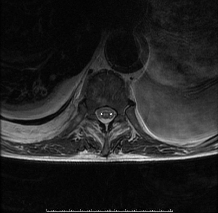
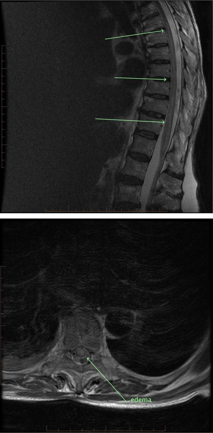
Bilateral symmetric T2 hyperintensities in the anterior horns, sparing dorsal columns — pathognomonic for ASA territory infarction.
Sagittal T2 shows longitudinal central cord hyperintensity:
Key Exam Points (Your Note Highlights)
- Dorsal horns & dorsal columns spared → preserved proprioception and vibration ✓
- UMN below lesion (corticospinal) → spasticity, hyperreflexia, +Babinski (as noted in your handwriting)
- LMN at level of lesion (anterior horn) → flaccid weakness at that segment
- Loss of pain & temp below lesion (spinothalamic) → bilateral
- Watershed = mid-thoracic, reinforced below T8 by artery of Adamkiewicz
- Classic cause = aortic aneurysm repair
Sources: Adams and Victor's Principles of Neurology, 12th ed. — "Anterior Myelopathy (Anterior Spinal Artery Syndrome)"
Kindly explain this paragraph in simple words
The user is referring to the paragraph in the image they already shared. Let me explain it in simple words.
ASA Occlusion — Explained Simply
The paragraph says:
"Spinal cord infarction sparing dorsal horns and dorsal columns. Watershed area is mid-thoracic ASA territory, as the artery of Adamkiewicz supplies ASA below T8. Can be caused by aortic aneurysm repair. Presents with UMN signs below the lesion (corticospinal tract), LMN signs at the level of the lesion (anterior horn), and loss of pain and temperature sensation below the lesion (spinothalamic tract)."
Breaking It Down Piece by Piece
🔴 "Spinal cord infarction sparing dorsal horns and dorsal columns"
Think of this like a heart attack, but in the spinal cord.
The front part of the spinal cord dies (no blood = no oxygen).
BUT the back part — the dorsal horns and dorsal columns — survive, because they have their own separate blood supply (posterior spinal arteries).
So the patient can still feel vibration and know where their limbs are (proprioception is intact), because those travel through the back (dorsal columns).
🔴 "Watershed area is mid-thoracic"
A watershed area is like the last house at the end of a water pipe — it gets water last, so if pressure drops, it runs dry first.
The mid-thoracic cord (around T4–T8) is the farthest point from the blood sources feeding the ASA. So when blood flow drops, this area infarcts first.
🔴 "The artery of Adamkiewicz supplies ASA below T8"
The ASA is one long artery running down the front of the cord. It gets reinforced (boosted) at the lower thoracic level by a branch called the artery of Adamkiewicz (usually coming off around T9–T12 on the left side).
- Above T8 → only getting blood from above (from the vertebral arteries)
- Below T8 → also getting blood from the artery of Adamkiewicz
So the area just above where Adamkiewicz joins in (T4–T8) is the most vulnerable — it's the furthest from both supply points. Classic watershed.
🔴 "Can be caused by aortic aneurysm repair"
During surgery to fix a large aortic aneurysm, surgeons clamp the aorta. This temporarily cuts off blood to all the arteries branching off it — including the artery of Adamkiewicz. If the cord is starved of blood too long → infarction.
This is one of the feared complications of aortic surgery.
🔴 "UMN signs BELOW the lesion (corticospinal tract)"
The corticospinal tract (the main motor highway from the brain) runs through the front of the cord — right in the ASA territory. When it's damaged:
- The brain can no longer properly control muscles below that level
- You get Upper Motor Neuron (UMN) signs: stiff muscles (spasticity), exaggerated reflexes, and a positive Babinski sign (big toe goes up when you scratch the foot)
Simple memory: UMN = stiff and brisk (like the brakes are jammed ON)
🔴 "LMN signs AT THE LEVEL of the lesion (anterior horn)"
The anterior horn contains the actual motor nerve cell bodies — the final neurons that directly tell your muscles to move. These are also in the ASA territory.
At the exact level of the infarct, these cells die:
- The nerve supply to that specific muscle group is completely cut
- You get Lower Motor Neuron (LMN) signs: weak and floppy muscles, wasted muscles, absent reflexes
Simple memory: LMN = floppy and wasted (like the brakes are cut)
🔴 "Loss of pain and temperature BELOW the lesion (spinothalamic tract)"
The spinothalamic tract (which carries pain and temperature signals up to the brain) also runs through the ASA territory. When it's damaged:
- Patient cannot feel pain or heat/cold below the level of the injury
- This is bilateral (both sides) because the ASA supplies both sides of the front cord
BUT — because the dorsal columns (vibration, proprioception) are spared — this creates a dissociated sensory loss: can't feel a pin prick, but can still feel a tuning fork. This dissociation is the hallmark.
The Big Picture in One Diagram
LEVEL OF LESION:
  ↳ Anterior horn destroyed → LMN signs (flaccid, areflexic) at that level

BELOW THE LESION:
  ↳ Corticospinal tract cut → UMN signs (spastic, hyperreflexic, Babinski)
  ↳ Spinothalamic tract cut → No pain, no temperature (bilateral)

PRESERVED (posterior spinal artery safe):
  ↳ Dorsal columns intact → Vibration & proprioception NORMAL
This pattern — UMN below + LMN at level + pain/temp loss + intact proprioception — is the textbook signature of ASA syndrome and a very common exam question!
This is a shared conversation. Sign in to Orris to start your own chat.