
Teach me femoral triangle in detail anatomy
Reading File
The femoral triangle is a key anatomical region in the upper thigh containing the femoral nerve, artery, and vein (lateral to medial: N-A-V). It's bounded by the inguinal ligament (superiorly), sartorius muscle (laterally), and adductor longus (medially). The floor is formed by iliopsoas and pectineus muscles, and the apex points downward into the adductor canal.
Searching sources...
Finding Sources
Finding Sources
Excellent results. Now let me read the detailed sections from Gray's Anatomy and the THIEME Atlas simultaneously.
Reading File
Reading File
Reading File
Great content. Now let me read the femoral sheath, femoral nerve, clinical sections, and fetch the key images in parallel.
Reading File
Reading File
Loading Image
Loading Image
Excellent content and superb images. Let me fetch two more key images - the femoral triangle contents and the femoral artery diagram.
Loading Image
Loading Image
Loading Image
I now have comprehensive textbook content and all the key diagrams. Let me compile the complete detailed answer.
Femoral Triangle - Complete Anatomy
Overview
The femoral triangle is a wedge-shaped (pyramid-shaped) depression located in the proximal anterior thigh, at the junction between the anterior abdominal wall and the lower limb. It acts as the main "gateway" through which neurovascular structures pass from the abdomen into the lower limb.
Boundaries
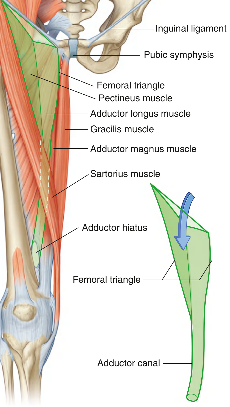
| Wall | Structure |
|---|---|
| Base (superior) | Inguinal ligament (from ASIS to pubic tubercle) |
| Lateral border | Medial margin of sartorius muscle |
| Medial border | Medial margin of adductor longus muscle |
| Apex | Where sartorius and adductor longus converge inferiorly - continuous with the adductor canal |
| Roof | Fascia lata + cribriform fascia (covering the saphenous opening) |
| Floor (lateral to medial) | Iliopsoas (lateral half) + Pectineus (medial half), and deeper: adductor longus |
The apex points inferiorly and opens into the adductor canal (Hunter's canal), which runs down the medial thigh and ends at the adductor hiatus in adductor magnus, leading into the popliteal fossa.
Contents (Lateral to Medial: NAVY or NAVeL)
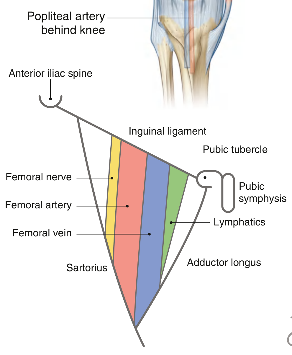
A useful mnemonic: N-A-V-eL (from lateral to medial):
- N - Femoral Nerve
- A - Femoral Artery
- V - Femoral Vein
- eL - Empty space (femoral canal) + Lymphatics
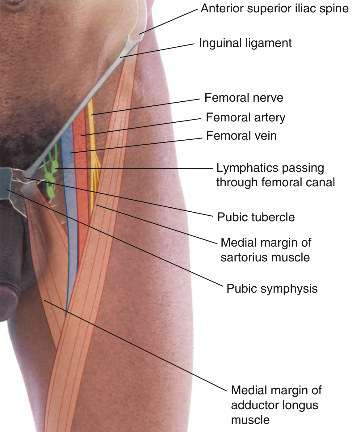
1. Femoral Nerve
- Largest branch of the lumbar plexus (L2, L3, L4)
- Descends through the muscular compartment (lacuna musculorum), lateral to the iliopectineal arch
- Lies lateral to the femoral artery in the femoral triangle
- NOT enclosed within the femoral sheath (this is a key exam point)
- Divides into multiple branches within the femoral triangle:
- Motor branches to quadriceps, sartorius, pectineus
- Cutaneous branches (intermediate and medial cutaneous nerves of the thigh)
- Gives rise to the saphenous nerve - the longest branch, which follows the femoral artery into the adductor canal
2. Femoral Artery
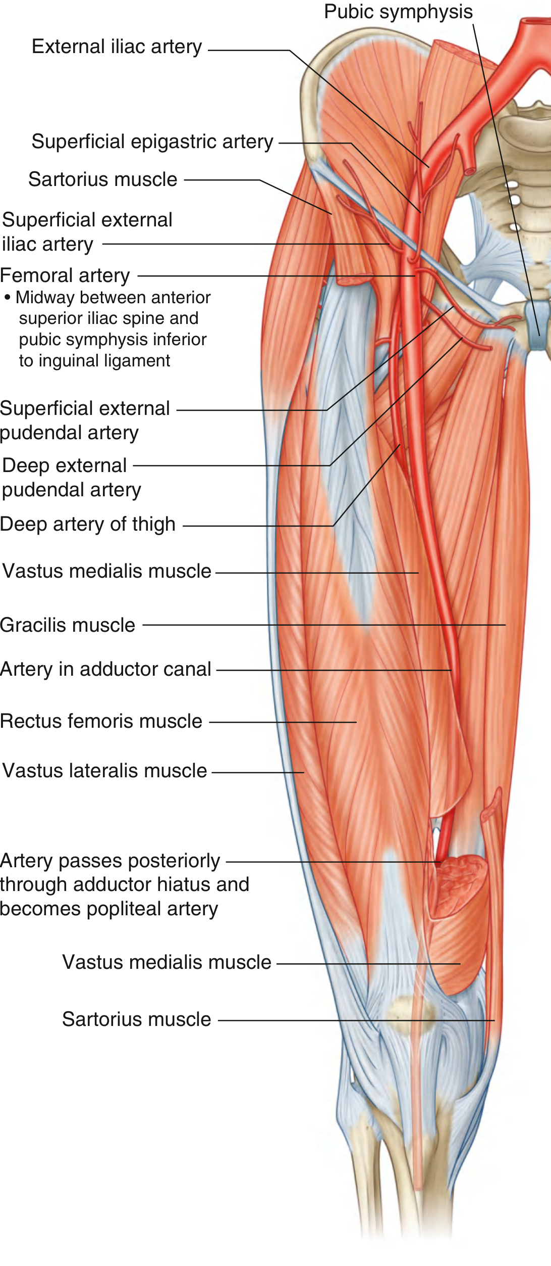
- Continuation of the external iliac artery, which becomes the femoral artery as it passes under the inguinal ligament
- Palpable just inferior to the inguinal ligament, midway between the ASIS and pubic symphysis (midpoint of the inguinal ligament)
- Passes vertically through the femoral triangle, then continues into the adductor canal
- Exits through the adductor hiatus to become the popliteal artery behind the knee
Branches in the Femoral Triangle
| Branch | Territory |
|---|---|
| Superficial epigastric artery | Lower anterior abdominal wall |
| Superficial circumflex iliac artery | Region near ASIS |
| Superficial external pudendal artery | Scrotum/labia majora |
| Deep external pudendal artery | Perineum |
| Deep artery of thigh (profunda femoris) | Main supply to the thigh muscles |
The profunda femoris (deep artery of the thigh) is the largest branch. It arises from the lateral side of the femoral artery in the femoral triangle, passes posteriorly between pectineus and adductor longus, then between adductor longus and adductor brevis. It gives:
- Medial circumflex femoral artery - main blood supply to the femoral head/neck
- Lateral circumflex femoral artery - ascending, descending, and transverse branches
- 3 perforating arteries - supply the posterior thigh muscles
3. Femoral Vein
- Continuation of the popliteal vein superiorly
- Lies medial to the femoral artery in the femoral triangle
- Receives the great saphenous vein through the saphenous opening (cribriform fascia) just inferior to the inguinal ligament
- Together with the femoral artery and lymphatics, it is enclosed in the femoral sheath
4. Lymphatics
- Lie in the most medial compartment of the femoral sheath - the femoral canal
- The Cloquet's node (or Rosenmüller node) is a deep inguinal lymph node lying at the femoral ring
- Drain lymph from the lower limb upward to the external iliac nodes
The Femoral Sheath
The femoral sheath is a funnel-shaped sleeve of fascia that surrounds the femoral artery, vein, and lymphatics (but NOT the femoral nerve) within the femoral triangle.
- Superiorly continuous with: transversalis fascia (anteriorly) and iliac fascia (posteriorly)
- Merges inferiorly with vessel adventitia about 4 cm below the inguinal ligament
- Divided into 3 compartments:
| Compartment | Position | Contents |
|---|---|---|
| Lateral | Lateral | Femoral artery |
| Intermediate | Middle | Femoral vein |
| Medial (femoral canal) | Most medial | Lymphatics + Cloquet's node |
Femoral Canal and Femoral Hernia
The superior opening of the femoral canal is the femoral ring. It is bounded by:
- Anteriorly: inguinal ligament
- Posteriorly: pectineus muscle + its fascia
- Medially: lacunar ligament (Gimbernat's ligament)
- Laterally: femoral vein
The femoral ring is a potential weak point in the lower abdomen and is the site of femoral hernias. Femoral hernias are more common in women (wider pelvis) and pass through the femoral ring into the femoral canal - they present as a lump below and lateral to the pubic tubercle (compared to inguinal hernias that are above and medial to the pubic tubercle).
Iliopectineal Arch - The Compartment Divider
The iliopectineal arch (a thickened band of iliac fascia) divides the space below the inguinal ligament into two compartments:
- Lacuna musculorum (lateral, muscular compartment): contains iliopsoas, femoral nerve, lateral femoral cutaneous nerve
- Lacuna vasorum (medial, vascular compartment): contains femoral artery, vein, lymphatics (all within femoral sheath), and femoral branch of genitofemoral nerve
Adductor Canal (Hunter's Canal) - Continuation from Apex
The adductor canal begins at the apex of the femoral triangle and runs down the medial thigh:
- Bounded by: vastus medialis (anterolateral), adductor longus then adductor magnus (posterior), sartorius (roof)
- Contains: femoral artery, femoral vein, saphenous nerve, nerve to vastus medialis, and medial femoral cutaneous nerve
- Ends at the adductor hiatus in adductor magnus → femoral vessels become popliteal vessels
Clinical Relevance
1. Femoral Pulse and Vascular Access
The femoral artery pulse is palpable midway between the ASIS and pubic symphysis, just below the inguinal ligament. This site is used for:
- Cardiac catheterization (coronary angiography, angioplasty)
- Peripheral vascular access (aortography, renal/gonadal vein cannulation)
- Femoral vein access: allows catheterization up to the right heart, pulmonary artery, superior vena cava, and renal veins
2. Femoral Hernia
- Passes through the femoral ring into the femoral canal
- More common in women
- Presents as a mass below and lateral to the pubic tubercle
- Risk of strangulation is high because the femoral ring has rigid bony/ligamentous boundaries
3. Femoral Nerve Block
Used for anesthesia of the anterior thigh and knee - needle placed just lateral to the femoral artery pulse
4. Deep Vein Thrombosis (DVT)
The femoral vein is a common site; femoral vein compression ultrasound is the first-line investigation
5. Femoral Nerve Injury
- Caused by trauma, pelvic surgery, or psoas hematoma
- Results in: weakness of knee extension (quadriceps), loss of knee jerk reflex (L3/L4), sensory loss over anterior thigh and medial leg (saphenous nerve territory)
6. Cruciate Anastomosis
A vital anastomosis around the hip joint formed by branches of the medial and lateral circumflex femoral arteries, the inferior gluteal artery, and the first perforating artery of the profunda femoris. This provides collateral circulation if the femoral artery is obstructed above the profunda femoris origin.
Summary Table
| Feature | Detail |
|---|---|
| Shape | Wedge/pyramid, apex pointing inferiorly |
| Superior base | Inguinal ligament |
| Lateral wall | Medial margin of sartorius |
| Medial wall | Medial margin of adductor longus |
| Floor (lateral) | Iliopsoas |
| Floor (medial) | Pectineus |
| Roof | Fascia lata + cribriform fascia |
| Contents (L→M) | Nerve, Artery, Vein, (Empty canal), Lymphatics |
| Femoral nerve | Lateral to artery; NOT in femoral sheath |
| Femoral sheath | Surrounds artery, vein, lymphatics only |
| Femoral canal | Most medial compartment; site of femoral hernia |
| Continues as | Adductor canal at apex |
Sources: Gray's Anatomy for Students, pp. 675-676, 703; THIEME Atlas of Anatomy - General Anatomy and Musculoskeletal System, p. 573
LAQ on KNEE JOINT
Reading File
The knee joint (tibiofemoral joint) is the largest and most complex joint in the body - a modified hinge joint between the femur, tibia, and patella. It has cruciate and collateral ligaments, two menisci, multiple bursae, and a large synovial cavity.
Searching sources...
Finding Sources
Finding Sources
Excellent. The best sources are Gray's Anatomy and S. Das. Let me read all the key sections in parallel.
Reading File
Reading File
Reading File
Reading File
Reading File
Reading File
Reading File
Loading Image
Loading Image
Reading File
Loading Image
Loading Image
I now have comprehensive content and excellent images. Let me compile the complete LAQ on the knee joint.
KNEE JOINT - Long Answer Question (LAQ)
1. Introduction
The knee joint is the largest and most complex synovial joint in the human body. It is a modified hinge joint (condylar type) formed by three articulations that share a single joint cavity:
- Medial tibiofemoral joint - between medial femoral condyle and medial tibial condyle
- Lateral tibiofemoral joint - between lateral femoral condyle and lateral tibial condyle
- Patellofemoral joint - between the patella and the anterior patellar surface of the femur
The patellofemoral articulation allows the pull of the quadriceps femoris to be directed anteriorly over the knee to the tibia without tendon wear.
2. Bones and Articular Surfaces
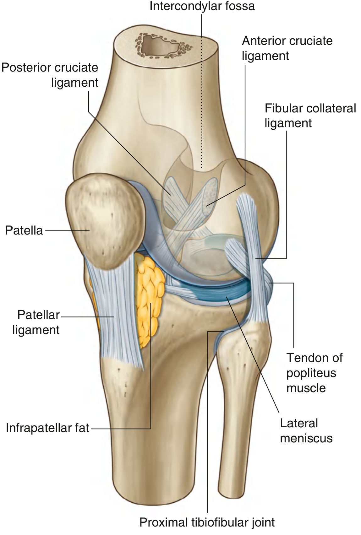
Femur (lower end)
- Two large condyles (medial and lateral) project posteriorly and inferiorly
- The articular surfaces on the posterior femoral condyles (curved and rounded) articulate with the tibia in flexion
- The surfaces on the inferior femoral condyles (broad and flat) articulate in full extension - this makes the joint more stable in extension
- The anterior patellar surface (V-shaped trochlear groove) articulates with the patella
- The intercondylar fossa (notch) separates the two condyles posteriorly and houses the cruciate ligaments
Tibia (upper end)
- Medial and lateral tibial condyles form the tibial plateau
- Medial plateau is oval, slightly concave
- Lateral plateau is more circular
- Between them: the intercondylar area where the cruciate ligaments and menisci attach
- The tibial tuberosity on the anterior surface is the attachment point for the patellar ligament
Patella
- Largest sesamoid bone in the body
- Embedded in the quadriceps femoris tendon
- Posterior surface articulates with the femoral trochlear groove
- Increases the mechanical advantage of the quadriceps by ~30%
3. Menisci
Two C-shaped fibrocartilaginous discs lying on the tibial plateau, deepening the shallow tibial articular surfaces.
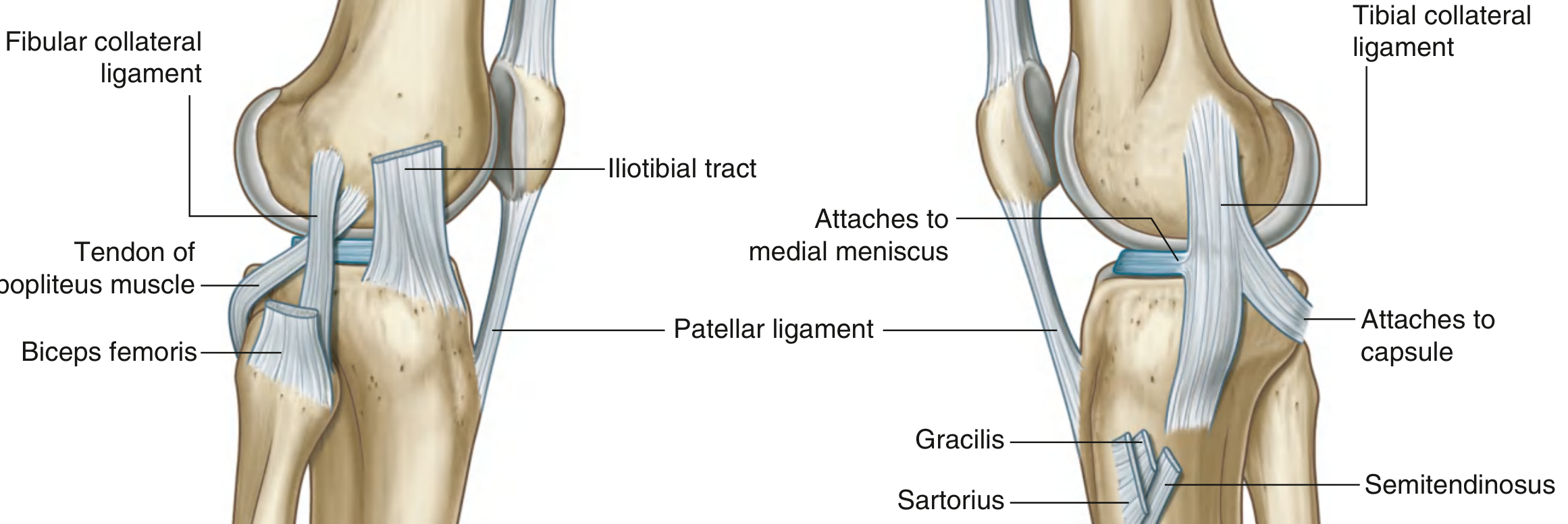
| Feature | Medial Meniscus | Lateral Meniscus |
|---|---|---|
| Shape | Larger, C-shaped (open C) | Smaller, more circular (nearly O-shaped) |
| Peripheral attachment | Firmly attached to joint capsule AND tibial collateral ligament | NOT attached to fibular collateral ligament; popliteus tendon passes between it and capsule |
| Mobility | Less mobile (tethered) | More mobile |
| Injury rate | More commonly torn | Less commonly torn |
| Special attachment | Anterior and posterior horns in intercondylar area | Attached via meniscofemoral ligaments (of Humphrey and Wrisberg) to PCL |
Functions of menisci:
- Deepen the articular surfaces (act as "shock absorbers")
- Distribute compressive load across a wider area
- Improve joint stability and lubrication
- Guide the rolling and gliding movements of the femoral condyles
4. Joint Capsule
The capsule is a fibrous sleeve enclosing the knee joint:
- Anteriorly: thin; attached to margins of the patella; reinforced by expansions from vastus medialis and lateralis (medial and lateral patellar retinacula), and the iliotibial tract anterolaterally
- Posteriorly: reinforced by the oblique popliteal ligament (expansion of semimembranosus tendon, running superolaterally) and the arcuate popliteal ligament
- The tendon of popliteus passes through an aperture in the posterolateral capsule
5. Ligaments
A. Patellar Ligament
- Continuation of the quadriceps femoris tendon below the patella
- Attached: apex of patella → tibial tuberosity
- Very strong; tested by the knee-jerk reflex (L3, L4)
B. Collateral Ligaments
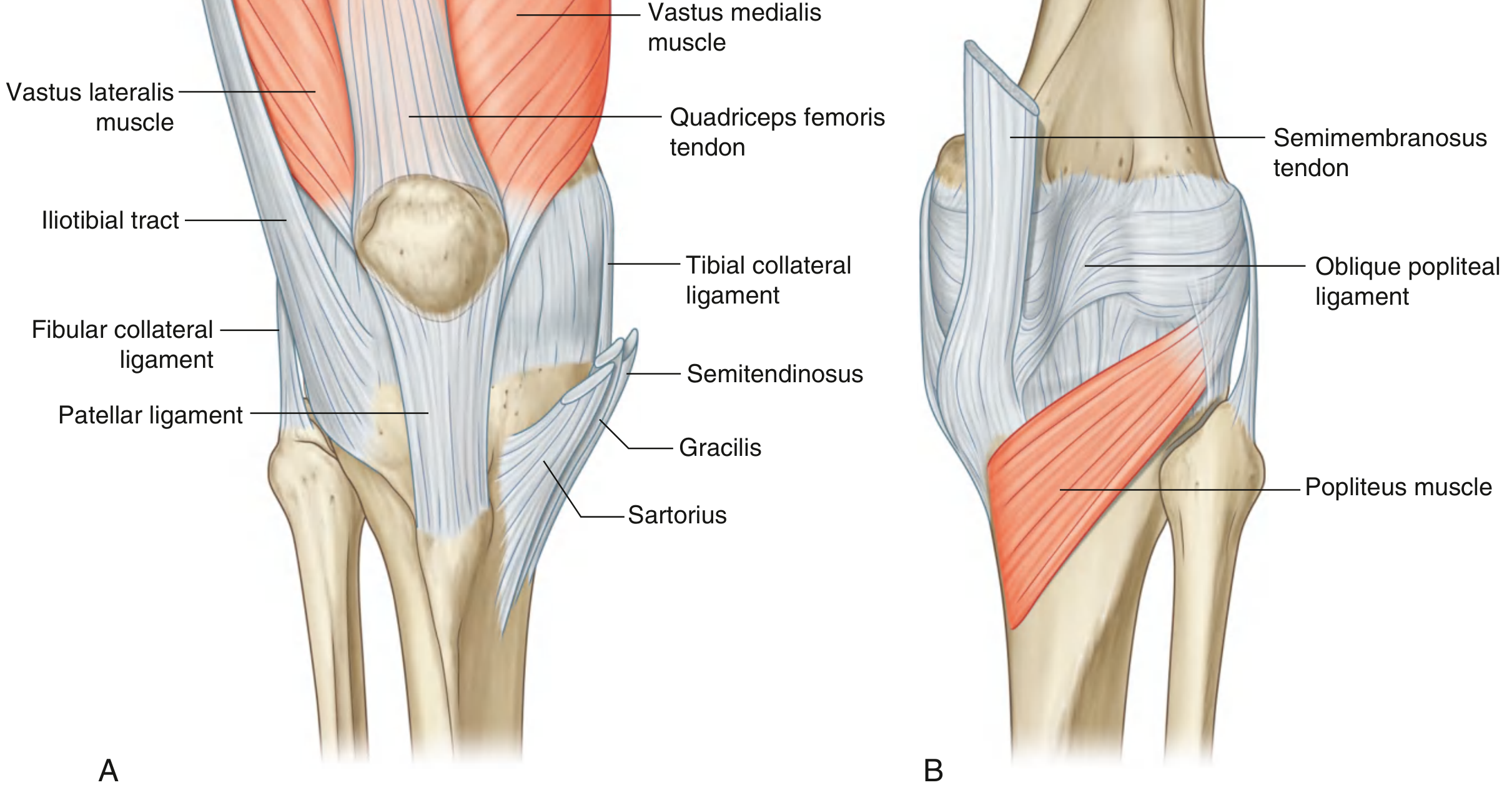
| Feature | Tibial (Medial) Collateral Ligament (TCL/MCL) | Fibular (Lateral) Collateral Ligament (FCL/LCL) |
|---|---|---|
| Shape | Broad and flat | Cord-like, round |
| Upper attachment | Medial femoral epicondyle (below adductor tubercle) | Lateral femoral epicondyle (above popliteus groove) |
| Lower attachment | Medial surface of tibia (behind pes anserinus) | Head of fibula (lateral surface) |
| Capsule relation | Fused with fibrous membrane; attached to medial meniscus | Separate from capsule; bursa between ligament and capsule |
| Taut in | Extension | Extension |
| Function | Resists valgus (abduction) stress | Resists varus (adduction) stress |
| Injury | From lateral blow (valgus force); part of unhappy triad | From medial blow (varus force); less common |
Both collateral ligaments are taut in extension and lax in flexion - this is why rotational movements of the knee are only possible in flexion.
C. Cruciate Ligaments
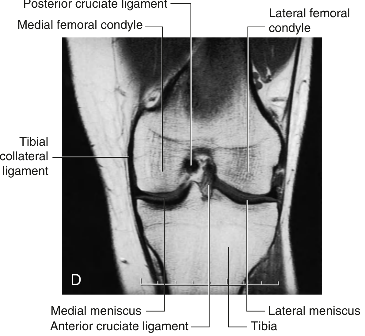
The cruciate ligaments are intracapsular but extrasynovial (covered by synovial membrane), located in the intercondylar region. They cross each other like an X (Latin: cruciate = cross-shaped).
| Feature | Anterior Cruciate Ligament (ACL) | Posterior Cruciate Ligament (PCL) |
|---|---|---|
| Tibial attachment | Anterior intercondylar area | Posterior intercondylar area |
| Femoral attachment | Posterior part of lateral wall of intercondylar fossa | Anterior part of medial wall of intercondylar fossa |
| Orientation | Passes upward, backward, laterally | Passes upward, forward, medially |
| Function | Prevents anterior displacement of tibia on femur; limits hyperextension; limits medial rotation | Prevents posterior displacement of tibia on femur; limits hyperflexion |
| Clinical test | Anterior drawer test; Lachman's test | Posterior drawer test; posterior sag sign |
| Blood supply | Rich blood supply → hemarthrosis when torn | Rich blood supply |
| Injury mechanism | Valgus force + external rotation (catching inner ski edge) | Dashboard injury (posterior force on flexed knee) |
Key fact: ACL injury causes immediate hemarthrosis because the ligament is intracapsular with a rich blood supply.
6. Synovial Membrane
The synovial membrane lines the inner surface of the fibrous capsule (but not the articular cartilage or menisci). It forms important folds:
- Suprapatellar bursa (pouch): extends 3-4 cm above the patella between the femur and quadriceps tendon - this is continuous with the main joint cavity and is the largest recess. Effusion here produces the "horseshoe swelling" above and around the patella
- Infrarapatellar fat pad: lies between the patellar ligament and the synovial membrane; contains the infrapatellar fold (ligamentum mucosum) which extends to the intercondylar fossa
- Alar folds flank the infrapatellar fat pad on each side
7. Bursae Around the Knee
There are approximately 13 bursae around the knee joint. The most clinically important:
| Bursa | Location | Clinical Significance |
|---|---|---|
| Suprapatellar bursa | Deep to quadriceps tendon, above patella | Communicates with joint; swells in effusion |
| Prepatellar bursa | Between patella and overlying skin | "Housemaid's knee" (friction bursitis) |
| Infrapatellar bursa (superficial) | Between patellar ligament and skin | "Clergyman's knee" |
| Infrapatellar bursa (deep) | Between patellar ligament and tibia | Does NOT communicate with joint |
| Popliteal (Baker's) cyst | Posterior joint, semimembranosus bursa | Communicates with joint; visible behind knee when standing |
| Pes anserinus bursa | Between TCL and pes anserinus tendons | Painful in overuse/OA |
8. Movements of the Knee Joint
| Movement | Range | Muscles |
|---|---|---|
| Flexion | 0° to ~140° (active), 160° (passive) | Hamstrings (biceps femoris, semitendinosus, semimembranosus), gastrocnemius, gracilis, sartorius, popliteus |
| Extension | 140° to 0° | Quadriceps femoris (rectus femoris, vastus medialis, lateralis, intermedius) |
| Medial rotation (tibia on femur) | ~10° in 90° flexion | Popliteus (also initiates unlocking), semitendinosus, semimembranosus, sartorius, gracilis |
| Lateral rotation | ~30° in 90° flexion | Biceps femoris |
Rotation is only possible when the knee is flexed (collateral ligaments become lax). It is completely prevented in full extension.
9. Locking and Unlocking Mechanism
This is a unique feature of the knee joint:
Locking (Extension → Full Extension)
During the last few degrees of extension:
- The medial femoral condyle has a larger articular surface than the lateral; after the lateral condyle exhausts its articular surface, the medial condyle continues to roll
- This causes medial rotation of the femur on the tibia (or lateral rotation of tibia if foot is free), "screwing" the joint tight
- All ligaments become taut
- The body's center of gravity falls anterior to the axis of the knee joint, passively maintaining extension
- Result: the joint is "locked" - requiring minimal muscular effort to stand
Unlocking
- Initiated by the popliteus muscle (the "key to unlock the knee")
- Popliteus laterally rotates the femur on the fixed tibia (or medially rotates tibia on fixed femur)
- This "unscrews" the joint, freeing the collateral ligaments and allowing flexion
10. Vascular Supply
An anastomotic network (genicular anastomosis) surrounds the knee joint, formed by:
- Descending genicular branch of the femoral artery
- 5 genicular branches of the popliteal artery (medial/lateral superior and inferior genicular + middle genicular)
- Circumflex fibular artery
- Anterior and posterior recurrent branches of the anterior tibial artery
- Descending branch of the lateral circumflex femoral artery
This rich anastomosis ensures the knee is supplied even if one vessel is compromised.
11. Nerve Supply
The knee joint is innervated according to Hilton's Law (a joint is supplied by nerves that also supply the muscles acting on it):
- Femoral nerve (and its branches - nerve to vastus medialis, saphenous nerve) - anterior and medial
- Obturator nerve - medial aspect
- Common peroneal nerve - lateral aspect (via its articular branch)
- Tibial nerve - posterior aspect
12. Relations
Anterior
- Quadriceps femoris tendon, patella, patellar ligament
- Prepatellar and infrapatellar bursae
Posterior (Popliteal Fossa)
- Popliteal artery (most anterior - closely related to posterior capsule, vulnerable in posterior dislocations)
- Popliteal vein (posterior to artery)
- Tibial nerve (most superficial)
- Common peroneal nerve
- Popliteal lymph nodes
- Small saphenous vein terminal part
Medial
- Tibial collateral ligament, sartorius, gracilis, semitendinosus (pes anserinus), semimembranosus
Lateral
- Fibular collateral ligament, biceps femoris, iliotibial tract, common peroneal nerve
13. Clinical Anatomy
Unhappy Triad (O'Donoghue's Triad)
A classic injury from a valgus + external rotation force (e.g., lateral tackle in football, skiing):
- Medial (tibial) collateral ligament tear
- Anterior cruciate ligament tear
- Medial meniscus tear
(The medial meniscus tears because it is attached to the MCL and cannot escape the force.)
Clinical Tests
| Test | Tests For | Positive Finding |
|---|---|---|
| Lachman's test | ACL integrity | Excessive anterior tibial translation at 20° flexion with soft endpoint |
| Anterior drawer test | ACL | Anterior tibial drawer at 90° flexion |
| Posterior drawer test | PCL | Posterior tibial drawer at 90° flexion |
| Valgus stress test | MCL | Pain/gapping at 0° and 30° flexion |
| Varus stress test | LCL | Pain/gapping at 0° and 30° flexion |
| McMurray's test | Meniscal tear | Pain/click on rotation with flexion/extension |
| Apley's grind test | Meniscal tear (prone position) | Pain with compression + rotation |
| Patellar tap (Ballottement) | Joint effusion | Patella bounces back after depression |
| Bulge sign | Small effusion | Fluid wave visible medially |
Joint Aspiration (Arthrocentesis)
Needle inserted: superolateral border of patella, directed inferiorly and medially into the suprapatellar pouch. Normal knee fluid is clear/viscous. Hemarthrosis (blood) suggests ACL or intraarticular fracture.
Patellofemoral Pain Syndrome
Pain from excessive lateral tracking of patella in trochlear groove. Aggravated by climbing stairs and prolonged sitting ("theatre sign").
Osteoarthritis of the Knee
- Most common joint affected in the body
- Loss of medial compartment cartilage → genu varum (bow leg)
- Loss of lateral compartment cartilage → genu valgum (knock knee)
- Findings: reduced joint space, subchondral sclerosis, osteophytes, cyst formation
14. Summary Table
| Feature | Detail |
|---|---|
| Type | Modified hinge (condylar synovial) |
| Bones | Femur, tibia, patella |
| Intraarticular structures | Menisci (medial + lateral), cruciate ligaments (ACL + PCL), fat pad |
| Extracapsular ligaments | Patellar, tibial collateral, fibular collateral |
| Intracapsular + extrasynovial | Cruciate ligaments |
| Locking muscle | Quadriceps + medial femoral rotation |
| Unlocking muscle | Popliteus (only muscle to unlock) |
| Nerve supply | Femoral, obturator, tibial, common peroneal |
| Blood supply | Genicular anastomosis (femoral + popliteal + anterior tibial) |
| Most common injury | ACL tear (followed by MCL, medial meniscus) |
| Effusion position | Moderate flexion (~15°) = "optimum position" |
Sources: Gray's Anatomy for Students, pp. 709-718, 779; S. Das Manual on Clinical Surgery 13th Ed., pp. 253-260
This is a shared conversation. Sign in to Orris to start your own chat.