
Langhans giant cell tuberculosis
Langhans giant cell tuberculosis granuloma histology
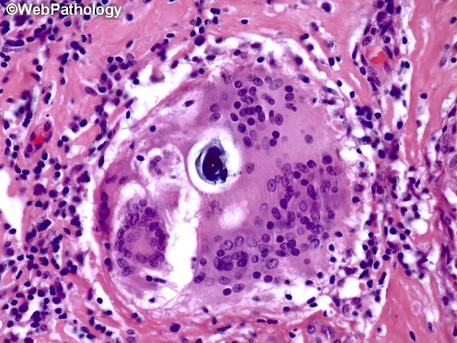
Histopathology image showing a granulomatous lesion composed of epithelioid histiocytes with a surrounding rim of lymphocytes and occasional Langhans-type multinucleated giant cells. Within a giant cell, lamellar inclusions known as Schaumann bodies are evident as concentric rings of protein and mineral material; calcium oxalate crystals may also be present. Asteroid bodies may be seen as stellate inclusions, though they are not required for diagnosis. The granuloma is noncaseating, lacking central necrosis, which favors sarcoidosis over infectious granulomatous processes such as tuberculosis. The tissue architecture shows well-formed granulomas with distinct borders, often adjacent to blood vessels in this section. The image is stained with Hematoxylin and Eosin (H&E), highlighting eosinophilic granulomatous material and basophilic nuclei within giant cells. Clinically, the presence of Schaumann bodies and Langhans giant cells supports a granulomatous inflammatory syndrome but is not pathognomonic for sarcoidosis. Differential considerations include berylliosis, hypersensitivity pneumonitis, fungal or mycobacterial infections, and foreign body reactions. Correlation with chest imaging, serum ACE levels, and clinical features is essential for diagnosis. This slide serves an educational role in recognizing granulomatous histology, giant cell inclusions, and lamellar calcific bodies, informing diagnostic workflows and teaching pathology trainees about sarcoidosis-related granulomatous disease.
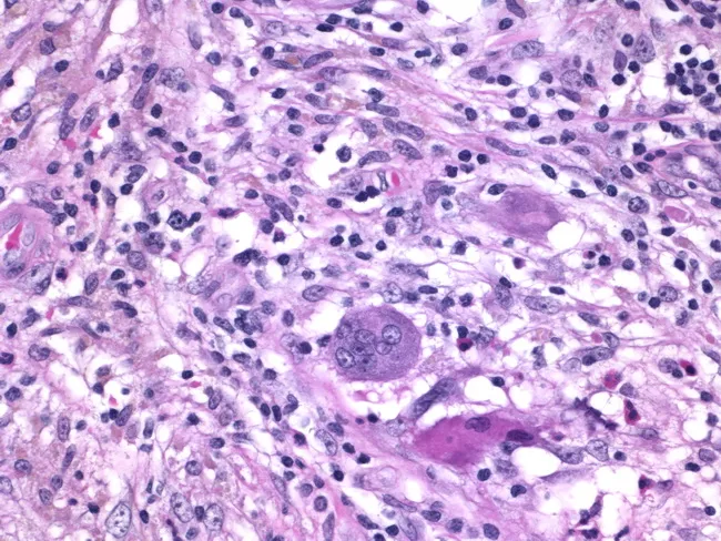
Histology of granulomatous mastitis on bright-field light microscopy. A dense inflammatory infiltrate forms granulomas centered on Langhans-type giant cells surrounded by epithelioid histiocytes, scattered lymphocytes, and occasional eosinophils. The image depicts a high-power view of the center of a granuloma within breast tissue biopsy; multinucleated giant cells with horseshoe-shaped nuclei are evident. The granulomatous architecture includes a peripheral rim of lymphocytes and histiocytes with minimal necrosis described. Staining by Hematoxylin and Eosin highlights basophilic nuclei and eosinophilic cytoplasm; pink collagenous stroma and adipose breast tissue may be visible in the background. Clinically, granulomatous mastitis can mimic infectious etiologies or neoplasia; the presence of Langhans giant cells strongly suggests a granulomatous process and warrants infectious workup for tuberculosis and fungal pathogens, as well as consideration of sarcoidosis or granulomatous reaction to foreign material. This image is diagnostic for granulomatous inflammation in breast tissue when correlated with clinical data and microbiologic studies; useful for education, differential diagnosis, and research on breast granulomatous disease. Additional educational value lies in distinguishing nonnecrotizing granulomas from caseating TB patterns, correlating histology with mammography and ultrasound, and guiding microbiologic testing and patient management in suspected granulomatous breast disease. The image supports teaching of granuloma biology and pathology.
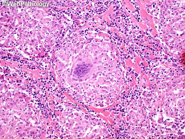
Histopathology via light microscopy of an H&E-stained lymph node biopsy demonstrates a granulomatous inflammatory response characterized by an epithelioid cell nodular formation with a surrounding lymphocytic infiltrate. Within the central core, a multinucleated Langhans-type giant cell is present, displaying nuclei arranged in a horseshoe pattern at the periphery. The granuloma is composed predominantly of well-differentiated epithelioid histiocytes with scant cytoplasm and tight cellular packing, producing a palisaded appearance that integrates with a mildly eosinophilic to translucent extracellular matrix. The surrounding paracortex contains a dense lymphoid crowding with scattered plasma cells and occasional eosinophils, consistent with chronic granulomatous inflammation. In this image, the granuloma lacks conspicuous caseous necrosis, a feature that supports noncaseating granulomatous processes; however, logical caveats apply since granulomas in sarcoidosis can mimic infectious etiologies. The presence of Langhans-type giant cells within nonnecrotizing granulomas is classically associated with sarcoidosis but can be seen in tuberculosis, atypical mycobacterial infections, fungal infections, berylliosis, and other granulomatous diseases. Additional diagnostic workup would include acid-fast bacilli staining and fungal stains, microbial cultures, and clinical correlation. Recognizing this histologic pattern guides differential diagnosis toward sarcoid-type granulomatous lymphadenitis and informs subsequent clinical management and exclusion of granulomatous infections. Correlation with imaging and laboratory data essential.
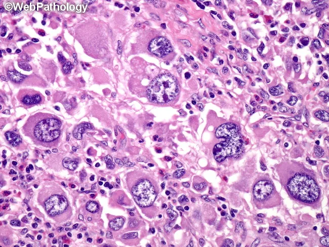
Histology image obtained with brightfield light microscopy on a hematoxylin and eosin stained tissue section. The specimen shows granulomatous inflammation characterized by aggregates of epithelioid macrophages (histiocytes) with elongated, pale to eosinophilic cytoplasm. Multinucleated giant cells, occasionally Langhans-type with peripheral, radiating nuclei, are interspersed among the epithelioid cells. Surrounding lymphocytes and a sparse fibrous stroma delineate nodular granulomas. In some fields there are crystalline or granular pink to eosinophilic deposits within the cytoplasm of histiocytes, and there may be mild necrosis at the center of larger granulomas, though necrosis is not uniformly present across the slide. The pattern is compatible with a granulomatous infectious or inflammatory process, requiring clinical correlation and microbiologic assessment. Differential diagnoses include mycobacterial infection (tuberculosis), fungal granulomas (histoplasmosis, blastomycosis), sarcoidosis, and foreign-body granulomatous reactions. The image emphasizes a macrophage-dominant infiltrate with discrete nodular architecture, preserving basic tissue planes while revealing cellular detail such as nuclear atypia is not prominent. This histologic phenotype is critical for guiding targeted tests (AFB stain, fungal stains, PCR) and therapeutic decisions, and it serves as an educational reference for recognizing granuloma formation in biopsy specimens. Correlation with clinical history and imaging is essential for diagnosis.
Langhans Giant Cell in Tuberculosis
Definition and Origin
- Histology: A Text and Atlas (Wheater/Ross), describing that "macrophages often fuse to form multinucleate giant cells called Langhans giant cells that isolate these pathogens from the body"
Formation - Mechanism
- Mycobacterium tuberculosis bacilli are phagocytosed by macrophages but resist intracellular killing
- Macrophages present mycobacterial antigens via MHC class II to helper CD4+ T lymphocytes
- T cells secrete IFN-γ, which activates macrophages into epithelioid cells
- Epithelioid macrophages undergo cell-to-cell fusion, producing the multinucleated Langhans giant cell
- The fusion is driven by cytokines (IL-4, M-CSF, GM-CSF, IFN-γ) and macrophage-macrophage adhesion molecules
Morphology - Key Features
| Feature | Detail |
|---|---|
| Size | Very large; 40-50 μm or more in diameter |
| Nuclei | 15-20+ nuclei arranged in a horseshoe or peripheral ring at the cell periphery |
| Cytoplasm | Abundant, pale, eosinophilic |
| Nucleus arrangement | Peripherally clustered - the defining hallmark |
| Stain | H&E shows eosinophilic cytoplasm with basophilic peripheral nuclei |
The Tuberculous Granuloma (Tubercle) - Structure
Outer zone: Lymphocytes & monocytes (rim)
          ↓
Middle zone: Epithelioid histiocytes (activated macrophages)
          ↓
Inner zone: Langhans giant cells
          ↓
Center: Caseous necrosis (amorphous, cheese-like, granular debris)
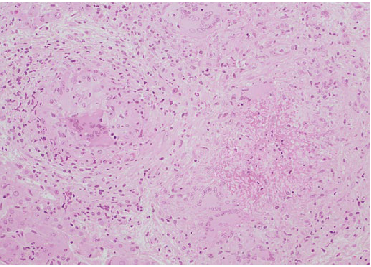
"Caseating granulomas (tubercles); foci of activated macrophages (epithelioid cells), rimmed by fibroblasts, lymphocytes; occasional Langhans giant cells; central necrosis with amorphous granular debris; acid-fast bacilli"
Histopathology Image
Langhans vs. Foreign Body Giant Cell
| Feature | Langhans Giant Cell | Foreign Body Giant Cell |
|---|---|---|
| Nuclear arrangement | Peripheral horseshoe/ring | Random/central scatter |
| Context | TB, leprosy, sarcoidosis, deep fungi | Sutures, talc, silica, cholesterol |
| Immune basis | T-cell mediated (type IV hypersensitivity) | Non-immune mechanical irritation |
| Associated necrosis | Caseous (in TB) | Usually absent |
Diseases Featuring Langhans Giant Cells
| Disease | Tissue Reaction |
|---|---|
| Tuberculosis | Caseating granulomas; epithelioid cells; Langhans cells; acid-fast bacilli |
| Leprosy | Acid-fast bacilli in macrophages; noncaseating granulomas |
| Sarcoidosis | Noncaseating granulomas; Langhans cells; Schaumann bodies; asteroid bodies |
| Crohn disease | Occasional noncaseating granulomas |
| Syphilis | Gumma with plasma cell infiltrate |
| Deep fungal infections | May mimic TB granuloma |
Important: Langhans giant cells are not pathognomonic for tuberculosis - they appear in any T-cell-mediated granulomatous disease. Caseating necrosis + AFB stain positivity is required to confirm TB specifically.
Clinical Relevance
- Diagnosis: Presence of Langhans giant cells on biopsy (lymph node, lung, liver, skin) prompts AFB staining, mycobacterial culture, and PCR for M. tuberculosis
- Host immunity indicator: Tuberculoid granuloma with Langhans cells = strong cell-mediated immunity (good host response); poorly formed granulomas = impaired immunity (as in HIV co-infection)
- PCR caveat: Mycobacterial DNA has been found in 16/20 sarcoidosis patients (Fitzpatrick's Dermatology), so histology alone cannot distinguish TB from sarcoidosis - clinical correlation is essential
- Fate of granuloma: May resolve, fibrose/calcify (Ghon complex), or liquefy and cavitate if caseous necrosis progresses