
Diffuse axonal injury
diffuse axonal injury MRI brain pathology
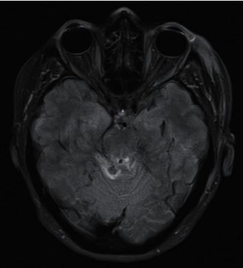
This diagnostic image is an axial Fluid-Attenuated Inversion Recovery (FLAIR) MRI of the brain at the level of the midbrain. The scan demonstrates characteristic features of Grade III Diffuse Axonal Injury (DAI). Key visual findings include multiple focal areas of hyperintense signal (hyperintensities) indicative of traumatic axonal shearing and localized edema. These lesions are specifically localized to the bilateral tectum of the midbrain and the periaqueductal gray matter. Additional hyperintense signal is visible extending into the superior cerebellar parenchyma and the superior cerebellar peduncles. The presence of these deep-seated lesions in the brainstem and cerebellar structures is a hallmark of severe traumatic brain injury (TBI). The surrounding cortical structures and ventricles are visible but secondary to the prominent brainstem pathology. This visual evidence serves as a critical educational tool for identifying the radiological progression and grading of shearing injuries in the setting of trauma neurology and neuroradiology.
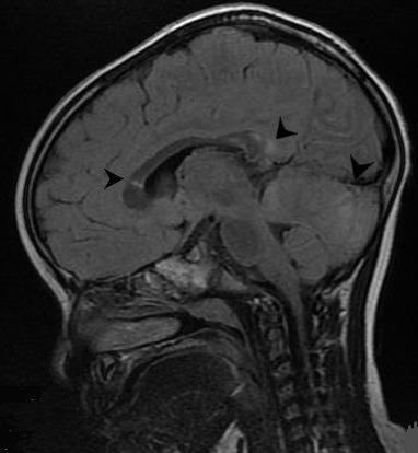
This diagnostic image is a sagittal T2-weighted Fluid-Attenuated Inversion Recovery (FLAIR) MRI scan of a pediatric patient's brain. The image highlights several hyperintense (bright) focal lesions, indicated by black arrowheads, which are characteristic of Diffuse Axonal Injury (DAI). These signal changes are located within critical neuroanatomical structures susceptible to shear-strain forces, including the splenium of the corpus callosum, the basal ganglia, and the cerebellar hemisphere. Morphologically, the lesions appear as small, rounded areas of high signal intensity, often referred to as 'axonal bulbs' or 'retraction balls' when viewed in the subacute phase of traumatic brain injury. The presence of these lesions in the corpus callosum and cerebellum is clinically significant as it correlates with secondary neurological manifestations such as tremor and dystonia following head trauma. This educational visual demonstrates the sensitivity of MRI over CT in detecting non-hemorrhagic DAI and provides a clear example of typical lesion distribution in traumatic axonal pathology.
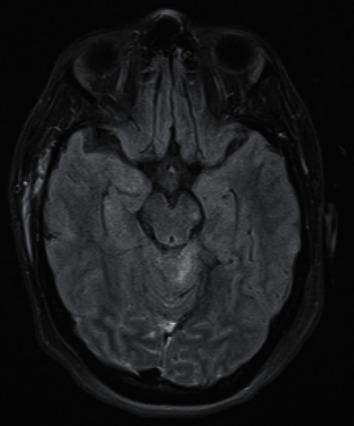
This diagnostic image is an axial Fluid-Attenuated Inversion Recovery (FLAIR) MRI of the human brain. The scan displays a cross-section at the level of the midbrain and upper pons. The image reveals Grade III Diffuse Axonal Injury (DAI), characterized by focal hyperintense (bright) lesions located in the left side of the midbrain and the pons. These signal abnormalities indicate edema or axonal shear injury against the darker, normal brain parenchyma. Surrounding anatomical structures include the temporal lobes, cerebral hemispheres with visible gyri and sulci, and the skull. The cerebrospinal fluid (CSF) within the ventricles and cisterns appears hypointense (dark), which is characteristic of the FLAIR sequence used to suppress water signal and highlight pathology. The image demonstrates the typical radiological appearance of severe traumatic brain injury affecting deep brainstem structures, which is clinically significant for assessing prognostic outcomes in trauma patients.
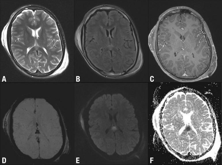
Six axial brain MRI sequences (A-F) demonstrate diagnostic features of Diffuse Axonal Injury (DAI) following Traumatic Brain Injury (TBI). The primary intra-axial lesion is located in the splenium of the corpus callosum. In T2-weighted (A) and FLAIR (B) images, the lesion appears hyperintense. It shows no enhancement on post-contrast T1-weighted imaging (C), suggesting an intact blood-brain barrier. Susceptibility Weighted Imaging (SWI, D) reveals a low signal intensity indicative of microhemorrhages. Diffusion-weighted imaging (DWI, E) shows a high signal, while the corresponding ADC map (F) shows low signal, confirming restricted diffusion characteristic of cytotoxic edema in acute axonal injury. Additionally, prominent extra-axial bilateral subgaleal hematomas are visible, appearing as fluid collections between the calvarium and scalp with variable signal across sequences. The image set illustrates the superiority of multi-parametric MRI over CT for identifying non-hemorrhagic and microscopic shear injuries in the white matter pathways.
"diffuse axonal injury" traumatic brain injury
Diffuse Axonal Injury (DAI)
Definition
- Robbins Pathologic Basis of Disease, p. 3913
- DiMaio's Forensic Pathology, p. 153
Mechanism of Injury
-
It is the duration of acceleration, not just its magnitude, that determines injury type: severe angular acceleration over a short time produces subdural hematomas; acceleration over a longer time produces DAI.
-
This explains why DAI is far more common in vehicular accidents (where absorbing materials prolong the time of force application) than in falls from standing height.
-
In theory, contact with an object is not required, but in practice human DAI almost always involves some head contact. DAI following blast injuries can occur without physical skull impact.
-
DAI is extremely unlikely from a fall from one's own height.
-
DiMaio's Forensic Pathology, pp. 152-153
Pathophysiology
- Primary axotomy - immediate mechanical disruption of axons at time of impact
- Secondary axotomy - apoptotic and degenerative cascade beginning 6-12 hours after injury, involving:
- Disruption of axoplasmic flow
- Calcium influx
- Cytoskeletal breakdown
- Sabiston Textbook of Surgery, p. 1331
Pathological Anatomy
- Gray-white matter junction
- Cerebral white matter
- Corpus callosum (especially the posterior body and splenium)
- Dorsolateral quadrant of the rostral brainstem
- Focal microhemorrhages in corpus callosum and dorsolateral brainstem (from simultaneous small vessel injury)
- Occasional laceration or transection of corpus callosum
- Gliding contusions of the cerebral cortex and hippocampi
-
Axons first appear dilated (sausage-link pattern) → then club-shaped → then round retraction balls by 18-24 hours
-
Retraction balls increase in number during the first week (because some axons are irreversibly injured but continue to function briefly before failing)
-
By 2-3 weeks, retraction balls decrease and microglial clusters become the dominant lesion
-
Later: astrocytosis and demyelination
-
Best demonstrated by silver impregnation or immunoperoxidase stains for amyloid beta-precursor protein (A-βPP) and alpha-synuclein
-
DiMaio's Forensic Pathology, pp. 153-154
-
Robbins Pathologic Basis of Disease, p. 3918
Grading
Pathological Grading (Adams Classification)
| Grade | Regions Involved |
|---|---|
| I | White matter of cerebral hemispheres (subcortical and deep) |
| II | Grade I + corpus callosum |
| III | Grade I + II + brainstem |
Clinical Grading (based on duration of coma)
| Severity | Coma Duration | Brainstem Signs |
|---|---|---|
| Mild | < 24 hours | Absent |
| Moderate | > 24 hours | Absent |
| Severe | > 24 hours | Present (or death) |
- DiMaio's Forensic Pathology, p. 154
Clinical Features
-
Immediate loss of consciousness at time of injury - a key distinguishing feature from other TBI types
-
Prolonged coma or persistent vegetative state
-
Neurological deficits that appear disproportionate to CT findings - a hallmark clinical clue
-
Up to 50% of individuals who develop coma shortly after trauma (without visible cerebral contusions) are thought to have DAI
-
DAI ranges from mild (akin to concussion) - with only axonal stretching and transient dysfunction - to severe, with complete axonal disruption
-
Should be on the differential in patients who fail to improve after surgical evacuation of hematoma, or remain unconscious despite no hemorrhage on CT
-
Robbins Pathologic Basis of Disease, p. 3913; Sabiston Textbook of Surgery, p. 1337
Imaging
CT Scan
- Often normal or minimal findings - the injury is radiographically insidious
- May show small hemorrhagic foci in deep white matter, corpus callosum, or brainstem
- Disproportionate clinical severity vs. CT appearance should prompt MRI
MRI (superior modality)
-
Shows multifocal hyperintense lesions at gray-white matter interfaces, corpus callosum, and brainstem
-
Best sequences:
- FLAIR - detects non-hemorrhagic lesions and edema
- Gradient echo (GRE) / Susceptibility-Weighted Imaging (SWI) - most sensitive for microbleeds
- DWI - shows restricted diffusion (cytotoxic edema) in acute phase
-
Visible lesions on MRI and CT are thought to be merely "the tip of the iceberg" of the true extent of injury
-
Grainger & Allison's Diagnostic Radiology, p. 1395; Sabiston Textbook of Surgery, p. 1333
Prognosis and Outcomes
- Leading cause of prolonged unconsciousness and persistent vegetative state following TBI
- The correlation between MRI lesion burden and neurological outcome is weak - clinical MRI has uncertain utility for predicting outcomes
- Secondary brain injury (hypoxia, hypotension, raised ICP) worsens outcomes and is the main target for treatment
- At least 3% of dementia cases in the general population are attributable to TBI, likely an underestimate
- A 2025 systematic review and meta-analysis in J Neurotrauma specifically evaluated the frequency and outcomes of DAI in severe TBI (PMID: 40485292)
Management
- Avoid hypotension and hypoxia - cerebral blood flow depends on adequate systemic perfusion
- ICP monitoring and control - targeting CPP (cerebral perfusion pressure)
- Neuroprotection research - agents under investigation include glibenclamide and xenon gas
- Neurocritical care: head-of-bed elevation, osmotherapy, temperature management
- Rehabilitation for survivors
Summary Table
| Feature | Detail |
|---|---|
| Mechanism | Rotational acceleration-deceleration (shearing) |
| Most common cause | High-velocity vehicular accidents |
| Key locations | Gray-white junction, corpus callosum, brainstem |
| Histological hallmark | Retraction balls (axonal swellings) |
| Staining | A-βPP immunoperoxidase, silver stain |
| Best imaging | MRI-SWI / FLAIR |
| CT appearance | Often normal; may show microhemorrhages |
| Clinical clue | Coma disproportionate to CT findings |
| Pathological grades | I (white matter) → II (+corpus callosum) → III (+brainstem) |
Hematoma
epidural hematoma subdural hematoma CT scan brain
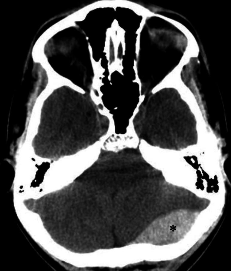
An axial non-contrast computerized tomography (CT) scan of the brain demonstrating an acute epidural (extradural) hematoma. The primary finding is a well-defined, biconvex (lens-shaped), hyperdense fluid collection located in the left occipital region, marked with an asterisk. This hyperdensity is characteristic of an acute hemorrhage within the epidural space, which typically does not cross cranial sutures. Secondary mass effect is visible, characterized by a mild left-to-right midline shift and compression of adjacent brain parenchyma. Anatomical landmarks include the skull base, posterior fossa structures, and the cerebellum. The image illustrates a classic neurosurgical emergency often associated with traumatic skull fractures and arterial bleeding, typically from the middle meningeal artery. This diagnostic image is intended for educational use in emergency medicine, radiology, and neurology to distinguish epidural hematomas from crescent-shaped subdural hematomas.
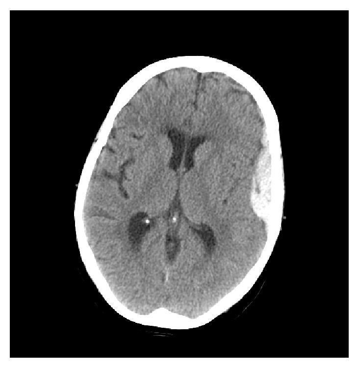
This diagnostic image is an axial non-contrast Computed Tomography (CT) scan of the head, demonstrating traumatic intracranial hemorrhage. The image reveals two distinct extra-axial collections in the left hemisphere. A hyperdense, biconvex (lens-shaped) epidural hematoma is localized in the left temporal region, exerting mass effect on the adjacent brain parenchyma. Anterior to this, a thin, hyperdense, crescent-shaped subdural hematoma is visible along the left frontal convexity, conforming to the cortical surface. The underlying cerebral tissue shows subtle signs of edema, indicated by a faint blurring of the grey-white matter interface in the left hemisphere. The lateral ventricles are visible and appear symmetric without significant compression, and there is no evident midline shift in this section. The calvarium appears intact without gross fractures. This scan serves as a clinical example of co-occurring epidural and subdural hematomas, highlighting the classic morphological differences between these two types of traumatic brain injuries.
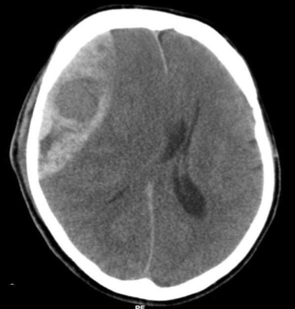
This diagnostic image is an axial non-contrast Computed Tomography (CT) scan of the brain, demonstrating a large, acute epidural (extradural) hematoma. The lesion is located on the right side of the skull and exhibits a classic biconvex or lens-shaped (lentiform) morphology, characterized by a hyperdense appearance relative to the brain parenchyma. The hematoma is clearly demarcated and constrained by the cranial sutures, positioned between the inner table of the skull and the dura mater. The image illustrates a significant mass effect, resulting in the effacement of adjacent sulci, partial compression of the right lateral ventricle, and a notable leftward midline shift of the falx cerebri. These findings are highly characteristic of an acute arterial bleed, typically involving the middle meningeal artery. This scan serves as a critical educational tool for identifying neurosurgical emergencies, understanding intracranial pressure dynamics, and recognizing the radiological hallmarks that differentiate epidural hematomas from crescent-shaped subdural hematomas.
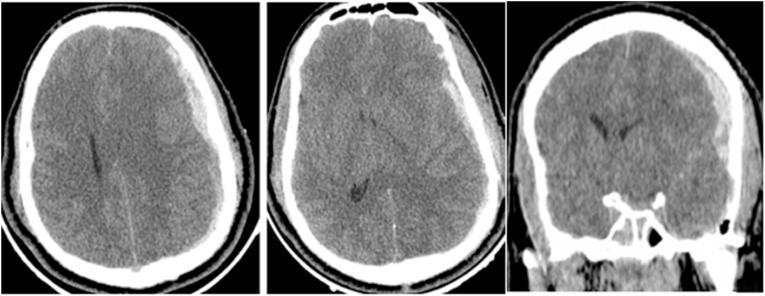
Non-contrast CT scan of the brain in axial and coronal planes demonstrating acute traumatic intracranial pathology. On the left side, there is a prominent acute subdural hematoma (ASDH) characterized by a hyperdense, crescent-shaped collection along the frontoparietal convexities. This mass effect causes significant midline shift toward the right and partial effacement of the ipsilateral lateral ventricle. Contralaterally, on the right side, a smaller hyperdense biconvex or lenticular collection is visible in the parieto-temporal region, consistent with an epidural hematoma (EDH). Additionally, a linear skull fracture is present on the right side of the skull vault. This diagnostic image illustrates the radiological features of mixed intracranial hemorrhages following severe head injury, highlighting the differential morphology between subdural (crescentic) and epidural (biconvex) blood collections and their secondary effects on brain parenchyma.
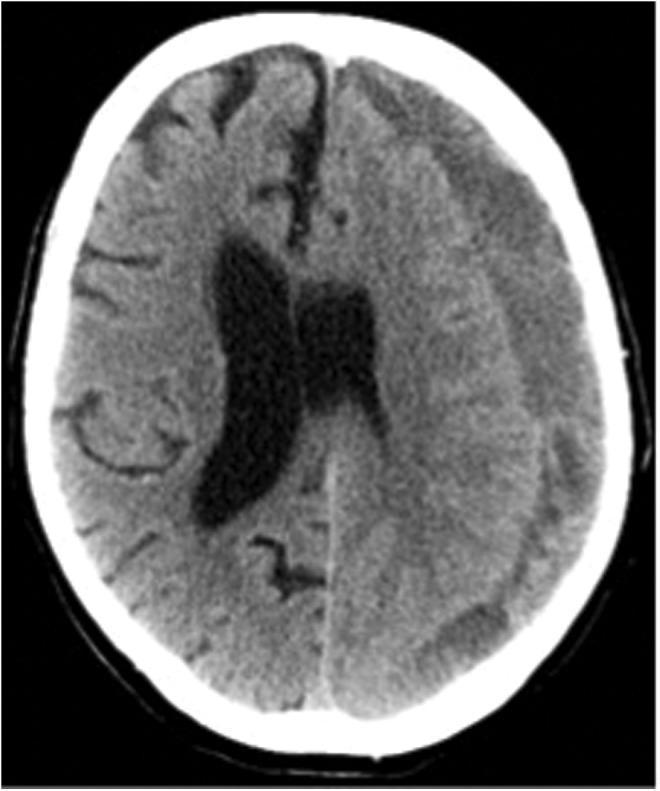
This diagnostic image is an axial non-contrast computed tomography (CT) scan of the brain demonstrating a chronic subdural hematoma (cSDH). The primary pathology is a large, hypodense, crescent-shaped collection located along the left cerebral hemisphere. This hypodensity indicates the hematoma is in a chronic stage, where liquefied blood products appear darker than the adjacent brain parenchyma. The collection extends across the coronal and lambdoid sutural margins, a hallmark of subdural versus epidural hemorrhages. Significant mass effect is evident, characterized by a marked midline shift toward the right side and compression of the left lateral ventricle. There is visible effacement of the cortical sulci on the ipsilateral side. This image is a classic example used in neurosurgery and radiology to illustrate the visual characteristics of chronic intracranial hemorrhage and its secondary effects on cerebral anatomy, such as subfalcine herniation and ventricular displacement.
Hematoma - Intracranial Types
- Epidural hematoma (EDH)
- Subdural hematoma (SDH) - acute, subacute, and chronic
- Subarachnoid hemorrhage (SAH)
- Intracerebral (intraparenchymal) hematoma (ICH)
Anatomical Overview
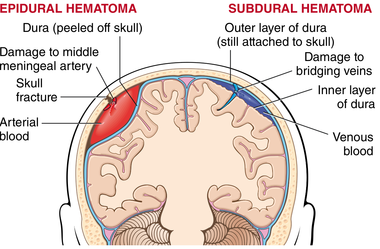
1. Epidural Hematoma (EDH)
Anatomy and Space
Mechanism
- Most commonly caused by temporal skull fracture crossing the middle meningeal artery groove - the fracture lacerates this artery
- Bleeding is arterial and high-pressure - it does not self-tamponade, and the hematoma expands rapidly
- If the ruptured vessel is venous, bleeding is slower and more similar to a subdural hematoma
- Rare non-traumatic causes: metastatic skull tumors, eosinophilic granuloma, craniofacial infections
Location
- Most common: lateral temporal region (middle meningeal artery territory)
- Can also be: frontal, occipital, vertex, or contrecoup
Classic Clinical Presentation
- Initial head trauma (may be brief or mild)
- "Lucid interval" - a period of relative clarity after the injury
- Rapid deterioration as hematoma expands
- Ipsilateral pupil dilation (early) → complete ophthalmoplegia → contralateral third nerve palsy as uncal herniation progresses
- Motor signs tend to appear late
- About 50% had only mild symptoms or no loss of consciousness initially - EDH can be picked up on CT with minimal symptoms
Imaging
- CT: Biconvex (lens-shaped / lentiform) hyperdense mass between skull and brain
- Does NOT cross suture lines (dura is fused at sutures)
- Convex on both surfaces (vs. SDH which is concave on the brain surface)
- Check for skull fracture crossing the middle meningeal groove
- MRI not required for diagnosis but useful for assessing underlying contusions
Management
- If causing brain displacement and consciousness impairment: emergency surgical evacuation
- Time from injury to treatment is the most critical determinant of prognosis
- Most patients operated on promptly recover, including ~1/3 of those with a fixed dilated pupil pre-operatively
- Only ~30% ultimately require surgical intervention (many detected incidentally on CT are managed conservatively)
- Plum and Posner's Diagnosis and Treatment of Stupor and Coma, pp. 245-247
- Robbins Pathologic Basis of Disease, pp. 3928-3943
2. Subdural Hematoma (SDH)
Anatomy and Space
Mechanism
- Caused by acceleration-deceleration injury tearing the bridging veins
- Bleeding is venous and low-pressure - typically self-tamponades unless coagulation is impaired
- Blood dissects through both dural layers to create the collection
- In elderly (atrophied brain) and infants, bridging veins are stretched or thin-walled, increasing susceptibility
Risk Factors for SDH
- Elderly (cerebral atrophy stretches bridging veins)
- Anticoagulants / antiplatelet agents (venous bleeding continues unchecked)
- Chronic alcoholism
- Hemodialysis
- Intracranial hypotension
- Infants < 2 years old (nonaccidental trauma)
Classification by Time
| Type | Timeframe | CT Appearance | Key Features |
|---|---|---|---|
| Acute | 0-14 days | Hyperdense (white), crescent-shaped | Severe trauma, often with contusions; coma common |
| Subacute | ~1-3 weeks | Isodense (hard to see on CT) | Use IV contrast CT or MRI |
| Chronic | > 2-3 weeks | Hypodense (dark) - iron metabolized | Elderly, often trivial/no remembered trauma |
Morphology (Acute → Chronic Progression)
- Freshly clotted blood along brain surface (no extension into sulci); subarachnoid space usually clear
- ~1 week: Lysis of clot
- ~2 weeks: Fibroblast ingrowth from dural surface
- 1-3 months: Hyalinized connective tissue forms
- Eventually: hematoma attached to inner dura by fibrous tissue; can retract to a thin "subdural membrane"
- Alternatively: thin-walled granulation tissue vessels re-bleed → chronic subdural hematoma
CT Shapes
- Crescent-shaped collection conforming to cortical surface
- Crosses suture lines (unlike EDH)
- Location: usually over the lateral cerebral hemispheres; bilateral in ~10-20%
Clinical Features
- No specific clinical syndrome - presentation is variable
- Acute: usually comatose immediately after severe injury; associated with underlying parenchymal damage → higher mortality
- Chronic: vague headaches, mental status changes (confusion, slowness), memory problems - often in elderly; no recalled injury in ~50%
- A useful clinical rule: "It could always be a subdural" - image even when focal signs are absent
Treatment
- Acute/subacute with mass effect: Surgical evacuation (emergency)
- Chronic: depends on size and symptoms
- Larger/symptomatic: Burr hole drainage
- Smaller/asymptomatic: can be managed conservatively
- Corticosteroids (e.g., dexamethasone) have been used; some evidence supports them as an alternative or adjunct to surgery for chronic SDH - the SUCRE trial is an ongoing randomized trial to clarify this
- Plum and Posner's Diagnosis and Treatment of Stupor and Coma, pp. 248-251
- Robbins Pathologic Basis of Disease, pp. 3947-3975
- Tintinalli's Emergency Medicine, pp. 1731-1732
3. Intracerebral Hematoma (ICH)
Mechanism
- Traumatic ICH: direct brain parenchyma injury causing intraparenchymal bleeding
- Can be spontaneous (hypertension, amyloid angiopathy, AVM, coagulopathy)
- Has been reported with all degrees of head trauma severity
Clinical Features
- Effects depend on size and location of bleeding and whether it is expanding
- > 50% of patients with ICH lose consciousness at time of impact
- Combined with contusions and perilesional edema, can produce significant mass effect and herniation
- Traumatic cerebellar hematoma presents similarly to other posterior fossa lesions
Management
- Conservative (ICP management) vs. surgical evacuation depending on size, location, and clinical trajectory
- Reverse anticoagulation urgently if applicable
4. Key Comparison Table
| Feature | Epidural | Acute Subdural | Chronic Subdural | Intracerebral |
|---|---|---|---|---|
| Space | Epidural (skull-dura) | Subdural (dura-arachnoid) | Subdural | Brain parenchyma |
| Vessel | Middle meningeal artery | Bridging veins | Bridging veins / membrane vessels | Cortical/perforating arteries |
| Blood type | Arterial | Venous | Venous | Mixed |
| CT shape | Biconvex (lens) | Crescent | Crescent (hypodense) | Irregular intraparenchymal |
| Crosses sutures? | No | Yes | Yes | N/A |
| CT density | Hyperdense | Hyperdense | Hypodense | Hyperdense |
| Lucid interval | Classic | Uncommon | N/A | Uncommon |
| Typical patient | Young adult, trauma | Severe TBI | Elderly, alcoholic | Any age |
| Urgency | Extreme emergency | Emergency | Less urgent | Variable |
| Treatment | Craniotomy / evacuation | Evacuation | Burr hole / steroids | Medical ± surgical |
5. Clinical Red Flags for Expanding Hematoma
- Decreasing GCS score - most reliable indicator of expanding lesion
- Worsening headache, focal neurologic signs, confusion progressing to coma
- Ipsilateral fixed dilated pupil (uncal herniation from temporal EDH/SDH)
- Decerebrate posturing (midbrain involvement)
- In elderly on anticoagulants: even a minor fall warrants CT
Key clinical pearl: In the elderly, brain atrophy creates more cranial vault space - significant hemorrhage can accumulate before obvious clinical deterioration. Always image elderly patients after head trauma regardless of apparent severity.
- Rosen's Emergency Medicine, p. 2411
- Plum and Posner's Diagnosis and Treatment of Stupor and Coma, pp. 245-254