
21 weeks pregnant with conus at leve of L3 to L4. is it normal in fetus
Find clinical guidelines on prenatal spinal cord assessment
jhhj
21 weeks pregnant with conus at leve of L3 to L4. is it normal in fetus
normal conus medullaris level fetal ultrasound 21 weeks gestation
conus medullaris level fetal ultrasound gestational age
PMID: 20582935
PMID: 36538102
PMID: 7933017
Short answer
What the evidence says
- Robbin, Filly & Goldstein (1994) — the classic sonographic study of fetal conus position — concluded that a termination level of L2-L3 or higher is normal, a level of L3 is indeterminate and needs neonatal re-evaluation, and a level of L3-L4 or lower is abnormal, warranting a careful search for associated anomalies (tethered cord, filum lipoma, occult dysraphism, etc.) (PMID 7933017).
- Perlitz et al. (2010), specifically studying fetuses at 20-24 weeks (the window that includes 21 weeks), found that 93% of normal fetuses had the conus ending at L2, the L2-3 interspace, or L3. They state that a conus found lower than L3 should prompt prenatal and postnatal surveillance (PMID 20582935).
- Sun et al. (2023), a large prospective cohort (1,202 fetuses, 17-40 weeks), built gestational-age-specific lower-limit reference tables and concluded the conus should continue moving cranially throughout pregnancy, and should not lie below L2 at birth. They created this precisely to help catch fetal tethered cord syndrome early (PMID 36538102).
- A standard neurology reference textbook notes the conus is around L4-L5 at 20 weeks, ascending to L3 by term (40 weeks), and reaching the adult L1-L2 level by about 2 months after birth (Localization in Clinical Neurology, 8th ed.). This is a more general teaching estimate; the dedicated fetal-ultrasound studies above (which directly measured 20-24 week fetuses) are more precise for your specific question and place the expected normal position a bit higher (around L2-L3) than this general textbook statement.
Putting it together for your case
- At 21 weeks, "L3-L4" falls at or past the point multiple studies classify as not clearly normal — it's in the range that should prompt closer look rather than reassurance.
- This does not automatically mean pathology. A single measurement at one time point, especially in the narrow window where the conus is actively migrating, can be affected by fetal position, image angle, and measurement technique (counting vertebrae from T12/last rib is technically difficult, and Perlitz's group could only reliably visualize it in ~71% of cases).
- What should happen next, per the literature and standard practice:
- Detailed anatomic survey / targeted spine ultrasound looking for associated findings: caudal regression, myelomeningocele/spina bifida, filum terminale thickening or lipoma, syrinx, sacral/vertebral segmentation anomalies, abnormal lower-extremity positioning or clubfoot (things that go along with a tethered cord).
- Follow-up ultrasound later in pregnancy to see whether the conus ascends appropriately as gestation advances (it should trend toward L2-L3 by term).
- Consider fetal MRI and maternal-fetal medicine/pediatric neurosurgery referral if the low position persists, other anomalies are seen, or the finding is isolated but the family wants further characterization.
- Postnatal spinal ultrasound (or MRI) is recommended if the conus remains low-lying near delivery, since tethered cord syndrome is a postnatal diagnosis of exclusion supported by serial imaging.
Bottom line
Create a summary document explaining findings to share with your obstetric team
mkdir -p /tmp/workspace/conus-medullaris-summary && ls -la /tmp/workspace/
~/conus-medullaris-summary/build_doc.py
from docx import Document
from docx.shared import Pt, Inches, RGBColor
from docx.enum.text import WD_ALIGN_PARAGRAPH
from docx.enum.table import WD_TABLE_ALIGNMENT
from docx.oxml.ns import qn
from docx.oxml import OxmlElement
import datetime

doc = Document()

# Default font
style = doc.styles["Normal"]
style.font.name = "Arial"
style.font.size = Pt(11)

DARK_BLUE = RGBColor(0x1F, 0x3B, 0x57)
GREY = RGBColor(0x55, 0x55, 0x55)

def set_cell_shading(cell, color_hex):
    tc_pr = cell._tc.get_or_add_tcPr()
    shd = OxmlElement('w:shd')
    shd.set(qn('w:val'), 'clear')
    shd.set(qn('w:color'), 'auto')
    shd.set(qn('w:fill'), color_hex)
    tc_pr.append(shd)

# ---------- Title ----------
title = doc.add_heading("Prenatal Ultrasound Finding: Conus Medullaris Position", level=0)
title.runs[0].font.color.rgb = DARK_BLUE

sub = doc.add_paragraph()
sub.alignment = WD_ALIGN_PARAGRAPH.LEFT
run = sub.add_run("Summary prepared for discussion with the obstetric / maternal-fetal medicine team")
run.italic = True
run.font.color.rgb = GREY
run.font.size = Pt(11)

date_p = doc.add_paragraph()
date_run = date_p.add_run(f"Date prepared: {datetime.date.today().strftime('%B %d, %Y')}")
date_run.font.size = Pt(10)
date_run.font.color.rgb = GREY

doc.add_paragraph()

# ---------- Case Summary ----------
doc.add_heading("Case Summary", level=1)
p = doc.add_paragraph()
p.add_run("Gestational age at scan: ").bold = True
p.add_run("21 weeks\n")
p2 = doc.add_paragraph()
p2.add_run("Finding: ").bold = True
p2.add_run("Conus medullaris (tapered end of the fetal spinal cord) visualized at the L3-L4 vertebral level on prenatal ultrasound.")

doc.add_paragraph(
    "This document summarizes what is known from the medical literature about normal conus "
    "medullaris position at this stage of pregnancy, to support discussion with the obstetric team "
    "about interpretation and appropriate follow-up. It is an educational summary, not a diagnosis."
)

# ---------- Background ----------
doc.add_heading("Background: Normal Conus Ascent in Pregnancy", level=1)
doc.add_paragraph(
    "During fetal development, the spinal column (vertebrae) grows faster than the spinal cord itself. "
    "As a result, the conus medullaris appears to migrate upward (\u201cascends\u201d) relative to the vertebral "
    "levels as pregnancy progresses, continuing to rise gradually until shortly after birth, when it "
    "reaches the adult position (around L1-L2)."
)

table = doc.add_table(rows=1, cols=2)
table.style = 'Light Grid Accent 1'
table.alignment = WD_TABLE_ALIGNMENT.CENTER
hdr = table.rows[0].cells
hdr[0].text = "Timepoint"
hdr[1].text = "Typical / Reference Conus Level"
for c in hdr:
    for para in c.paragraphs:
        for r in para.runs:
            r.bold = True
    set_cell_shading(c, "1F3B57")
    for para in c.paragraphs:
        for r in para.runs:
            r.font.color.rgb = RGBColor(0xFF, 0xFF, 0xFF)

rows_data = [
    ("~19-24 weeks (2nd trimester)", "Most normal fetuses: L2, L2-3 interspace, or L3 (per Perlitz et al., 2010, studying 20-24 week fetuses)"),
    ("General teaching estimate at 20 weeks", "~L4-L5 (per standard neurology reference texts; less precise than dedicated fetal ultrasound studies)"),
    ("By term / 40 weeks", "~L3, continuing to trend toward L2-L3"),
    ("~2 months after birth (adult level)", "L1-L2"),
]
for label, val in rows_data:
    row = table.add_row().cells
    row[0].text = label
    row[1].text = val

doc.add_paragraph()

# ---------- Interpretation ----------
doc.add_heading("Interpretation of an L3-L4 Finding at 21 Weeks", level=1)
doc.add_paragraph(
    "Based on the dedicated fetal-ultrasound literature (rather than general anatomy teaching points), "
    "a conus level of L3-L4 at 21 weeks falls at or below the range most studies classify as reassuring "
    "for this gestational age:"
)

bullets = [
    ("Robbin, Filly & Goldstein (1994) - classic sonographic reference study",
     "L2-L3 or higher = normal. L3 = indeterminate, needs neonatal re-evaluation. "
     "L3-L4 or lower = abnormal; warrants a careful sonographic search for associated anomalies "
     "(tethered cord, filum lipoma, occult spinal dysraphism)."),
    ("Perlitz et al. (2010) - 20-24 week fetuses",
     "93% of normal fetuses had the conus ending at L2, the L2-3 interspace, or L3. "
     "A conus lower than L3 should prompt prenatal and postnatal surveillance."),
    ("Sun et al. (2023) - large prospective cohort, 1,202 fetuses, 17-40 weeks",
     "Conus position should continue moving cranially throughout pregnancy and should not lie below "
     "L2 at birth. Gestational-age-specific reference tables were developed to help identify fetal "
     "tethered cord syndrome early."),
]
for source, finding in bullets:
    p = doc.add_paragraph(style="List Bullet")
    r1 = p.add_run(source + ": ")
    r1.bold = True
    p.add_run(finding)

doc.add_paragraph()
note = doc.add_paragraph()
note.add_run("Key point: ").bold = True
note.add_run(
    "This single measurement is not necessarily abnormal on its own, but it is in a range that the "
    "literature treats as needing a closer look rather than automatic reassurance. Ultrasound "
    "measurement of the conus is technically difficult (reliably visualized in roughly 70% of "
    "fetuses in published series) and can be affected by fetal position, image angle, and vertebral "
    "counting method."
)

# ---------- Suggested next steps ----------
doc.add_heading("Suggested Discussion Points / Next Steps", level=1)
steps = [
    "Detailed targeted spine ultrasound: look for associated findings such as caudal regression, "
    "spina bifida/myelomeningocele, filum terminale thickening or lipoma, syringomyelia, sacral or "
    "vertebral segmentation anomalies, or abnormal lower-limb positioning (e.g., clubfoot) that can "
    "accompany a tethered cord.",
    "Repeat/follow-up ultrasound later in pregnancy to confirm the conus ascends appropriately over "
    "time, as expected physiologically.",
    "Consider fetal MRI and maternal-fetal medicine or pediatric neurosurgery referral if the low "
    "position persists, other anomalies are seen, or further characterization is desired.",
    "Postnatal spinal ultrasound (or MRI) if the conus remains low-lying near delivery, since tethered "
    "cord syndrome is generally confirmed postnatally with serial imaging and clinical follow-up.",
]
for s in steps:
    doc.add_paragraph(s, style="List Number")

# ---------- References ----------
doc.add_heading("References", level=1)
refs = [
    "Robbin ML, Filly RA, Goldstein RB. The normal location of the fetal conus medullaris. "
    "J Ultrasound Med. 1994;13(7):541-546. PMID: 7933017.",
    "Perlitz Y, Izhaki I, Ben-Ami M. Sonographic evaluation of the fetal conus medullaris at 20 to 24 "
    "weeks' gestation. Prenat Diagn. 2010. PMID: 20582935.",
    "Sun M, Chen M, Gao G, Tao B, Shang A. Gestational age-specific reference standards of low-lying "
    "conus medullaris level in fetuses: a prospective cohort study. Childs Nerv Syst. 2023. "
    "PMID: 36538102.",
    "Localization in Clinical Neurology, 8th Edition - Spinal Cord chapter (general reference on "
    "conus medullaris development).",
]
for r in refs:
    doc.add_paragraph(r, style="List Bullet")

doc.add_paragraph()
disclaimer = doc.add_paragraph()
d_run = disclaimer.add_run(
    "This summary is intended to support - not replace - discussion with your obstetrician or "
    "maternal-fetal medicine specialist, who can interpret this finding alongside the full ultrasound "
    "images and your complete clinical picture."
)
d_run.italic = True
d_run.font.size = Pt(9)
d_run.font.color.rgb = GREY

out_path = "/tmp/workspace/conus-medullaris-summary/Conus_Medullaris_Summary_for_Obstetric_Team.docx"
doc.save(out_path)
print("Saved:", out_path)
cd /tmp/workspace/conus-medullaris-summary && python3 build_doc.py
Conus Medullaris Summary for Obstetric Team
Word Document · DOCX
- Case Summary – 21 weeks gestation, conus medullaris at L3-L4
- Background – table showing how the conus normally ascends over gestation (2nd trimester → term → 2 months postnatal)
- Interpretation – summarized findings from the three key studies (Robbin 1994, Perlitz 2010, Sun 2023), noting that L3-L4 at 21 weeks falls at/below the range considered reassuring, though it isn't automatically abnormal
- Suggested discussion points/next steps – targeted spine ultrasound for associated anomalies, follow-up scan later in pregnancy, possible fetal MRI/MFM or pediatric neurosurgery referral, postnatal imaging if it remains low
- References with PMIDs for the team to pull up directly
- A closing note framing this as a discussion aid, not a diagnosis
1 and 2
fetal targeted spine ultrasound tethered cord associated findings filum lipoma sonographic markers
timing follow-up ultrasound low-lying fetal conus medullaris third trimester recommendation
1. Detailed/Targeted Spine Ultrasound - What It Looks For
- Filum terminale thickness - a thickened, echogenic filum (>2 mm) is a classic marker of a tight/fatty filum causing cord tethering, and often accompanies a low-lying conus.
- Filum terminale lipoma or fibrolipoma - a fatty mass at the end of the thecal sac; can tether the cord even when the conus otherwise looks reasonably positioned.
- Vertebral/sacral segmentation anomalies - hemivertebrae, sacral agenesis, or caudal regression, which are associated with abnormal cord position.
- Open or closed spinal dysraphism - spina bifida, myelomeningocele, or a skin-covered mass over the lower spine.
- Syringomyelia - fluid-filled cavity within the cord itself.
- Overlying skin/soft tissue markers - subcutaneous lipoma, hairy patch (hypertrichosis), or a skin dimple/sinus tract over the lower back - these are the same "cutaneous stigmata" pediatricians look for on newborns with suspected tethered cord.
- Lower limb positioning - clubfoot or other lower-extremity deformities can co-occur with tethered cord.
- Associated brain findings - ventriculomegaly or Chiari II-type changes if there's any suspicion of an open neural tube defect.
2. Follow-Up (Repeat) Ultrasound Later in Pregnancy
- Why: the conus continues to migrate cranially throughout gestation, so a repeat scan checks whether it's ascending as expected (trending toward L2-L3 by term) or staying persistently low relative to the gestational-age norms.
- Typical timing: most published series that follow low-lying conus cases reassess in the third trimester (commonly cited around 28-32 weeks, sometimes closer to 32-34 weeks), giving enough time to see a meaningful ascent while still leaving time to plan delivery/postnatal workup if needed. Your MFM specialist will pick the exact timing based on your anatomy scan quality and institutional protocol.
- What reassures vs. what doesn't:
- Reassuring: conus has ascended toward L2-L3, no new anomalies, normal filum thickness.
- Persistent concern: conus remains at or below L3-L4, thickened filum, or new associated findings - this raises the case for fetal MRI and a planned postnatal ultrasound/MRI plus pediatric neurosurgery involvement.
- After birth: if the conus is still low near delivery, postnatal spinal ultrasound is typically done in the first few months (evaluation window roughly 3-10 days after delivery for a baseline neuro exam, with imaging follow-up through infancy), since ultrasound is easiest to interpret before the vertebral bodies ossify, generally before about 4-6 months of age. MRI is used if ultrasound is inconclusive or surgery is being considered.
Find clinical images showing normal vs. low-lying conus on fetal ultrasound
second trimester ultrasound fetal spine conus medullaris L2-L3 normal position
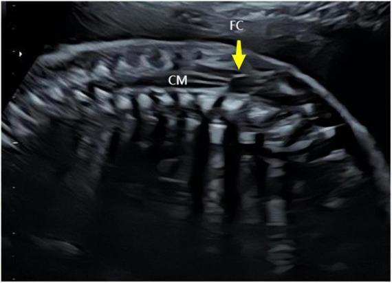
Normal Conus Medullaris (with an incidental filar cyst)
Low-Lying Conus / Tethered Cord at 21 Weeks (directly comparable to your case)
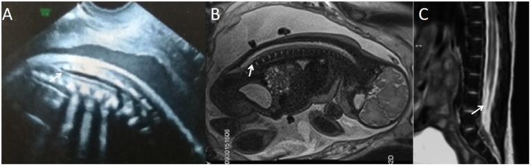
- Panel A - prenatal ultrasound at ~21 weeks. The arrow marks the conus medullaris as a tapering hypoechoic structure ending clearly lower/more caudal than expected.
- Panel B - fetal MRI at the same 21 weeks, giving better soft-tissue contrast and a clearer view of exactly where the cord terminates relative to the vertebral bodies.
- Panel C - postnatal T2-weighted MRI at 22 days old, confirming the pathologically low-lying conus extending well below the expected L1-L2 level, consistent with tethered cord syndrome.
Why this comparison matters for your finding
- Normal: cord tapers into the conus at or above roughly L2-L3 by mid-second trimester, with a clean, smooth termination (the cyst in image 1 is incidental and not a marker of pathology).
- Abnormal/low-lying: the cord fails to taper at the expected level and extends further down the canal (as in image 2), which on ultrasound alone can be subtle, which is exactly why a low-normal ultrasound finding (like your L3-L4 measurement) often prompts either a repeat scan or fetal MRI, since MRI (Panel B) resolves the termination level more precisely than ultrasound (Panel A) can.
Search for recent research on outcomes of prenatally-diagnosed low conus
prenatal low-lying conus medullaris outcome
fetal tethered cord syndrome prognosis
recent outcomes prenatally diagnosed low-lying conus medullaris tethered cord 2023 2024 2025
PMID: 39999507
PMID: 35879383
PMID: 36719707
PMID: 33485624
PMID: 36564726
PMID: 34687566
Recent Research on Outcomes After Prenatal Diagnosis of a Low Conus
Key findings
- Isolated CM anomaly, no other findings: live birth rate 94.6%, and CM ascended above L3 in 89.2% of these fetuses on follow-up.
- CM anomaly + other structural/genetic anomalies: live birth rate only 22.1% (reflecting terminations for major associated anomalies), and CM ascended above L3 in only 19.1%.
- A CM below L4 was identified as the statistical cutoff that best predicted true lumbosacral spinal anomalies (ROC analysis).
- On 1-5 year follow-up, nearly all live-born children in both groups grew and developed normally with no tethered cord symptoms, aside from 3 cases with adverse outcomes overall.
- Bottom line from this study: an isolated low CM above L4, with no other anomalies, generally carries a favorable prognosis. Caution is warranted specifically when CM sits at or below L4 (PMID 39999507).
- Diagnosed at an average of 23-24 weeks (close to your 21-week timing).
- 24.3% of these low CM cases had an associated spinal dysraphism (open spina bifida or tethered cord syndrome).
- Proposed cutoff: CM at or below the L4.25 level predicted spinal dysraphism with 96% specificity (66.7% sensitivity, AUC 0.877).
- Of the 6 tethered cord cases that continued to live birth, 5 required surgical release, but short-term postnatal prognosis for live births overall was good (PMID 36564726).
- Confirms the conus reaches its normal birth-level position by around 26 weeks' gestation, with the most significant ascent happening between roughly 13-18 weeks and 26-32 weeks.
- This matters for your case: at 21 weeks, the conus is still actively migrating, so an L3-L4 finding now is not the final word, a genuine reassessment around or after 26 weeks is when the "settled" position becomes more meaningful (PMID 35879383).
- Refines the risk-stratification bands: at the start of the third trimester, CM above the L2/3 disc = normal; CM at L3 = physiological but needs follow-up; CM below L3 = suggestive of tethered cord syndrome.
- By term, the conus normally migrates to the L1/2 disc level (PMID 34687566).
- Only 4 of 26 cases (15%) were "solitary" tethered cord with no other findings; the remaining 22 had associated anomalies: spina bifida occulta/aperta, hydrocephalus, VACTERL association, cloacal exstrophy, or chromosomal abnormalities.
- Chromosomal abnormality rate was 23.1% in this cohort, reinforcing why a detailed anomaly scan (and possibly genetic counseling) is a standard next step when a low CM is found (PMID 36719707).
- In 38 fetuses with suspected tethered cord/low spinal cord, MRI outperformed ultrasound, catching additional diagnoses (including 2 cases of diastematomyelia missed on ultrasound) and providing more information for counseling and management decisions (PMID 33485624).
How this updates the picture for your case
- Your finding of L3-L4 at 21 weeks sits right at the boundary multiple recent studies flag as needing closer follow-up (L4 cutoff in Zhang 2025; L4.25 cutoff in Jing 2022; "L3 = needs follow-up" in Sun 2021).
- It is not in the range these studies associate with a high probability of true pathology (that's more consistently below L4-L5), but it's also not in the clearly reassuring range (above L2-L3).
- Timing works in your favor: significant conus ascent normally continues until about 26 weeks, so your team will likely want a repeat scan around/after 26 weeks rather than drawing conclusions now.
- If the follow-up scan shows the conus has ascended above L3 with no other anomalies, the 2025 outcome data (94.6% live birth, largely normal development at 1-5 years) is genuinely reassuring.
- If it stays low or other findings appear, the literature consistently recommends a detailed anomaly scan, consideration of fetal MRI (more sensitive than ultrasound), and genetic counseling, given the meaningfully higher rate of associated structural and chromosomal findings in that scenario.
how many sacral vertebrae are seen at 21 weeks
How many sacral vertebrae can u count at 21 weeks scan
How many sacral vertebrae can u count at 21 weeks scan