
Anisocoria , pathophysiology , causes and management
Reading File
Finding Sources
Finding Sources
Searching PubMed
"anisocoria"[MeSH Terms] OR anisocoria
Reading File
Reading File
Reading File
Searching Images
anisocoria differential diagnosis flowchart Horner syndrome third nerve palsy
Anisocoria
Anisocoria means the two pupils are unequal in size. The first job is to decide which pupil is the abnormal one. The second is to decide whether the cause is dangerous, such as a third nerve palsy from an aneurysm or a Horner syndrome from carotid dissection or an apical lung tumor.
Pathophysiology
Pupil size depends on two opposing muscles in the iris.
| Pathway | Muscle | Effect | Neurotransmitter | Course |
|---|---|---|---|---|
| Parasympathetic | Sphincter pupillae | Miosis | Acetylcholine | Edinger-Westphal nucleus, CN III, ciliary ganglion, short ciliary nerves |
| Sympathetic | Dilator pupillae | Mydriasis | Norepinephrine | Hypothalamus (1st order), then ciliospinal center C8-T2 (2nd order), then superior cervical ganglion, then along the carotid to the eye (3rd order) |
Anisocoria arises when one side of this system fails or is pharmacologically blocked.
- Parasympathetic failure gives a large pupil that reacts poorly to light. Causes include CN III lesions, ciliary ganglion damage (Adie pupil), iris sphincter damage, or anticholinergic drugs.
- Sympathetic failure gives a small pupil with dilation lag. This is Horner syndrome.
- Iris pathology (trauma, inflammation, ischemia, synechiae) can make the pupil either small or large.
- Afferent lesions (retina, optic nerve) do not cause anisocoria. As Kanski notes, "in afferent (sensory) lesions, the pupils are equal in size. Anisocoria implies disease of the efferent (motor) nerve or the iris itself" - Kanski's Clinical Ophthalmology, 10th ed.
Which pupil is abnormal? Compare light and dark
This is the key bedside step.
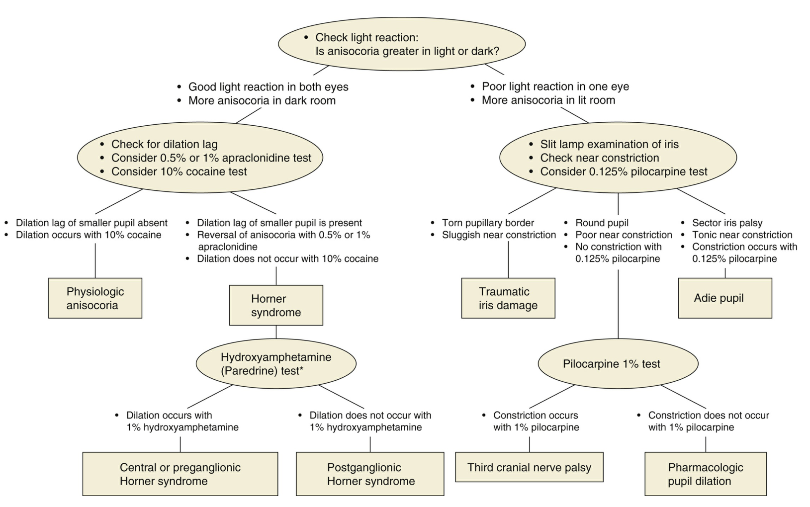
- Anisocoria greater in bright light: the large pupil is abnormal. It fails to constrict. Think parasympathetic lesion, sphincter injury, or a mydriatic drug.
- Anisocoria greater in the dark: the small pupil is abnormal. It fails to dilate. Think Horner syndrome, an inflamed iris, or a miotic drug.
- Same size in all lighting: this suggests physiologic (simple) anisocoria.
Bradley and Daroff add that physiologic anisocoria is often slightly greater in the dark, so a small difference in the dark does not by itself mean Horner syndrome (Bradley and Daroff's Neurology in Clinical Practice, Examination of the Pupils). Adams and Victor summarize it as "light exaggerates the anisocoria caused by a third-nerve lesion, and darkness accentuates the anisocoria in the case of Horner syndrome" (Adams and Victor's Principles of Neurology, 12th ed, p. 300).
Causes
1. Physiologic (simple) anisocoria
- About 20% of normal people have a difference of 0.3 to 0.5 mm or more (Adams and Victor, p. 300).
- The difference is the same in light and dark, and it can vary from day to day.
- Both pupils react normally. There is no ptosis and no ocular motility deficit.
- It needs no treatment.
2. Abnormal pupil is small (miosis)
- Horner syndrome: mild ptosis, dilation lag, and sometimes anhidrosis. The cause may be central, preganglionic, or postganglionic. Examples are brainstem or lateral medullary stroke, Pancoast tumor, carotid dissection, cavernous sinus lesions, and cluster headache.
- Iritis: pain, redness, and cells and flare in the anterior chamber.
- Miotic drugs: pilocarpine, opioids, and others.
- Argyll Robertson pupils: these are bilateral, irregular, and show light-near dissociation, classically in neurosyphilis. Mild anisocoria is common.
- Long-standing Adie pupil: it may become small over time.
3. Abnormal pupil is large (mydriasis)
- Third nerve palsy: ptosis and/or ophthalmoplegia are present. A dilated pupil should prompt urgent exclusion of a posterior communicating artery aneurysm. Other causes are uncal herniation, ischemia, tumor, and trauma.
- Adie (tonic) pupil: it reacts poorly to light, slowly and tonically to accommodation, and is hypersensitive to dilute pilocarpine (0.125%).
- Iris sphincter damage from trauma or surgery: the pupillary margin is torn and the iris shows transillumination defects.
- Pharmacologic mydriasis: atropinic or sympathomimetic agents, such as scopolamine patches, nebulized ipratropium, or accidental contact. This is common in healthcare workers.
- Acute angle-closure glaucoma: a mid-dilated, fixed pupil with a painful red eye.
- Episodic anisocoria: migraine-associated, benign episodic mydriasis, or seizure-related.
(Sources: The Wills Eye Manual, 10.1 Anisocoria, pp. 635-636; Rosen's Emergency Medicine, Anisocoria.)
Danger signs. In a comatose patient, a unilateral blown pupil suggests herniation. In a patient with neck pain or headache, think carotid dissection (Horner syndrome) or aneurysm (third nerve palsy).
Evaluation
- History: onset, duration (old photographs help), pain, headache, trauma, eye surgery, drug or eye drop exposure, and medications.
- Resting pupil size in light and dark. Also check eyelid position, globe position (to exclude proptosis), and extraocular motility. The Wills Eye Manual states that these "MUST be evaluated."
- Light reflex (direct and consensual), then near response. Look for light-near dissociation. Test for a relative afferent pupillary defect.
- Slit lamp exam: look for sphincter tears, synechiae, and iritis.
- Pharmacologic testing (1-2 drops in each eye, then re-examine at 30-45 minutes):
| Suspected problem | Test | Interpretation |
|---|---|---|
| Large pupil from drug blockade | Pilocarpine 1% | The pupil fails to constrict, so the iris sphincter is blocked or damaged. A pupil from third nerve palsy or Adie pupil will constrict. |
| Adie pupil | Pilocarpine 0.125% | The affected pupil constricts and the normal pupil does not (cholinergic denervation supersensitivity). |
| Horner syndrome | Apraclonidine 0.5-1% | The small pupil dilates, so miosis reverses (supersensitivity). Cocaine 4-10% is the older test: the Horner pupil dilates little or not at all. |
| Localizing Horner syndrome | Hydroxyamphetamine 1% | It must be given at least 24 to 48 hours after cocaine or apraclonidine. A pupil that fails to dilate suggests a postganglionic lesion. One that does dilate suggests a central or preganglionic lesion. |
Apraclonidine and cocaine responses can occur with a lesion anywhere along the sympathetic chain. Localization otherwise relies on associated signs (Adams and Victor, p. 300).
- Imaging, guided by the diagnosis:
- Third nerve palsy: urgent CT angiography or MR angiography to look for an aneurysm.
- Horner syndrome: imaging from the brain to the chest. Add neck vessel imaging (MRA or CTA) if there is acute pain or neck trauma.
Management
Treatment depends on the cause.
| Cause | Management |
|---|---|
| Physiologic anisocoria | Reassure the patient. No workup is needed if the examination is otherwise normal. |
| Pharmacologic mydriasis or miosis | Stop the agent. The pupil recovers over hours to days. |
| Third nerve palsy | This is an emergency when the pupil is involved. Arrange immediate neurovascular imaging and neurosurgical or neurology input. Manage microvascular risk factors if the pupil is spared. Treat the underlying cause. |
| Horner syndrome | Treat the underlying cause: carotid dissection (antithrombotic therapy), tumor (oncologic care), stroke, or other. Acute painful Horner syndrome needs urgent vascular imaging. |
| Adie (tonic) pupil | It is benign. Use dilute pilocarpine (0.0625-0.125%) for photophobia or near blur, or reading glasses. Consider a systemic cause if there is hyporeflexia (Holmes-Adie syndrome) or dysautonomia. |
| Iritis | Topical steroids and cycloplegics. Look for an underlying systemic disease. |
| Angle-closure glaucoma | Emergency treatment to lower intraocular pressure, plus laser peripheral iridotomy. |
| Traumatic iris injury | Examine for hyphema, lens injury, and open globe. Refer to ophthalmology. Cosmetic or glare problems may need an iris prosthesis or surgical repair. |
| Argyll Robertson pupils | Test for syphilis (serology) and treat with penicillin. |
Disposition. Admit or urgently image if any of these are present:
- acute onset with headache or neck pain
- ptosis or ophthalmoplegia with a dilated pupil
- altered consciousness
- new neurologic deficit
Otherwise, long-standing, isolated, and reactive anisocoria with normal lids and motility can be followed as an outpatient.
PubMed check
A search of recent systematic reviews (last 2 years) found nothing that changes this approach. One 2026 review looked at pupil changes as a marker of autonomic dysregulation after spinal cord injury [PMID 42812636], but it does not alter the evaluation above.
This is a shared conversation. Sign in to Orris to start your own chat.