
in detail about gbs
I'll look up Guillain-Barré Syndrome in the medical library for a detailed, textbook-quality answer.
Searching sources...
Reading File
Now let me search the medical library for GBS content in parallel:
Finding Sources
Finding Sources
Searching PubMed
"Guillain-Barre syndrome"[MeSH Terms] AND treatment
Good. The best sources are Bradley and Daroff's Neurology, Goldman-Cecil Medicine, Miller's Anesthesia, and Robbins Pathology. Let me read the neurology and medicine textbooks in parallel:
Reading File
Reading File
Reading File
Excellent content. Let me now read more from Bradley's Neurology (treatment and management) and Goldman-Cecil (management/prognosis) in parallel:
Reading File
Reading File
I have extensive content. Let me now read the treatment section from Goldman-Cecil and also read the GBS prognosis/ICU aspects from Bradley's Neurology:
Reading File
Reading File
I now have comprehensive textbook content. Let me read the GBS treatment section from Goldman-Cecil and the prognosis/management from Bradley's neurology:
Reading File
Reading File
I now have comprehensive content from multiple authoritative textbooks. I also have a great diagram image URL. Let me compile the full detailed answer:
Loading Image
I now have all the information needed for a comprehensive answer. Here is the full detailed overview of Guillain-Barré Syndrome:
Guillain-Barré Syndrome (GBS) - A Detailed Overview
1. Definition
Guillain-Barré syndrome refers to a group of acquired, inflammatory polyradiculoneuropathies that share:
- Acute onset (maximum deficit by 4 weeks)
- Elevated CSF protein with low cell counts (cytoalbuminologic / albuminocytological dissociation)
- A monophasic course
First described in 1916 by Guillain, Barré, and Strohl, the key clinical features recognized were motor weakness, areflexia, paresthesias with minor sensory loss, and raised CSF protein without pleocytosis. With the eradication of poliomyelitis, GBS has become the leading cause of acute paralytic disease in Western countries.
- Goldman-Cecil Medicine, p. 4065
- Bradley and Daroff's Neurology in Clinical Practice, p. 2663
2. Epidemiology
| Parameter | Data |
|---|---|
| Annual incidence | 1-2 per 100,000 |
| AIDP incidence (Europe/North America) | 0.6-1.9 per 100,000 |
| Male:Female ratio | 1.4-1.5:1 |
| Age distribution | All ages; incidence rises with age (0.8 in <18 yrs, 3.2 in >60 yrs) |
| Preceding illness | 60% have prior respiratory or GI infection |
Notable epidemiological associations:
-
Campylobacter jejuni gastroenteritis is the most common preceding infection, especially in axonal variants
-
Zika virus is associated with a significantly increased risk of all GBS forms
-
Hepatitis E is a trigger in Belgium and Netherlands (5-10% of cases)
-
SARS-CoV-2 infection or most vaccines are NOT substantially associated with GBS (except a slight increase with ChADOx1nCoV-19 vaccine: 0.6 cases/100,000 doses)
-
GBS incidence actually declined during the COVID-19 pandemic due to reduced spread of usual infectious triggers
-
Goldman-Cecil Medicine, p. 4065-4066
3. Classification / Subtypes
Common Subtypes
| Subtype | Key Feature | Geography |
|---|---|---|
| AIDP (Acute Inflammatory Demyelinating Polyradiculoneuropathy) | Demyelination of nerve roots and peripheral nerves | 97% of cases in North America and Europe |
| AMAN (Acute Motor Axonal Neuropathy) | Pure motor; axonal injury; no sensory loss | Summer epidemics in China; children/young adults |
| AMSAN (Acute Motor Sensory Axonal Neuropathy) | Both motor and sensory axons; more severe; poor recovery | Less common |
Rare Variants
-
Miller-Fisher Syndrome (MFS): Classic triad of ophthalmoplegia, ataxia, and areflexia. Facial weakness, ptosis, and pupillary abnormalities may occur. NCS velocities are normal. Anti-GQ1b antibodies in >85% of cases. Accounts for 6% of GBS in Western countries, 18% in Taiwan.
-
Pharyngeal-cervical-brachial variant
-
Facial diplegia with paresthesias
-
Multiple cranial neuropathy variant
-
Paraparetic variant
-
Acute pandysautonomia
-
Bradley and Daroff's Neurology in Clinical Practice, p. 2663-2664
4. Pathobiology / Pathogenesis
All forms of GBS result from postinfectious molecular mimicry: the immune system attacks peripheral nerve antigens because they structurally resemble antigens presented by microbes, particularly C. jejuni.
In AIDP: Multifocal inflammatory demyelination of spinal roots and peripheral nerves, with mononuclear cell infiltrates rich in macrophages. Injury is most extensive in nerve roots and proximal nerve segments. Both T-cell-mediated and antibody-mediated responses are involved (T cells considered dominant).
In AMAN (shown in diagram below):
- The HS/0:19 serotype of C. jejuni shares GM1-like epitopes with axonal membranes
- This triggers production of anti-GM1 antibodies
- Activated complement components (C3d, C5b-9) and immunoglobulin deposit at nodes of Ranvier
- Macrophages are recruited to targeted nodes and invade the periaxonal space
- This leads to Wallerian degeneration
- Lack of blood-nerve barrier at motor nerve terminals makes distal axons especially vulnerable to circulating GM1 antibodies
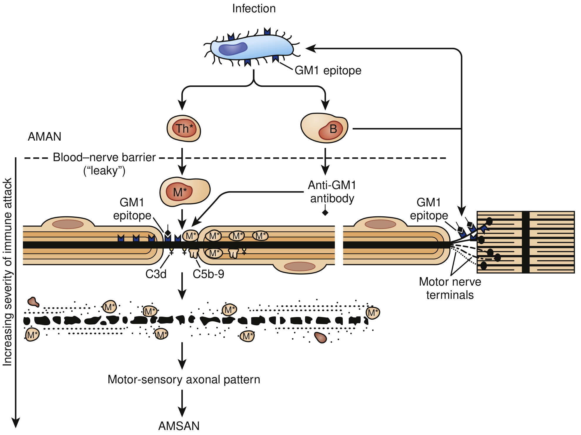
Fig: Immune Injury to Nerve Fibers in AMAN. Molecular mimicry of GM1-like epitopes between C. jejuni lipopolysaccharide and axonal membranes drives autoimmune attack. Increasing severity leads from AMAN to AMSAN. (Bradley and Daroff's Neurology, Fig. 106.18)
- Robbins & Kumar Basic Pathology, p. 809
- Goldman-Cecil Medicine, p. 4066
5. Clinical Features
Classic Presentation
- Weakness is the most common initial symptom - can range from mild difficulty walking to total quadriplegia with respiratory failure
- Classic ascending paralysis: leg weakness spreading to arms over hours to days
- Symmetrical weakness; proximal weakness is very common
- Areflexia/Hyporeflexia: invariable (may be absent early in disease)
- Symptoms progress over days to a maximum of 4 weeks (plateau then recovery)
Sensory Features
- Sensory loss is not prominent - often limited to distal vibration sense impairment
- Pain is underappreciated: moderate-to-severe pain in extremities, interscapular area, or back in ~70% during acute phase; may persist for a year in one-third
Cranial Nerve Involvement (45-75% of cases)
- Bilateral facial paresis in at least 50% of patients
- Extraocular muscle and lower cranial nerve involvement (less common)
- Occasional facial myokymia
- 5% present with isolated cranial nerve involvement that then descends
Autonomic Dysfunction (65% of cases)
- Cardiac arrhythmias (tachycardia, bradycardia)
- Blood pressure lability (hypertension and hypotension)
- Urinary retention
- Ileus
- Pupillary dysfunction
- Autonomic dysfunction is a major cause of mortality in ICU patients
Respiratory Involvement
-
Respiratory failure requiring mechanical ventilation in 9-30% of cases (increases with age)
-
Predictors of respiratory failure:
- Rapid disease progression (onset to admission <7 days)
- Severity of limb weakness
- Facial weakness
- Bulbar weakness
-
Goldman-Cecil Medicine, p. 4066
-
Bradley and Daroff's Neurology in Clinical Practice, p. 2663-2665
6. Diagnostic Criteria
Features Required for Diagnosis (Asbury & Cornblath criteria)
- Progressive weakness of both legs and arms
- Areflexia or hyporeflexia
Clinical Features Supportive of Diagnosis
- Progression over days to 4 weeks
- Relative symmetry
- Mild sensory symptoms or signs
- Bifacial palsies
- Autonomic dysfunction
- Absence of fever at onset
- Recovery beginning 2-4 weeks after progression ceases
Laboratory Features Supportive of Diagnosis
- Elevated CSF protein with <10 cells/µL (albuminocytologic dissociation)
- CSF protein may be normal in the first 7-10 days and remains normal in up to 10% of cases
- CSF WBC >50/mL suggests HIV or infection (Lyme disease)
- Electrodiagnostic features of nerve conduction slowing or block
- AIDP: reduced conduction velocities, conduction blocks
- AMAN/AMSAN: reduced compound muscle action potential (CMAP) amplitudes with relatively preserved conduction velocities
Antibodies
-
Anti-GQ1b: present in >85% of Miller-Fisher syndrome cases
-
Anti-GM1: present in AMAN, associated with C. jejuni infection
-
Bradley and Daroff's Neurology in Clinical Practice, Box 106.10, p. 2663
-
Goldman-Cecil Medicine, p. 4066-4067
7. Differential Diagnosis
Key conditions to exclude:
| Category | Conditions |
|---|---|
| Neuromuscular junction | Myasthenia gravis crisis, botulism, Lambert-Eaton |
| Spinal cord | Transverse myelitis, neuromyelitis optica, vascular myelopathy, cord compression |
| Anterior horn cell | Acute poliomyelitis, West Nile virus, enteroviruses |
| Toxic neuropathy | Arsenic, thallium, organophosphates, tick paralysis |
| Metabolic | Severe hypophosphatemia, hypermagnesemia, hypokalemia |
| Myopathy | Immune-mediated necrotizing myopathy (elevated CK), critical illness myopathy |
| Other neuropathies | Critical illness polyneuropathy, acute porphyria, diphtheria, vasculitic neuropathy |
Warning signs that suggest an alternative diagnosis:
-
Asymmetric weakness
-
Persistent bladder/bowel dysfunction
-
CSF cell count >50/mL
-
Fever at onset
-
Sensory level (suggests spinal cord)
-
Sharp sensorimotor level on examination
-
Goldman-Cecil Medicine, p. 4067
-
Bradley and Daroff's Neurology in Clinical Practice, p. 2666
8. Treatment
Supportive Care (Mainstay)
All GBS patients with worsening disease should be hospitalized. ICU admission is indicated for those with:
- Any predictors of respiratory failure
- Evidence of dysautonomia
- Signs of respiratory insufficiency
Respiratory Monitoring - "20-30-40 Rule":
- Vital capacity < 20 mL/kg (or 30% decline from baseline)
- Maximal inspiratory pressure < 30 cm H₂O
- Maximal expiratory pressure < 40 cm H₂O
Elective intubation should be performed when:
- FVC falls below 12-15 mL/kg (or <18 mL/kg with severe oropharyngeal weakness)
- Arterial PO₂ falls below 70 mmHg on room air
- Tracheostomy is required when ventilatory support is needed for >2 weeks
Monitor: pulse oximetry, heart rate, blood pressure, FVC every 4-6 hours when awake.
Immunotherapy - Two Equivalent First-Line Treatments
| Treatment | Dose | Mechanism | Notes |
|---|---|---|---|
| Intravenous Immunoglobulin (IVIG) | 0.4 g/kg/day x 5 days (total 2 g/kg) | Suppresses immune response (exact mechanism unclear) | Preferred for most patients; easier to administer |
| Plasmapheresis (plasma exchange) | 5 exchanges over 2 weeks | Removes offending antibodies from circulation | Equivalent to IVIG; not combined with IVIG |
Important notes:
- IVIG and plasmapheresis are equally effective - neither is clearly superior
- Corticosteroids are NOT effective in GBS and are not recommended
- Combining IVIG + plasmapheresis offers no additional benefit
- Treatment is most effective when started within 4 weeks of symptom onset
Supportive Measures
-
DVT prophylaxis (enoxaparin + compression stockings)
-
Pain management (gabapentin, carbamazepine for neuropathic pain; opioids for severe acute pain)
-
Urinary catheterization if needed
-
Bowel management for ileus
-
Cardiac monitoring for arrhythmia
-
Nutritional support (NG or PEG feeding in prolonged cases)
-
Rehabilitation (physiotherapy, occupational therapy)
-
Bradley and Daroff's Neurology in Clinical Practice, p. 2668-2670
-
Goldman-Cecil Medicine, p. 4067
-
Robbins & Kumar Basic Pathology, p. 809
9. Prognosis
| Timeframe | Data |
|---|---|
| Reach maximum disability by 2 weeks | 50% of patients |
| Reach maximum disability by 3 weeks | 75% of patients |
| Reach maximum disability by 4 weeks | >90% of patients |
| Mortality at 6 months (with modern care) | ~3% (was 33% before positive-pressure ventilation) |
| Full or near-full recovery | ~80% of patients |
| Persistent disability | ~20% |
Poor prognostic indicators:
- Older age
- Preceding diarrheal illness (especially C. jejuni)
- Axonal variants (AMAN, AMSAN)
- Severe weakness at nadir
- Reduced CMAP amplitudes in upper extremities (indicating axonal loss)
- Need for mechanical ventilation
After the acute phase, slow recovery occurs over weeks to months. Pain and fatigue can persist for a year or more in a significant minority.
- Goldman-Cecil Medicine, p. 4067
- Bradley and Daroff's Neurology in Clinical Practice, p. 2670
10. GBS vs. CIDP - Key Distinction
| Feature | GBS | CIDP |
|---|---|---|
| Time course | Acute - maximum deficit ≤4 weeks | Chronic - progresses >2 months; relapsing-remitting |
| Course | Monophasic | Chronic or relapsing |
| Treatment | IVIG or plasmapheresis (short course) | IVIG, plasmapheresis, or corticosteroids (long-term) |
| Steroids | Ineffective | Effective |
| Prognosis | Usually good recovery | Variable; may cause long-term disability |
Summary
GBS is an acute immune-mediated polyradiculoneuropathy triggered most commonly by Campylobacter jejuni infection through molecular mimicry. It presents as ascending weakness with areflexia, reaching maximum disability within 4 weeks. Respiratory failure (needing ventilation in ~30%) and autonomic instability are the life-threatening complications. Diagnosis rests on clinical features, CSF albuminocytologic dissociation, and nerve conduction studies. Treatment with IVIG or plasmapheresis is equally effective; steroids are contraindicated. With modern ICU care, mortality has fallen to ~3%, and ~80% of patients make a good recovery.
Recent evidence note: A 2026 systematic review (PMID: 41038424) confirms that physical exercise programs benefit fatigue and functional capacity in GBS and CIDP during rehabilitation - supporting active physiotherapy in recovery.
how to examine cns
Reading File
Searching sources...
Finding Sources
Finding Sources
Reading File
Finding Sources
Excellent - rich content from Bradley and Daroff's Neurology. Let me continue reading for deeper detail on each component:
Reading File
Finding Sources
I got the wrong section. Let me search specifically for cranial nerve examination, motor examination, sensory examination, and reflexes sections:
Finding Sources
Finding Sources
Reading File
Reading File
Excellent content from Harrison's and Goldman-Cecil. Let me read the motor, sensory, reflex, and coordination examination sections:
Reading File
Reading File
Now let me read the Harrison's motor and sensory examination details, plus the mental status section:
Reading File
I now have comprehensive textbook content from Harrison's and Bradley/Daroff's covering all components of the CNS examination. I have enough to write the complete detailed answer.
How to Examine the Central Nervous System (CNS)
The neurological examination is a systematic assessment of every region of the nervous system. It begins the moment you meet the patient - observing facial expression, posture, gait, and speech. Below is a structured, comprehensive guide based on standard clinical neurology practice.
Overview - Components of the CNS Examination
| Component | What It Tests |
|---|---|
| 1. Mental Status | Cortex, limbic system, frontal/temporal lobes |
| 2. Cranial Nerves I-XII | Brainstem, cranial nerve nuclei and pathways |
| 3. Motor System | Corticospinal tract, basal ganglia, LMN, muscle |
| 4. Coordination & Cerebellar | Cerebellum, spinocerebellar tracts |
| 5. Sensory System | Dorsal columns, spinothalamic tract, cortex |
| 6. Reflexes | UMN/LMN, spinal cord segments |
| 7. Gait & Balance | Integration of motor, sensory, cerebellar |
The experienced clinician uses a focused examination guided by the history, plus a screening examination of everything else. More complex functions are tested first - a patient who tandem walks normally does not have a significant cerebellar or proprioceptive problem.
- Bradley and Daroff's Neurology in Clinical Practice, p. 28
1. Mental Status Examination
Begin during history-taking - observe the patient's level of consciousness, attention, language, memory, and behaviour.
Consciousness & Alertness
- Note whether the patient is alert, confused, lethargic, or comatose
- Use the Glasgow Coma Scale (GCS) if impaired: Eye (1-4) + Verbal (1-5) + Motor (1-6)
Orientation
- Ask: person (name), place, date, year
Attention
- Ask to recite months backward, or serial 7s (subtract 7 from 100 repeatedly)
- Digit span forward (normal ≥6) and backward (normal ≥4)
Language (4 components)
- Fluency: Is speech fluent or non-fluent?
- Comprehension: Follow a 2-3 step command ("Pick up the paper with your right hand, fold it, and put it on the table")
- Repetition: Repeat a phrase ("No ifs, ands, or buts")
- Naming: Name common objects (watch, pen, knuckle)
- Note: Non-fluent aphasia + impaired comprehension = Broca's area lesion (left frontal); Fluent aphasia + impaired comprehension = Wernicke's area (left temporal)
Memory
- Immediate recall: Repeat 3 words
- Short-term: Recall 3 words after 5 minutes
- Long-term: Ask about past events
Visuospatial Function
- Ask to copy intersecting pentagons or draw a clock face
- Impaired in right parietal lobe lesions (constructional apraxia)
Higher Functions
- Praxis: Can patient mime brushing teeth or combing hair?
- Calculation: Serial 7s, simple arithmetic
- Abstraction: Interpret a proverb
- Judgment & insight
Quick Bedside Cognitive Tools
- Mini-Mental State Examination (MMSE): 30 points; <24 suggests cognitive impairment
- Montreal Cognitive Assessment (MoCA): More sensitive for mild cognitive impairment
Observational clues: hypomimia suggests parkinsonism; a worried/astonished expression may suggest progressive supranuclear palsy; ptosis may indicate myasthenia gravis or a brainstem lesion; the pattern of speech may reveal dysarthria or aphasia from the outset.
- Bradley and Daroff's Neurology, p. 28
2. Cranial Nerve Examination
Test cranial nerves in numerical order (group III, IV, VI together).
CN I - Olfactory Nerve
- Test when: spontaneous loss of smell, suspected Parkinson's disease, or head injury
- Method: With eyes closed, ask the patient to sniff a mild familiar odour (coffee, toothpaste) and identify it
- Anosmia: inferior frontal lobe lesion (e.g., meningioma), Parkinson's disease, head trauma
CN II - Optic Nerve
- Visual acuity: Test each eye separately using a Snellen chart (with glasses on)
- Visual fields by confrontation: Face the patient at ~60 cm; compare their fields to yours; move a finger in each quadrant (inferior then superior); test both eyes simultaneously first, then separately
- Bitemporal hemianopia: optic chiasm lesion (e.g., pituitary adenoma)
- Homonymous hemianopia: optic tract/radiation or occipital cortex lesion
- Pupillary light reflex / RAPD (Swinging flashlight test): Shine light alternately into each eye; relative afferent pupillary defect (RAPD) = optic nerve lesion on affected side
- Fundoscopy: Examine the optic disc (color, margins, cup-disc ratio), vessels, and retina
- Papilloedema: raised intracranial pressure
- Optic atrophy: previous optic neuritis or compression
CN III, IV, VI - Oculomotor, Trochlear, Abducens
- Pupil size and symmetry: Note anisocoria (unequal pupils)
- Large unreactive pupil: CN III palsy (compression, herniation)
- Small pupil + ptosis + anhidrosis: Horner syndrome (sympathetic pathway lesion)
- Pinpoint pupils: pontine lesion
- Accommodation reflex: Ask patient to follow finger moving to nose - pupil should constrict
- Extraocular movements (EOM): Ask patient to follow finger in an "H" pattern; look for:
- Paresis (limited movement)
- Diplopia in any direction (ask the patient directly)
- Nystagmus: fast and slow phases; note direction and type
- Horizontal nystagmus assessed at 45°, not at extreme gaze
- CN III palsy: Ptosis, "down and out" eye, dilated unreactive pupil
- CN IV palsy: Vertical diplopia; head tilt; hypertropia
- CN VI palsy: Esotropia; failure of abduction; horizontal diplopia
CN V - Trigeminal Nerve
- Sensory: Test light touch and pinprick in all three divisions (V1: forehead/scalp; V2: cheek/upper lip; V3: lower jaw/chin) on each side
- Corneal reflex: Lightly touch the cornea with a wisp of cotton (not conjunctiva); normal = blink bilaterally (afferent = CN V; efferent = CN VII)
- Motor (V3): Ask to clench teeth - palpate masseters; test jaw deviation against resistance (pterygoids)
- Jaw deviates toward side of lesion (weak pterygoid)
CN VII - Facial Nerve
- Inspect face at rest for asymmetry
- Test:
- Raise eyebrows (frontalis)
- Forcefully close eyes (orbicularis oculi) - try to open them
- Show teeth / smile (orbicularis oris)
- Puff out cheeks
- Key distinction:
- UMN (central) facial palsy: Lower two-thirds weakness only (forehead spared) - contralateral hemisphere/cortex lesion
- LMN (peripheral) facial palsy: Entire ipsilateral face weak including forehead - e.g., Bell's palsy, CN VII compression
CN VIII - Vestibulocochlear Nerve
- Hearing: Rub fingers near each ear; ask patient to detect whisper at ~60 cm
- Rinne test: Vibrating tuning fork (512 Hz) on mastoid until no longer heard, then at ear - normally air > bone (Rinne positive); if bone > air = conductive loss
- Weber test: Vibrating fork on forehead midline - normal lateralizes to neither side; lateralizes to bad ear in conductive loss, to good ear in sensorineural loss
- Vestibular function: Head impulse test, Dix-Hallpike maneuver for BPPV, nystagmus assessment
CN IX & X - Glossopharyngeal & Vagus
- Ask patient to say "Ahh" - palate should rise symmetrically in midline
- Unilateral palsy: uvula deviates away from lesion side
- Gag reflex: Touch posterior pharynx with tongue depressor (afferent CN IX; efferent CN X)
- Listen for hoarseness (recurrent laryngeal nerve, CN X)
- Assess swallowing
CN XI - Spinal Accessory Nerve
- Trapezius: Ask patient to shrug shoulders against resistance
- Sternocleidomastoid: Ask patient to turn head to the opposite side against resistance
CN XII - Hypoglossal Nerve
-
Ask patient to protrude tongue
-
Tongue deviates toward the side of an LMN lesion (weak side)
-
Look for fasciculations or wasting (LMN lesion)
-
Tongue deviates away from an UMN (contralateral cortical) lesion
-
Harrison's Principles of Internal Medicine 22E, p. 3423-3425
-
Goldman-Cecil Medicine, p. 464-480
3. Motor Examination
Step 1: Appearance (Inspection & Palpation)
- Look for muscle wasting/atrophy (LMN lesion, disuse)
- Look for fasciculations at rest (LMN - anterior horn cell disease, e.g., ALS)
- Look for involuntary movements:
- Resting tremor (Parkinson's disease - "pill-rolling")
- Postural tremor (essential tremor)
- Intention tremor (cerebellar disease)
- Chorea, athetosis, dystonia, myoclonus, tics
Step 2: Tone
Test resistance to passive movement with the patient relaxed (distract them if needed):
| Type of Tone Change | Character | Lesion |
|---|---|---|
| Spasticity | Velocity-dependent resistance; "clasp-knife" | UMN (corticospinal tract) |
| Rigidity | Uniform resistance in all directions; "lead pipe" | Extrapyramidal (basal ganglia) |
| Cogwheel rigidity | Ratchet-like jerky resistance | Parkinsonism |
| Paratonia | Fluctuating resistance | Frontal lobe or normal difficulty relaxing |
| Hypotonia / Flaccidity | Reduced resistance | LMN or peripheral nerve lesion, cerebellar |
- Upper limbs: rapid pronation/supination, wrist flexion/extension
- Lower limbs (supine): hands under knees, raise rapidly - normal heel drags; increased tone = heel lifts immediately
Step 3: Power (MRC Scale)
| Grade | Description |
|---|---|
| 0 | No movement |
| 1 | Flicker/trace contraction, no joint movement |
| 2 | Movement only with gravity eliminated |
| 3 | Movement against gravity, not against resistance |
| 4- | Movement against mild resistance |
| 4 | Movement against moderate resistance |
| 4+ | Movement against strong resistance |
| 5 | Full power |
UMN pattern of weakness: Extensors > flexors in upper limb; flexors > extensors in lower limb
LMN pattern: Distal weakness, wasting, fasciculations, hyporeflexia
Key screening tests for power:
-
Pronator drift: Both arms extended with eyes closed for 10 seconds - weak arm drifts downward and pronates (UMN lesion)
-
Upper limb: wrist/finger extensors (radial nerve, C7), grip, finger abduction
-
Lower limb: hip flexors, knee extensors, ankle dorsiflexion (L4/5), toe extension
-
Harrison's Principles of Internal Medicine 22E, p. 3424
4. Coordination & Cerebellar Examination
Limb Coordination
- Finger-to-nose test: Ask patient to touch their nose, then your finger, repeatedly; look for intention tremor and dysmetria (past-pointing)
- Heel-to-shin test: Ask patient to run one heel down the opposite shin repeatedly
- Rapid alternating movements (dysdiadochokinesis): Alternating pronation/supination of the hand rapidly; slowed, irregular = cerebellar dysfunction
Signs of Cerebellar Disease (DANISH mnemonic)
- Dysdiadochokinesis
- Ataxia (limb and gait)
- Nystagmus (horizontal, towards lesion)
- Intention tremor
- Scanning (slurred, staccato) speech
- Hypotonia
Note:
Limb incoordination from a cerebellar lesion will be associated with: ataxia, dysdiadochokinesis, nystagmus, and normal sensation.
If coordination is impaired with normal cerebellar signs but loss of proprioception, the cause is a sensory ataxia (dorsal column lesion).
5. Sensory Examination
Test primary modalities and higher cortical sensory functions.
Primary Modalities
| Modality | Pathway Tested | Method |
|---|---|---|
| Light touch | Dorsal columns (mainly) | Cotton wool on skin |
| Vibration | Dorsal columns | 128 Hz tuning fork on bony prominences (great toe, medial malleolus, tibial tuberosity) |
| Joint position sense (proprioception) | Dorsal columns | Hold toe/finger laterally; move up or down; patient identifies direction with eyes closed |
| Pain (pinprick) | Spinothalamic tract | Disposable pin; compare sides and proximal vs distal |
| Temperature | Spinothalamic tract | Hot and cold tubes on skin |
Testing strategy: Start distally; if abnormal, map the level proximally. Compare both sides.
Patterns of sensory loss:
- Stocking-glove distribution: Peripheral neuropathy (GBS, diabetes)
- Sensory level (below a dermatome): Spinal cord lesion
- Dissociated sensory loss (pain/temperature lost but vibration/proprioception intact): Anterior cord syndrome or syringomyelia
- Hemisensory loss: Contralateral hemisphere or thalamic lesion
- Crossed (face one side, body opposite): Brainstem lesion
Higher Cortical Sensory Functions (parietal lobe)
- Graphesthesia: Trace a number on the palm with eyes closed - patient identifies it
- Stereognosis: Patient identifies object placed in hand (coin, key) with eyes closed
- Two-point discrimination: Minimum distance to distinguish two simultaneous points
- Sensory extinction: Two areas touched simultaneously - parietal lesion causes extinction of one stimulus (usually contralateral)
6. Reflexes
Deep Tendon Reflexes (DTRs)
| Reflex | Level | Method |
|---|---|---|
| Biceps | C5, C6 | Tap biceps tendon at elbow |
| Brachioradialis | C5, C6 | Tap radius 5 cm above wrist |
| Triceps | C7, C8 | Tap triceps tendon above olecranon |
| Knee (patellar) | L3, L4 | Tap patellar tendon; knee jerk |
| Ankle (Achilles) | S1, S2 | Tap Achilles tendon; plantar flexion |
Grading:
- 0 = Absent
- 1+ = Diminished
- 2+ = Normal
- 3+ = Brisk (may be normal)
- 4+ = Clonus present (pathological; UMN lesion)
UMN lesion = Hyperreflexia, clonus, Babinski sign
LMN lesion = Hyporeflexia or areflexia
Plantar Response (Babinski Sign)
- Stroke the lateral sole with a blunt object from heel toward little toe, then curve medially
- Normal: Plantar flexion of toes (downgoing)
- Abnormal (Babinski positive / extensor): Big toe dorsiflexion ± fanning of other toes = UMN lesion above S1
- Present normally in infants up to 2 years (myelination not complete)
Superficial Reflexes
- Abdominal reflexes: Stroke each quadrant toward umbilicus; umbilicus moves toward stimulus; absent = UMN lesion ipsilaterally (T8-T12)
- Cremasteric reflex: Stroke inner thigh - testicle rises (L1, L2); absent = UMN or LMN lesion
- Anal reflex (S3, S4): Touch perianal skin; sphincter contracts
Primitive Reflexes (release signs = frontal lobe disease)
- Grasp reflex: Stroke patient's palm - fingers grasp examiner's fingers
- Palmomental reflex: Stroke thenar eminence - chin muscle twitches ipsilaterally
- Snout/pout reflex: Tap philtrum - pursing of lips
Special Reflexes
- Hoffman's sign: Flick terminal phalanx of middle finger; thumb and index flex = hyperreflexia (UMN lesion in cervical cord or above)
- Clonus: Rapidly dorsiflex the foot; rhythmic oscillations = UMN lesion
7. Gait and Balance
Observe gait spontaneously - note stance, base width, cadence, arm swing, and stride length.
| Gait Type | Characteristics | Lesion |
|---|---|---|
| Hemiparetic | One arm not swinging; leg circumducts | Contralateral hemisphere (stroke) |
| Spastic scissor | Legs cross over each other; stiff | Bilateral UMN (spinal cord or bilateral cortical) |
| Parkinsonian (festinant) | Shuffling, small steps, stooped, en bloc turns, reduced arm swing | Basal ganglia |
| Cerebellar (ataxic) | Wide base, irregular, staggering, worse eyes open AND closed | Cerebellum |
| Sensory ataxic | Wide-based, positive Romberg, worse eyes closed | Dorsal columns / peripheral neuropathy |
| Steppage | High-stepping; foot drop; slaps the ground | Common peroneal nerve or L4/5 |
| Waddling | Bilateral hip abductor weakness | Myopathy, bilateral hip disease |
| Antalgic | Reduced weight-bearing on one side | Pain |
Romberg Test
- Patient stands with feet together, arms at sides:
- Eyes open: stable
- Eyes closed: if the patient falls = Romberg positive
- Positive Romberg = loss of proprioception (dorsal column or peripheral sensory neuropathy) - the patient compensates visually; removing vision unmasks the deficit
- In cerebellar ataxia, the patient is unsteady with both eyes open and closed (Romberg is negative)
Tandem Gait (Heel-to-Toe Walking)
- Walk heel-to-toe along a straight line
- Fails with cerebellar midline disease or significant sensory loss
Postural Stability (Pull Test)
- Stand behind patient; pull back on shoulders briskly
- Normal: 1-2 corrective steps
- Abnormal (retropulsion): Parkinson's disease, PSP
8. Localisation Summary
Use findings to localise the lesion (essential step in neurological diagnosis):
| Level | Key Signs |
|---|---|
| Cortex/Hemisphere | Cognitive impairment, aphasia, seizures, contralateral hemiparesis, hemianopia, cortical sensory loss |
| Brainstem | Cranial nerve palsies + crossed motor/sensory signs (ipsilateral face, contralateral body) |
| Cerebellum | Ipsilateral limb ataxia, dysarthria, nystagmus, hypotonia |
| Spinal cord | Sensory level, UMN signs below level, sphincter dysfunction, spared head |
| Anterior horn / LMN | Flaccid weakness, wasting, fasciculations, areflexia |
| Peripheral nerve | Stocking-glove sensory loss, hyporeflexia |
| Neuromuscular junction | Fatigable weakness (myasthenia), spared sensation |
| Muscle | Proximal weakness, spared sensation, no fasciculations |
- Harrison's Principles of Internal Medicine 22E, Table 433-1
- Bradley and Daroff's Neurology in Clinical Practice, p. 28-30
Quick Reference - Screening Neurological Examination (Bradley & Daroff)
| Component | What to Test |
|---|---|
| Mental status | Assessed during history |
| CN I | Smell (when indicated) |
| CN II | Acuity, fields, RAPD, fundoscopy |
| CN III/IV/VI | Pupils, EOM, nystagmus |
| CN V | Facial sensation, corneal reflex, jaw power |
| CN VII | Eye closure, smile, forehead wrinkling |
| CN VIII | Whisper test, Rinne/Weber if indicated |
| CN IX/X | Palate elevation, gag |
| CN XI | Shoulder shrug |
| CN XII | Tongue protrusion |
| Motor | Involuntary movements, muscle bulk, tone, power (all groups), pronator drift |
| Coordination | Finger-nose, heel-shin, rapid alternating movements |
| Reflexes | Biceps, triceps, brachioradialis, knee, ankle, plantars |
| Sensation | Pinprick/light touch on hands and feet, joint position in hallux and index finger, vibration at ankle |
| Gait & Balance | Spontaneous gait, tandem walk, Romberg test, pull test |
This structured approach ensures every part of the CNS is assessed, abnormalities are localised accurately, and appropriate investigations are ordered.
examine
Could you clarify what you'd like me to examine?
Asking for Preferences
What would you like me to examine in detail?
Searching sources...
Reading File
Finding Sources
Finding Sources
Reading File
Reading File
Reading File
I now have all the detail needed. Here is the full clinical examination of a GBS patient:
Clinical Examination of a GBS Patient
Examining a patient with suspected or confirmed GBS is a structured, head-to-toe neurological assessment with special attention to the hallmark findings: ascending weakness, areflexia, autonomic instability, and respiratory compromise. The examination also monitors for progression and complications.
Step 1: General Inspection (Before Touching the Patient)
As soon as you enter the room, observe:
| What to Look For | Significance in GBS |
|---|---|
| Facial expression | Bilateral facial weakness (bilateral CN VII palsy in ~50%) - flat, expressionless face |
| Posture | Inability to hold upright posture; proximal weakness |
| Breathing pattern | Shallow, laboured breathing; use of accessory muscles = impending respiratory failure |
| Limb position | Flaccid limbs lying flat; no spontaneous movement |
| Nasogastric/PEG tube | Suggests bulbar involvement with dysphagia |
| Tracheostomy/ventilator | Severe disease with respiratory failure |
| Cardiac monitor | Autonomic instability being monitored |
Ask the patient about: preceding illness (fever, diarrhoea, URTI 1-4 weeks ago), time of symptom onset, rate of progression, and any breathing difficulty or swallowing problems.
Step 2: Vital Signs - CRITICAL in GBS
Monitoring vitals is not routine - it is life-saving in GBS:
| Parameter | What to Assess | Significance |
|---|---|---|
| Respiratory rate | Count over 1 minute | Tachypnoea = early respiratory failure |
| SpO₂ | Pulse oximetry | Desaturation = respiratory compromise |
| Forced Vital Capacity (FVC) | Spirometry at bedside | <20 mL/kg = critical; monitor every 4-6 hours |
| NIF (Negative Inspiratory Force) | Bedside manometer | <-30 cmH₂O = impending failure ("20-30-40 rule") |
| Blood pressure | Lying and standing | Wide swings (hypo + hypertension) = autonomic dysfunction |
| Heart rate | Rate and rhythm | Tachycardia/bradycardia/arrhythmias = autonomic dysfunction |
| Temperature | Look for infection | Fever at onset should make you reconsider the diagnosis |
The "20-30-40 Rule" for intubation risk:
- Vital capacity < 20 mL/kg
- Maximum inspiratory pressure < 30 cmH₂O
- Maximum expiratory pressure < 40 cmH₂O
If all 3 are met → immediate ICU transfer and elective intubation.
Step 3: Motor Examination - The Core of GBS
3a. Inspect
- Look for muscle wasting (usually absent early; may appear after prolonged disease)
- Look for fasciculations (rare in GBS; their presence suggests anterior horn cell disease - reconsider diagnosis)
- Note posture of limbs: flaccid, lying flat
3b. Tone
- Hypotonia / Flaccidity in all affected limbs - the hallmark of a lower motor neuron/peripheral nerve lesion
- In GBS: tone is reduced or absent (not spastic)
- Test by passive wrist flexion/extension, knee lift with heel drag
3c. Power - Test Systematically Using MRC Scale (0-5)
Test each muscle group bilaterally and note asymmetry:
Upper Limbs:
| Muscle Group | Nerve | Root |
|---|---|---|
| Shoulder abduction (deltoid) | Axillary | C5 |
| Elbow flexion (biceps) | Musculocutaneous | C5-C6 |
| Elbow extension (triceps) | Radial | C7 |
| Wrist extension | Radial | C6-C7 |
| Finger extension | Radial (posterior interosseous) | C7 |
| Grip / finger flexion | Median/Ulnar | C8 |
| Finger abduction | Ulnar | T1 |
Lower Limbs:
| Muscle Group | Nerve | Root |
|---|---|---|
| Hip flexion (iliopsoas) | Femoral | L2-L3 |
| Knee extension (quadriceps) | Femoral | L3-L4 |
| Knee flexion (hamstrings) | Sciatic | L5-S1 |
| Ankle dorsiflexion (tibialis anterior) | Deep peroneal | L4-L5 |
| Ankle plantarflexion (gastrocnemius) | Tibial | S1 |
| Great toe extension | Deep peroneal | L5 |
Pattern in GBS:
- Typically symmetrical
- Proximal and distal weakness (unlike polyneuropathy which is usually distal-predominant)
- Classically ascending: legs > arms > respiratory/bulbar muscles
- In severe disease: near-total quadriplegia
Pronator Drift Test: Arms extended in supination with eyes closed - weak arm drifts and pronates. In GBS this may be negative or abnormal due to flaccidity rather than UMN drift.
Head lift test: Ask the patient to lift their head off the pillow - inability to do so is a predictor of needing mechanical ventilation.
Step 4: Reflexes - The Most Consistent Finding
Deep Tendon Reflexes (DTRs):
| Reflex | Normal | In GBS |
|---|---|---|
| Biceps (C5-C6) | 2+ | Absent or diminished |
| Brachioradialis (C5-C6) | 2+ | Absent or diminished |
| Triceps (C7) | 2+ | Absent or diminished |
| Knee/Patellar (L3-L4) | 2+ | Absent - most consistent finding |
| Ankle/Achilles (S1) | 2+ | Absent - most consistent finding |
Areflexia or hyporeflexia is the invariable feature of GBS - though it may be absent very early in the disease course. If reflexes are brisk or hyperreflexia is present, seriously question the diagnosis.
Plantar Response (Babinski):
- In GBS: typically flexor (normal/downgoing) - as expected with an LMN/peripheral nerve lesion
- An extensor plantar (Babinski positive) in a suspected GBS patient suggests a concurrent UMN (spinal cord/central) lesion - investigate urgently
Step 5: Sensory Examination
Sensory loss in GBS is not prominent but present in most patients - it is motor-predominant.
What to Test:
| Modality | Method | Finding in GBS |
|---|---|---|
| Pinprick | Disposable pin, compare distally to proximally | Mild distal loss; glove-and-stocking pattern |
| Light touch | Cotton wool | Mild distal impairment |
| Vibration | 128 Hz tuning fork at great toe, medial malleolus | Distally reduced - most common sensory finding |
| Joint position sense (proprioception) | Move great toe/finger up or down | Usually relatively preserved in early GBS |
| Temperature | Cold/warm tubes | May be mildly impaired distally |
Sensory pattern in GBS:
- Distal > proximal (glove-and-stocking)
- Mild to moderate - motor deficits dominate
- Vibration sense is the most commonly affected modality
- Pain/paresthesias (burning, tingling) present in ~70% of patients - often severe
Pain Assessment:
- Ask specifically about back pain (interscapular/lumbar - a common early complaint in GBS, sometimes misdiagnosed as musculoskeletal)
- Dysesthetic pain: burning, tingling of limbs
- Deep aching limb pain (muscle/joint pain)
Important: if there is a sensory level on the trunk (sensation normal above a dermatome, absent below), this suggests a spinal cord lesion, NOT GBS - arrange urgent MRI spine.
Step 6: Cranial Nerve Examination
Cranial nerves are affected in 45-75% of GBS patients:
| CN | Test | Expected Finding in GBS |
|---|---|---|
| CN II | Visual acuity, visual fields, fundoscopy | Usually normal; papilloedema if ICP raised (rare) |
| CN III/IV/VI | Extraocular movements, pupil size | Ophthalmoplegia (in Miller-Fisher variant); check for ptosis |
| CN V | Facial sensation, corneal reflex | May be affected; corneal reflex loss |
| CN VII (KEY) | Raise brows, close eyes, show teeth | Bilateral facial weakness in ~50% - symmetric "facial diplegia" |
| CN IX/X | Palate elevation, gag reflex, voice | Bulbar weakness: nasal voice, dysphagia, impaired gag reflex |
| CN XI | Shoulder shrug | Neck flexor/extensor weakness |
| CN XII | Tongue protrusion | Tongue weakness (less common) |
Miller-Fisher Variant triad (if suspected):
- Ophthalmoplegia - eyes don't move fully
- Ataxia - unsteady gait/limbs
- Areflexia - absent reflexes (often without limb weakness)
Step 7: Coordination
- Finger-nose and heel-shin tests
- In classic GBS: incoordination is mainly due to weakness, not true cerebellar dysfunction
- In Miller-Fisher variant: true cerebellar-type ataxia is present
- Differentiate sensory ataxia (proprioception loss) from cerebellar ataxia using Romberg test
Step 8: Gait Assessment
Observe the patient walking (if they can):
| Gait Finding | Interpretation |
|---|---|
| Wide-based, unsteady | Sensory ataxia (proprioceptive loss) or cerebellar (MFS variant) |
| Steppage gait | Foot drop from distal weakness |
| Unable to walk at all | Moderate-severe disease |
| Unable to rise from chair without arms | Proximal lower limb weakness |
Functional grading (Hughes disability scale) - assess at each visit:
| Grade | Status |
|---|---|
| 0 | Healthy |
| 1 | Minor symptoms; runs normally |
| 2 | Walks 5 m unaided |
| 3 | Walks 5 m with aid |
| 4 | Bedridden/wheelchair |
| 5 | Requires ventilation |
| 6 | Death |
Step 9: Autonomic Assessment
Autonomic dysfunction in 65% of GBS patients - a major cause of mortality:
| System | What to Assess | Findings |
|---|---|---|
| Cardiovascular | HR and rhythm; BP lying/standing | Tachycardia/bradycardia; postural hypotension; hypertension |
| Bowel | Bowel sounds; last bowel movement | Ileus; constipation |
| Bladder | Urinary retention; catheter needed? | Urinary retention (neurogenic bladder) |
| Pupils | Size, reactivity | Iridoplegia (dilated unreactive pupils) |
| Sweating | Skin | Anhidrosis or episodic diaphoresis; acral vasoconstriction |
"Vagal spells": sudden bradycardia, heart block, or asystole triggered by tracheal suction or straining - watch for these in ICU patients. ECG changes (T-wave, ST, QT prolongation) can occur.
Step 10: Look for Complications
| Complication | Clinical Sign |
|---|---|
| Respiratory failure | Tachypnoea, accessory muscle use, SpO₂ drop, low FVC |
| DVT/PE | Calf swelling, tachycardia, pleuritic chest pain |
| Pneumonia/aspiration | Fever, crepitations on chest auscultation |
| Pressure sores | Inspect skin over bony prominences |
| Pain | Assess severity (VAS); often undertreated |
| Depression/anxiety | Mental status; mood |
| Urinary infection | Suprapubic tenderness, fever (from catheterization) |
Summary of Key Examination Findings in GBS
| Domain | Classic GBS Finding |
|---|---|
| Tone | Flaccid (hypotonia) |
| Power | Symmetric weakness, ascending, proximal + distal |
| Reflexes | Areflexia (most consistent sign) |
| Plantar response | Flexor (downgoing - LMN pattern) |
| Sensation | Mild distal loss; reduced vibration; glove-stocking |
| Cranial nerves | Bilateral facial weakness; bulbar; ophthalmoplegia (MFS) |
| Autonomic | BP/HR instability, urinary retention, ileus |
| Coordination | Impaired due to weakness (not cerebellar); ataxia in MFS |
| Gait | Weak, steppage, or unable to walk |
| Respiratory | FVC, NIF - monitor closely for failure |
Red Flags That Suggest Another Diagnosis (NOT GBS)
| Finding | Alternative Diagnosis to Consider |
|---|---|
| Fever at onset | Meningitis, encephalitis, Lyme, HIV |
| Brisk reflexes / Babinski +ve | Spinal cord lesion (myelopathy) |
| Sensory level | Transverse myelitis, cord compression |
| Bowel/bladder symptoms very early | Spinal cord disease |
| Asymmetric weakness | Stroke, mononeuritis, compressive lesion |
| CSF pleocytosis (>50 WBC) | HIV seroconversion, Lyme disease, CMV |
| Normal NCS early, ascending course | Consider polio, botulism, tick paralysis |
| Ophthalmoplegia + unreactive pupils | Botulism (no sensory loss, dry mouth) |
- Bradley and Daroff's Neurology in Clinical Practice, p. 2663-2670
- Neuroanatomy through Clinical Cases 3rd Edition, p. 407
- Goldman-Cecil Medicine, p. 4066-4067
anything more about gbs
Searching sources...
Reading File
Finding Sources
Finding Sources
Finding Sources
Searching PubMed
"Guillain-Barre syndrome"[MeSH Terms] AND diagnosis
Reading File
Reading File
Reading File
Loading Image
Reading File
I now have comprehensive additional detail on GBS. Here is a focused deep-dive covering areas not fully addressed before:
Additional GBS - Deeper Details
1. Electrodiagnostic Studies (NCS / EMG) - The Cornerstone of Subtype Classification
Nerve Conduction Studies (NCS) and EMG are essential in GBS - they confirm the diagnosis, classify the subtype (demyelinating vs axonal), and help predict prognosis.
AIDP (Demyelinating) - NCS Findings
| Parameter | Finding |
|---|---|
| Motor conduction velocity | Reduced (<70-80% of lower limit of normal) |
| Distal motor latency | Prolonged |
| F-wave latency | Prolonged or absent (early sign of root demyelination) |
| Compound Motor Action Potential (CMAP) | Reduced amplitude or conduction block |
| Sensory Nerve Action Potential (SNAP) | Abnormal (reduced or absent) |
| Conduction block | Present (hallmark of AIDP) |
| H-reflex | Absent (early, even before other changes) |
Conduction block = CMAP amplitude drops >50% from distal to proximal stimulation - indicates focal demyelination. This is what causes clinical weakness disproportionate to axonal damage.
AMAN (Axonal Motor) - NCS Findings
| Parameter | Finding |
|---|---|
| Motor conduction velocity | Normal |
| Distal motor latency | Normal |
| CMAP amplitude | Reduced or absent (axonal loss) |
| SNAP | Normal (pure motor - sensory spared) |
| Conduction block | Usually absent |
| Needle EMG | Fibrillations and positive sharp waves (denervation) appear weeks later |
AMSAN (Axonal Motor + Sensory) - NCS Findings
- Same as AMAN but SNAPs also reduced or absent
- Needle EMG: abundant fibrillations; persistently inexcitable motor nerves
- Worst prognosis of all subtypes
Miller-Fisher Syndrome (MFS) - NCS Findings
- SNAPs reduced or absent (sural sparing pattern in 1/3)
- Motor NCS usually normal
- F-waves and needle EMG usually normal
- CSF: elevated protein in ~70% at 1 week
Important pitfall: NCS may be normal in the first 7-10 days of GBS. If the clinical picture is strongly suggestive but NCS is normal, repeat in 1 week (see management flowchart below).
- Bradley and Daroff's Neurology, p. 2666-2670
2. Serological / Antibody Testing
| Antibody | Subtype / Variant | Clinical Value |
|---|---|---|
| Anti-GQ1b (IgG) | Miller-Fisher syndrome, Bickerstaff brainstem encephalitis | Present in 95-98% of MFS - highly specific |
| Anti-GM1 (IgG) | AMAN | Associated with C. jejuni; predicts poor prognosis |
| Anti-GD1a (IgG) | AMAN | Associated with C. jejuni |
| Anti-GD1b | AMSAN, sensory GBS | Sensory nerve involvement |
| Anti-GalNAc-GD1a | AMAN (China) | Motor-specific |
| Anti-ganglioside antibodies (AIDP) | None specific | NOT clinically useful in AIDP |
| Campylobacter serology | AMAN, AIDP | Identifies trigger; predicts axonal risk |
| CMV/EBV/VZV/Mycoplasma serology | AIDP | Identifies trigger only |
Ganglioside antibody testing is only clinically useful for MFS (anti-GQ1b) and axonal variants (anti-GM1). Testing is not recommended routinely in classic AIDP.
3. CSF Analysis - The "Albuminocytologic Dissociation"
| CSF Parameter | Classic Finding in GBS | Notes |
|---|---|---|
| Protein | Elevated (>45 mg/dL; often 100-1000 mg/dL) | Rises due to radiculitis and inflammation |
| Cell count (WBC) | <10 cells/µL - near normal | KEY: high protein with low cells = dissociation |
| Glucose | Normal | |
| Opening pressure | Normal | |
| Appearance | Clear |
Timing caveat: CSF protein may be normal in the first 7-10 days (remains normal in up to 10% of all cases). In early GBS with normal CSF, repeat LP after 1 week if suspicion is high.
Red flag - when to question the diagnosis:
- WBC > 50/µL → consider HIV seroconversion, Lyme disease, CMV radiculitis, lymphomatous meningitis
- WBC > 10 but < 50 may occasionally occur in GBS with concurrent HIV or CMV
4. MRI Findings
- Brain MRI: usually normal in classic GBS
- MRI spine with gadolinium: may show nerve root enhancement of the cauda equina - particularly in children. This reflects inflammation at the roots.
- MRI is primarily used to exclude alternative diagnoses (myelopathy, cord compression, brainstem lesion)
- In MFS: MRI brain occasionally shows brainstem lesions or gadolinium enhancement of ocular motor nerves
5. Subtype Deep-Dive
AMAN - Acute Motor Axonal Neuropathy
- First reported in epidemic proportions in children/young adults in northern China (summer months)
- Now the most common GBS subtype in Asia
- Pure motor: no sensory involvement clinically or electrophysiologically
- Normal SNAPs; reduced CMAP; no conduction slowing
- Pathology: macrophage intrusion between axon and myelin at nodes of Ranvier, leading to membrane-attack complex formation → conduction failure
- Anti-GM1 and anti-GD1a IgG antibodies (molecular mimicry with C. jejuni lipopolysaccharide)
- Paradox: most patients improve as rapidly as AIDP despite axonal pattern - because conduction failure is often reversible without true axonal degeneration
AMSAN - Acute Motor-Sensory Axonal Neuropathy
- Fulminant, rare, and severe
- Maximum deficit within <7 days from onset
- Profound quadriparesis, severe muscle wasting, prolonged ventilation
- Absent CMAPs + absent SNAPs; no conduction slowing
- Abundant fibrillations on needle EMG
- Extensive wallerian degeneration in ventral AND dorsal roots
- Poor prognosis - recovery is delayed and often incomplete
Miller-Fisher Syndrome (MFS)
- Classic triad: Ophthalmoplegia + Ataxia + Areflexia
- Diplopia is often the first symptom
- Ocular signs: from complete ophthalmoplegia (dilated unreactive pupils) to partial external ophthalmoparesis ± ptosis
- Ataxia: due to peripheral mismatch between proprioceptive input from muscle spindles and kinesthetic information from joint receptors
- Motor strength: characteristically preserved - but ~5% overlap with classic GBS and develop quadriparesis
- Anti-GQ1b IgG: 95-98% positive (most sensitive and specific test in neurology)
- Precipitants: C. jejuni (20%), H. influenzae (8%)
- Prognosis: excellent - most recover fully within weeks to months
- Related condition: Bickerstaff Brainstem Encephalitis (BBE) - also anti-GQ1b positive, presents with ophthalmoplegia + ataxia + altered consciousness (CNS involvement)
6. Management Flowchart
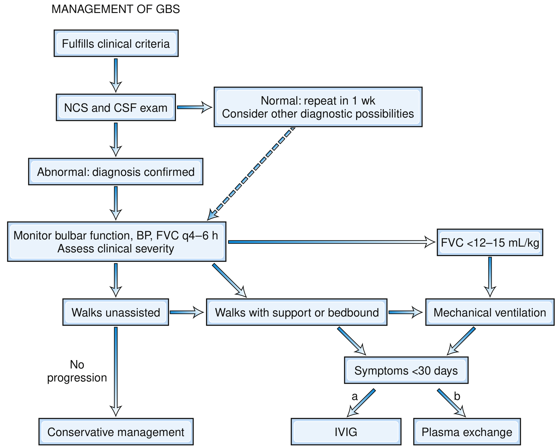
Fig. GBS Management Pathway (Bradley and Daroff's Neurology, Fig. 106.19)
Key steps from the flowchart:
- Patient fulfills clinical criteria → proceed to NCS + CSF
- If NCS/CSF normal → repeat in 1 week; consider other diagnoses
- If abnormal → confirmed diagnosis
- Monitor bulbar function, BP, FVC every 4-6 hours; assess severity
- Walks unassisted, no progression → Conservative management (observe)
- Walks with support OR bedridden AND symptoms <30 days → IVIG or plasma exchange
- FVC <12-15 mL/kg → Mechanical ventilation
7. IVIG vs Plasmapheresis - Comparative Details
| Feature | IVIG | Plasma Exchange (PE) |
|---|---|---|
| Dose | 0.4 g/kg/day × 5 days (total 2 g/kg) | 5 sessions over 10-14 days (200-250 mL/kg total) |
| Efficacy | Equal to PE | Equal to IVIG |
| Preferred? | Yes - easier, safer, more accessible | Alternative if IVIG unavailable/contraindicated |
| Mechanism | Suppresses immune response; Fc receptor blockade; anti-idiotype antibodies | Removes circulating antibodies, complement, cytokines |
| Contraindications | IgA deficiency (anaphylaxis risk); renal failure | Haemodynamic instability; sepsis; poor venous access |
| Side effects | Headache, fever, rash, renal failure (sucrose-containing), thrombosis, aseptic meningitis | Hypotension, line complications, coagulopathy |
| Combination with each other | NO benefit; not recommended | Same |
| Corticosteroids combined | No added benefit; may worsen | Same |
Timing matters: Treatment within 2 weeks of onset is most effective; treatment up to 4 weeks is still beneficial. After 4 weeks, immunotherapy is unlikely to help.
8. Pain Management in GBS (Often Undertreated)
Pain is present in ~70% of GBS patients and persists in 1/3 for up to a year:
| Pain Type | Best Treatment |
|---|---|
| Neuropathic (burning, dysesthetic) | Gabapentin, carbamazepine, pregabalin |
| Deep aching/musculoskeletal | NSAIDs, opioids |
| Severe acute pain | Opioids (morphine, tramadol) |
| Epidural pain | Epidural infusion (in ventilated patients) |
9. Rehabilitation in GBS (Post-Acute Phase)
A 2026 systematic review (PMID 41824781) confirms rehabilitation is essential in GBS recovery:
- Physiotherapy: progressive resistance and aerobic exercise; reduces fatigue and improves functional capacity
- Occupational therapy: activities of daily living retraining
- Speech therapy: bulbar and swallowing rehabilitation
- Respiratory physiotherapy: breathing exercises, weaning protocols
- Psychological support: depression and anxiety are common; quality of life is significantly affected
- Orthoses: ankle-foot orthoses for foot drop; wrist splints
10. GBS vs Acute-Onset CIDP - A Critical Distinction
Some patients initially diagnosed with GBS actually have acute-onset CIDP - this matters because CIDP responds to steroids but GBS does not.
Suspect acute-onset CIDP (not GBS) if:
- Disease progresses or deteriorates beyond 8 weeks
- >2 treatment-related fluctuations (improvement then worsening after IVIG/PE), especially beyond 1 month
- Prominent sensory symptoms dominate
- Multifocal enlargement of peripheral nerves on ultrasound
- >3 treatment-related fluctuations is almost diagnostic of CIDP
GBS recurrence: Up to 5% of GBS patients have a true recurrence after complete recovery - this is rare and distinct from treatment-related fluctuations.
11. Prognostic Scoring - EGOS / mEGOS
The Erasmus GBS Outcome Score (EGOS) predicts the probability of walking independently at 6 months:
| Predictor of Poor Outcome | Details |
|---|---|
| Age >60 years | Higher score = worse |
| Preceding diarrhoeal illness | C. jejuni link |
| MRC sum score at admission | Lower = worse |
| Rapid progression (<7 days to peak) | Suggests axonal |
| Low distal CMAP amplitudes (<20% LLN) or inexcitable nerves | Axonal loss |
| Preceding CMV infection | Typically more severe |
| Ventilatory support needed | Severe disease |
| Hyponatremia | Autonomic/SIADH |
Higher CSF neurofilament light chain (NfL) and tau levels at admission predict worse motor and functional outcomes - emerging biomarkers for severity stratification.
12. Long-Term Outcomes
| Outcome | Proportion |
|---|---|
| Complete or near-complete recovery | ~80% |
| Recovery complete by 12 months | ~70% |
| Recovery complete by 24 months | ~82% |
| Persistent disability at 1 year | ~20% |
| Mortality (modern ICU care) | 2-5% |
| Recurrence | ~5% |
Causes of death in GBS:
- Respiratory failure / ventilator complications
- Autonomic instability (cardiac arrhythmias, sudden cardiac death)
- Pulmonary embolism
- Sepsis / pneumonia (aspiration)
- Acute hypoxic-ischaemic encephalopathy
13. GBS in Special Populations
GBS in Children
- More common form: AMAN (particularly in Asia after C. jejuni)
- MRI spine may show gadolinium enhancement of cauda equina
- Generally better prognosis than adults
- Pain and back pain are prominent early features
GBS in Pregnancy
- Rare but serious
- Physiological changes increase risk of respiratory failure and aspiration
- IVIG is preferred over plasma exchange (haemodynamic safer)
- Does not cross placenta; fetus generally unaffected
- Succinylcholine is contraindicated (risk of hyperkalaemia and cardiac arrest due to upregulated ACh receptors)
GBS in the Elderly
- Higher incidence (3.2 per 100,000 in >60 years)
- More severe disease; greater respiratory failure risk
- Poor prognosis; higher mortality
- Autonomic complications more dangerous
Summary Table: GBS Subtypes at a Glance
| Feature | AIDP | AMAN | AMSAN | MFS |
|---|---|---|---|---|
| Frequency (West) | 97% | Rare | Rare | 5-6% |
| Frequency (Asia) | Less common | Most common | Less common | 18% (Taiwan) |
| Motor weakness | Yes | Yes | Yes | Usually spared |
| Sensory loss | Yes (mild) | No | Yes | Ataxia |
| Ophthalmoplegia | Rare | Rare | Rare | Yes |
| Reflexes | Absent | Absent | Absent | Absent |
| Motor NCS | Slowed, blocks | Normal CV, low CMAP | Normal CV, low CMAP | Normal |
| SNAP | Abnormal | Normal | Absent | Reduced |
| Key antibody | None specific | Anti-GM1/GD1a | Anti-GD1b | Anti-GQ1b |
| Pathology | Demyelination | Motor axon/node | Motor + sensory axon | Sensory axon |
| Prognosis | Good | Good (often) | Poor | Excellent |
- Bradley and Daroff's Neurology in Clinical Practice, p. 2663-2675
- Goldman-Cecil Medicine, p. 4065-4068
patient history taking in detail
Searching sources...
Reading File
Finding Sources
Finding Sources
Finding Sources
Finding Sources
Finding Sources
Finding Sources
Reading File
Finding Sources
Bradley and Daroff's Chapter 1 has the exact content I need. Let me read the full history-taking section:
Reading File
Excellent - this has very rich content. Let me now read the Harrison's principles section on history taking as well:
Finding Sources
Reading File
Reading File
Reading File
I have all the content I need. Let me now read the diagnostic flowchart image and compile the full answer:
Loading Image
I now have comprehensive textbook content from Bradley and Daroff's Neurology. Here is the complete, detailed guide to patient history taking:
Patient History Taking - A Detailed Guide
The history is the single most important step in clinical medicine. In neurology especially, a well-taken history provides the localization of the lesion, the likely pathophysiology, and a focused differential diagnosis - all before a single examination finding is elicited.
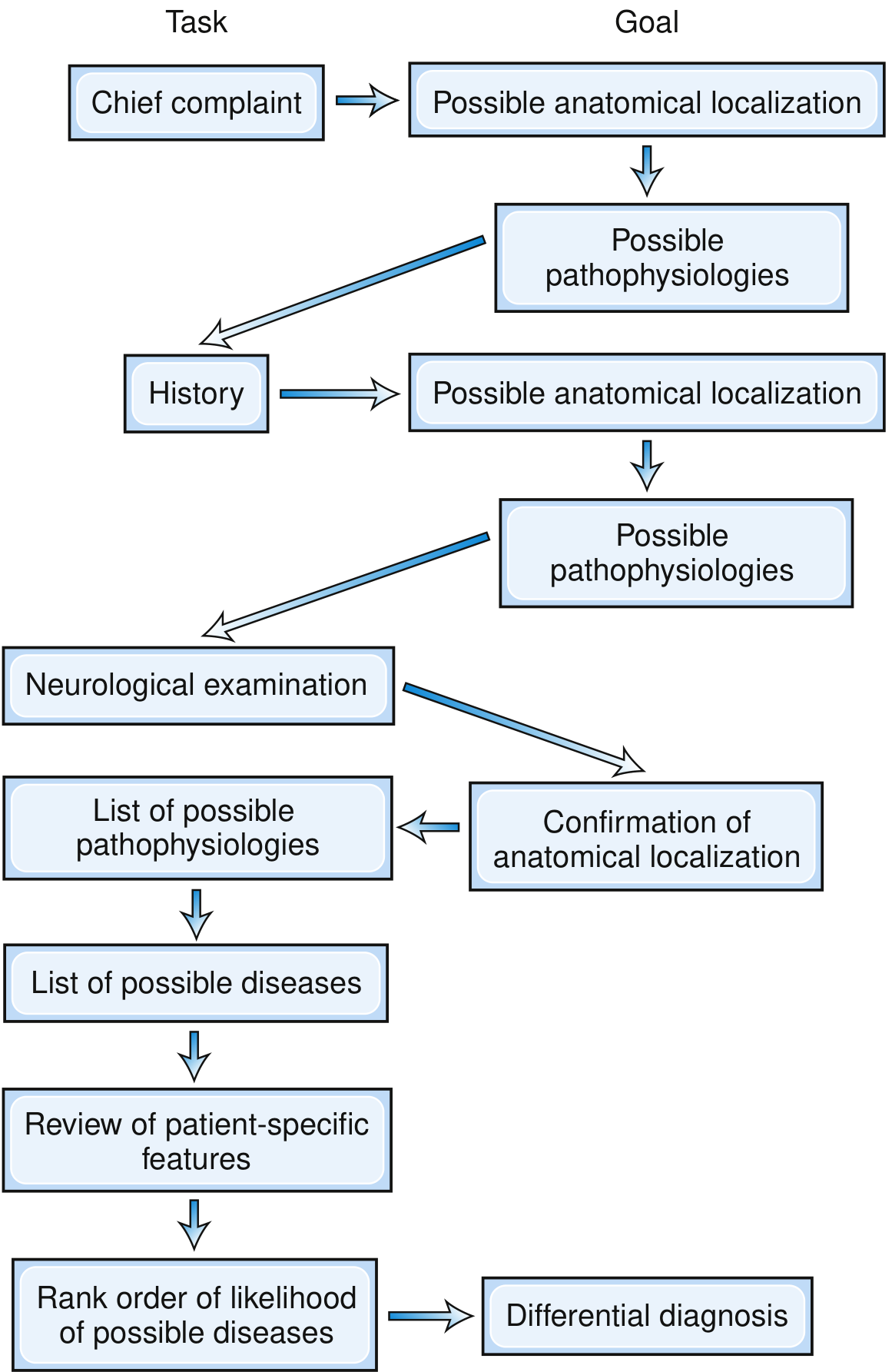
The Structure of a Complete Patient History
A full medical history has 8 components, each serving a distinct purpose:
| Component | Abbreviation | Purpose |
|---|---|---|
| 1. Introductory/Demographic Data | - | Who the patient is |
| 2. Chief Complaint | CC | Why they came today |
| 3. History of Present Illness | HPI | Full story of the current problem |
| 4. Review of Patient-Specific Information | - | Background factors shaping disease risk |
| 5. Review of Systems | ROS | Symptoms not yet volunteered |
| 6. Past Medical and Surgical History | PMH/PSH | Previous illnesses, operations |
| 7. Family History | FH | Hereditary and shared-environment diseases |
| 8. Social History | SH | Lifestyle, occupation, habits, relationships |
Step 0: The Environment and Opening (Before Any Questions)
Before starting the interview, create the right conditions:
- Introduce yourself: name and role
- Ensure privacy: close the curtain or door
- Ensure comfort: patient seated comfortably; you at eye level (never standing over them)
- Establish rapport: a few social pleasantries first - "What type of work have you done most of your life?" / "How old are you?" / "Are you right- or left-handed?"
- Open the interview gently: "How can I be of service?" / "What brings you to see me today?" / "What is bothering you the most?"
- Empathetic demeanor: the patient may be anxious about a potentially serious diagnosis; acknowledge this
- Establish expectations: ask what the patient is hoping to learn or have confirmed from the visit
- At a follow-up visit: "How have you been since we last met?" / "Has anything changed?"
Never leap directly into questioning. The opening exchange establishes trust, sets the patient at ease, and motivates honest disclosure.
- Bradley and Daroff's Neurology, p. 24-25
Step 1: Demographic Data (Biographic Information)
Record at the outset:
| Data | Clinical Value |
|---|---|
| Full name | Identity, family tree |
| Age | Age-specific disease likelihood (e.g., GBS incidence ↑ with age) |
| Sex / Gender | Sex-linked diseases; hormonal influences |
| Handedness | Language dominance (left hemisphere = dominant in 95% of right-handers) |
| Occupation | Toxic exposures, physical demands, stress |
| Referring physician | For correspondence and continuity of care |
| Informant | Who gave the history (patient, relative, caregiver) + reliability assessment |
Step 2: Chief Complaint (CC)
The chief complaint is the patient's own words describing their main problem - one to three sentences maximum.
- Record it verbatim in quotation marks: "My legs have been getting weak for the past two weeks"
- It focuses the entire subsequent history
- It provides the first clue to anatomical localisation and aetiology
- It also reveals the patient's understanding of their own condition
Key questions at this stage:
- "What is your main problem today?"
- "What brought you to the hospital/clinic?"
- "What worries you most?"
The mode of onset is critically important. A sudden onset of headache, clumsiness, and diplopia suggests a vertebrobasilar stroke. The same symptoms over weeks suggest a posterior fossa tumour. Exacerbations and remissions point to multiple sclerosis. The chief complaint, plus the onset, starts narrowing the differential immediately.
- Bradley and Daroff's Neurology, p. 25
Step 3: History of Present Illness (HPI)
This is the most detailed and important section. It tells the complete story of the current illness from first symptom to present day.
The Core Framework - SOCRATES (for each symptom)
| Letter | Stands For | Questions to Ask |
|---|---|---|
| S | Site | "Where exactly is it?" / "Can you point to it?" |
| O | Onset | "When did it start?" / "What were you doing?" / "Was it sudden or gradual?" |
| C | Character | "What does it feel like?" / "How would you describe it?" (burning, stabbing, dull, electric, cramping) |
| R | Radiation | "Does it spread anywhere?" / "Does it go down your arm/leg?" |
| A | Associations | "Any other symptoms at the same time?" (nausea, vomiting, weakness, vision change) |
| T | Time course | "How has it changed over time? Constant or intermittent? Getting better or worse?" |
| E | Exacerbating and relieving factors | "What makes it worse?" / "What makes it better?" / "Does anything help?" |
| S | Severity | "On a scale of 0 to 10, how severe is it?" / "Does it affect your daily life?" |
The Temporal-Severity Profile (Most Important in Neurology)
The timeline of a symptom is the single most powerful diagnostic clue:
| Time Course | Likely Mechanism | Example |
|---|---|---|
| Sudden onset, maximal from the start (seconds to minutes) | Vascular (ischaemia, haemorrhage); seizure | Stroke, subarachnoid haemorrhage |
| Rapid progression (hours to days) | Inflammation, infection, demyelination | GBS, encephalitis, transverse myelitis |
| Subacute progression (days to weeks) | Inflammatory, autoimmune, metabolic | MS relapse, tumour |
| Slowly progressive (weeks to months) | Neoplasm, degenerative, hereditary | Brain tumour, Parkinson's disease, ALS |
| Relapsing-remitting (episodes with recovery between) | Demyelination, vascular TIAs, paroxysmal | Multiple sclerosis, migraine, epilepsy |
| Episodic / paroxysmal (brief attacks, normal between) | Epilepsy, migraine, paroxysmal dyskinesias | Seizures, migraine with aura |
| Fluctuating (day-to-day variation) | Neuromuscular junction (myasthenia); metabolic | Myasthenia gravis |
Clarifying the Patient's Language
Patients and doctors often use the same word to mean different things - always clarify:
| Patient says | May actually mean | Ask to clarify |
|---|---|---|
| "Numb" | Weakness, paralysis, or anaesthesia | "Can you still feel things? Can you still move it?" |
| "Dizziness" | Vertigo, lightheadedness, presyncope, confusion | "Does the room spin? Do you feel faint? Unsteady?" |
| "Blurred vision" | Diplopia, visual field loss, or true blurring | "Is it double? Does covering one eye help?" |
| "Blackouts" | Syncope, seizure, or confusion | "Did you lose consciousness? For how long?" |
| "Pounding headache" | Not necessarily pulsatile | "Describe exactly what you feel" |
| "Weakness" | Fatigue, sensory loss, or true motor weakness | "Can you lift your arm? Your leg?" |
Questions to Ask in the HPI
Onset:
- "When did you first notice this problem?"
- "Were there any warning signs before it started?"
- "Were you doing anything in particular when it began?"
Progression:
- "Has it changed since it started? Got better, worse, or stayed the same?"
- "Is it there all the time or does it come and go?"
- "At its worst, how does it affect your daily activities?"
- "Can you still walk? Work? Care for yourself?"
Spread:
- "Has it spread to involve other parts of your body?"
- "Did it start in one place and move?" (e.g., ascending paralysis in GBS)
Triggers and relieving factors:
- "Does anything bring it on? Activity, posture, eating, stress, heat?"
- "Does rest help? Does medication help? Which ones?"
Associated symptoms (always ask):
- Headache, visual changes, hearing loss
- Weakness, numbness, tingling
- Speech or swallowing difficulty
- Bladder/bowel dysfunction
- Fits or blackouts
- Falls or gait disturbance
- Memory or personality change
- Nausea, vomiting, fever
How to Handle an Unclear or Unreliable History
- Get a collateral history from a relative, partner, or witness (essential for seizures, dementia, childhood illness, unconscious patient)
- Be aware of secondary gain (disability claims, lawsuits) affecting reliability
- Suspect somatoform/psychogenic disorder if: symptoms started suddenly after trauma; clinical features are incongruous with organic disease; multiple organ systems are involved simultaneously
- Note the patient's intelligence, memory, language function, and educational level - these all affect the quality of the history
- In children: history is always a composite of the child + parent; ask the child separately if abuse or substance use is a concern
Never accept previous doctors' opinions or test results without critical reappraisal. Take a fresh history every time. Mistakes in the prior history lead to errors in diagnosis that persist for years.
Step 4: Review of Patient-Specific Information
This section captures background factors that contextualise the illness:
Medications
- All current medications (prescription, over-the-counter, vitamins, herbal, oral contraceptives, supplements)
- Patients frequently omit OTC drugs and supplements
- Always ask about drugs that cause neurological side effects:
- Isoniazid → peripheral neuropathy
- Ethambutol → optic neuropathy
- Lithium → tremor, ataxia, nystagmus
- Neuroleptics → Parkinson-like syndrome, tardive dyskinesia
- Chemotherapy agents → peripheral neuropathy
- Metronidazole, nitrofurantoin → neuropathy
- Ask about allergies (drug, food, contrast dye) and nature of reaction
Occupation and Exposures
- Current and previous occupations
- Chemical exposures at work or home (heavy metals, solvents, pesticides)
- Organophosphates, lead, arsenic, mercury all cause neurological disease
- Agricultural workers, factory workers, painters - high-risk occupations
Substance Use
- Alcohol: quantity (units per week), duration, last drink
- Alcohol causes cerebellar degeneration, peripheral neuropathy, Wernicke's encephalopathy
- Tobacco: pack-year history (lung cancer → brain metastases)
- Illicit drugs: type, route, frequency
- Prescription drug misuse
Stress and Psychosocial Background
- Recent stressful life events: bereavement, divorce, job loss
- Domestic situation: who lives at home, support network
- Any hint of physical or emotional abuse
- Mental health: anxiety, depression, previous psychiatric history
Sexual History
- Important when considering HIV, syphilis (neurosyphilis), and STI-related neurological disease
- Ask sensitively; may need privacy from relatives or parents
Step 5: Review of Systems (ROS)
A systematic checklist of symptoms not already covered in the HPI. This catches diagnoses that might have been missed.
Neurological ROS (Ask Specifically)
| System | Questions |
|---|---|
| Cognition/Personality | Memory problems? Confusion? Personality change? Difficulty with work/home tasks? |
| Mood | Depression? Anxiety? Hallucinations? Delusions? |
| Seizures/Consciousness | Fits? Blackouts? Episodes of confusion or unresponsiveness? |
| Headache | Location, character, frequency, severity, triggers, associated symptoms (nausea, photophobia, phonophobia) |
| Vision | Blurring, double vision, loss of vision (partial or complete), flashing lights, floaters |
| Hearing | Loss, tinnitus, vertigo |
| Speech and Language | Slurred speech? Difficulty finding words? Understanding speech? |
| Swallowing | Choking on liquids/solids? Nasal regurgitation? |
| Smell and Taste | Reduced or altered? |
| Weakness | Any limb? Distribution? Proximal or distal? |
| Sensory symptoms | Numbness, tingling, burning, pins and needles? Where? |
| Pain | Location, character, radiation |
| Involuntary movements | Tremor? Twitching? Jerking? |
| Coordination | Clumsiness, difficulty with fine tasks (buttons, writing)? |
| Gait/Balance | Falls? Unsteadiness? Needing a stick or wall support? |
| Autonomic | Dizziness on standing (orthostatic hypotension)? Sweating abnormalities? Palpitations? |
| Bladder/Bowel | Urinary urgency, retention, incontinence? Constipation? |
| Sexual function | Erectile dysfunction? Loss of libido? |
| Sleep | Insomnia? Excessive daytime sleepiness? Restless legs? |
General Systems ROS
| System | Key Symptoms |
|---|---|
| Cardiovascular | Chest pain, palpitations, dyspnoea, leg swelling, claudication |
| Respiratory | Cough, breathlessness, haemoptysis, wheeze |
| Gastrointestinal | Abdominal pain, change in bowel habit, weight loss, jaundice, dysphagia |
| Genitourinary | Dysuria, haematuria, frequency, discharge |
| Musculoskeletal | Joint pain, swelling, stiffness, rash |
| Endocrine | Thirst, polyuria, heat/cold intolerance, weight change, fatigue |
| Haematological | Easy bruising, bleeding, lymph node swelling, anaemia symptoms |
| Skin | Rash, ulcers, pigmentation changes |
A positive response in the ROS can unlock the diagnosis. Example: A patient complaining of ataxia and hemiparesis who also admits to unilateral deafness should raise suspicion for an acoustic neuroma. A patient with paraparesis plus headaches points toward a parasagittal meningioma rather than a spinal cord lesion.
Step 6: Past Medical and Surgical History (PMH/PSH)
Record all previous illnesses, operations, and hospitalisations with dates.
Medical History
Ask specifically about:
- Hypertension, diabetes, hyperlipidaemia (cerebrovascular disease risk)
- Cardiac disease: AF (embolic stroke), cardiomyopathy, valvular disease
- Autoimmune diseases: SLE, rheumatoid arthritis, vasculitis, sarcoidosis
- Malignancy: any history of cancer (brain metastases, paraneoplastic syndromes)
- Infections: TB, HIV, syphilis, viral meningitis, Lyme disease
- Metabolic: thyroid disease, renal failure, liver disease (hepatic encephalopathy), vitamin B12 deficiency (subacute combined degeneration)
- Previous neurological events: prior strokes, seizures, head trauma, meningitis
- Psychiatric history: depression, bipolar disorder, schizophrenia
Always ask about gastric surgery (→ vitamin B12 deficiency → subacute combined degeneration of cord). Never assume the patient will volunteer it.
Surgical History
- All operations with dates
- Anaesthetic complications
- Post-operative neurological events
- Head trauma, fractures, spinal surgery
Obstetric/Gynaecological History (in women)
- Pregnancies, deliveries, miscarriages
- Oral contraceptive use (→ cerebral venous thrombosis risk)
- Menstrual cycle and neurological correlates (catamenial epilepsy, menstrual migraine)
Paediatric Patients - Perinatal History
- Pregnancy complications (infections, hypertension, drugs)
- Birth history (gestational age, mode of delivery, birth asphyxia, APGAR scores)
- Neonatal problems (jaundice, seizures, feeding difficulties)
- Developmental milestones: sitting, walking, talking, social development
- School performance; learning difficulties
Step 7: Family History (FH)
Many neurological disorders are inherited or familial:
- Construct a three-generation pedigree if a hereditary condition is suspected
- Ask about each first-degree relative: parents, siblings, children
- Ask specifically:
- "Has anyone else in the family had similar symptoms?"
- "Any neurological conditions, muscle diseases, or epilepsy in the family?"
- "Any sudden cardiac deaths or strokes at a young age?"
- "Are your parents related to each other?" (consanguinity → autosomal recessive disorders)
Conditions Where Family History is Critical
| Condition | Inheritance |
|---|---|
| Huntington's disease | Autosomal dominant |
| Hereditary motor and sensory neuropathy (Charcot-Marie-Tooth) | AD/AR/X-linked |
| Duchenne/Becker muscular dystrophy | X-linked recessive |
| Friedreich's ataxia | Autosomal recessive |
| Myotonic dystrophy | Autosomal dominant |
| Familial amyloid polyneuropathy | AD (transthyretin mutations) |
| Familial epilepsy syndromes | Various |
| Migraine | Strong familial tendency |
| Wilson's disease | Autosomal recessive |
Patients may be unaware of a family history if relatives were misdiagnosed, died early, or never sought medical attention. Ask about relatives who "walked strangely," "had shaky hands," or "died young."
Step 8: Social History (SH)
The social history reveals the context of the patient's life and how illness fits into it:
| Domain | Questions |
|---|---|
| Living situation | "Who do you live with?" / "Do you live alone?" / "Is your home accessible (stairs, bathroom)?" |
| Support network | "Who helps you at home?" / "Do you have a carer?" |
| Occupation | "What is/was your job?" - note current employment status, physical demands, stress |
| Education | Highest level - important for interpreting cognitive tests |
| Financial concerns | Disability claims; litigation; any secondary gain (affects reliability) |
| Diet and nutrition | Vegetarian/vegan (B12 risk); alcohol-related malnutrition |
| Travel history | Recent travel abroad (tropical infections, Zika, Japanese encephalitis) |
| Sexual history | HIV, neurosyphilis, STI risk |
| Exercise and activities | Baseline function; activities affected by illness |
| Driving | Does the patient drive? (crucial in epilepsy, syncope, vision problems) |
Closing the Interview
At the end of the history:
- Summarise back to the patient: "So if I've understood correctly, you've had..."
- Ask: "Is there anything else you'd like to tell me or ask me?" - often the most important symptom comes out here
- Explain what happens next: examination, investigations, plan
- Check understanding: "Do you have any questions?"
- Express concern and engagement: "I can see this has been very difficult for you."
The Diagnostic Logic - What the History Achieves
After taking a full history, the experienced clinician should be able to answer:
| Question | How the History Answers It |
|---|---|
| Where is the lesion? | Temporal profile + symptom distribution → anatomical localisation |
| What is the mechanism? | Speed of onset, course, triggers → vascular / inflammatory / degenerative / neoplastic |
| What is the cause? | Risk factors, exposures, family history, prior illness → aetiology |
| How severe is it? | Functional impact, disability, ADL limitation |
| What is the patient's expectation? | Patient's own words and concerns |
In neurology, the principle is unique: the history localises the lesion AND suggests the mechanism simultaneously. The examination then confirms what the history predicted. If the examination findings are inconsistent with the history, one of them is wrong - and the history usually wins.
- Bradley and Daroff's Neurology in Clinical Practice, p. 24-30
Quick Reference - Complete History Template
1. DEMOGRAPHICS: Name, Age, Sex, Handedness, Occupation, Informant

2. CHIEF COMPLAINT: [Patient's own words] - [Duration]

3. HISTORY OF PRESENT ILLNESS (SOCRATES):
   - Onset: when, how (sudden/gradual), circumstances
   - Character: quality, severity (0-10)
   - Location and radiation
   - Time course: constant vs. intermittent; progressive vs. stable vs. improving
   - Aggravating and relieving factors
   - Associated symptoms
   - Effect on daily activities

4. PATIENT-SPECIFIC INFORMATION:
   - Current medications (all of them, including OTC/herbal)
   - Allergies
   - Occupational exposures
   - Substance use (alcohol, tobacco, drugs)
   - Psychosocial stressors

5. REVIEW OF SYSTEMS:
   - Neurological (cognition, mood, seizures, headache, vision, hearing, speech,
     swallowing, weakness, sensation, coordination, gait, autonomic, bladder/bowel)
   - Cardiovascular, Respiratory, GI, GU, MSK, Endocrine, Haematological, Skin

6. PAST MEDICAL HISTORY:
   - Medical conditions (with dates)
   - Surgical procedures (with dates)
   - Previous hospitalizations
   - Childhood illnesses / developmental history (paeds)

7. FAMILY HISTORY:
   - Three generations; specific neurological, cardiac, psychiatric, metabolic diseases
   - Consanguinity

8. SOCIAL HISTORY:
   - Living situation and support
   - Occupation and education
   - Lifestyle: diet, exercise, travel
   - Driving status
   - Sexual history (if relevant)
- Bradley and Daroff's Neurology in Clinical Practice, Chapter 1, p. 24-30
history taking in detail
I just covered patient history taking in comprehensive detail in my previous response. Let me know if you'd like me to focus on a specific aspect:
Asking for Preferences
Which aspect of history taking would you like to go deeper on?
Searching sources...
Finding Sources
Finding Sources
Finding Sources
Finding Sources
Finding Sources
Finding Sources
Reading File
Reading File
Reading File
Finding Sources
Reading File
I now have rich system-specific history content from Goldman-Cecil Medicine and Bradley/Daroff's Neurology. I have everything needed to write a comprehensive system-by-system guide to general medical history taking.
General Medical History Taking - Complete System-by-System Guide
The history is the foundation of every clinical encounter. It should be structured, systematic, and patient-centred. In all disciplines of medicine, the history provides the diagnosis in 70-80% of cases before any test is ordered.
The Structure of a General Medical History
1. Demographic Data / Identifying Information
2. Chief Complaint (CC)
3. History of Present Illness (HPI) — SOCRATES
4. Systemic Enquiry (Review of Systems — ROS)
5. Past Medical and Surgical History (PMH / PSH)
6. Drug and Allergy History
7. Family History (FH)
8. Social History (SH)
The Environment and Opening
Before any questioning:
- Introduce yourself (name and role)
- Ensure privacy - draw the curtain, close the door
- Sit at eye level - never stand over the patient
- Establish rapport: "How can I help you today?" / "What is bothering you the most?"
- Clarify the informant - is the patient the historian, or is it a relative/carer?
- Assess reliability - consider the patient's level of consciousness, cognitive function, language barrier, and potential secondary gain (litigation, disability claims)
Allow the patient to speak first in their own words before asking focused questions. Never interrupt within the first 60 seconds.
1. Demographic / Identifying Information
| Item | Clinical Relevance |
|---|---|
| Name, age, sex | Age- and sex-specific disease prevalence |
| Occupation | Toxic exposures, physical demands, stress |
| Ethnicity | Disease prevalence differs (e.g., SCD in Africans; TB in South Asians) |
| Referral source | Who sent them; what they already know |
| Informant | Patient / relative / carer - note reliability |
2. Chief Complaint (CC)
Record in the patient's own words, in quotation marks, with duration.
"My legs have been getting weak for the past two weeks."
Key at this stage:
- The CC focuses all subsequent questioning
- It reveals the patient's understanding of their illness
- It is the first clue to location and aetiology
Opening questions:
- "What is your main problem today?"
- "What brought you to the hospital?"
- "What worries you most?"
3. History of Present Illness (HPI)
The richest and most diagnostically important section. Take this for each symptom using the SOCRATES framework.
SOCRATES Framework
| Letter | Domain | Questions |
|---|---|---|
| S - Site | Where is it? | "Where exactly is it?" / "Can you point to it with one finger?" |
| O - Onset | When and how did it start? | "When did you first notice it?" / "Did it come on suddenly or gradually?" / "What were you doing?" |
| C - Character | What does it feel like? | "Describe it in your own words" / "Is it sharp, dull, burning, cramping, pressing, tight?" |
| R - Radiation | Does it spread? | "Does it go anywhere?" / "Does it travel down your arm or leg?" |
| A - Associations | What comes with it? | "Any other symptoms at the same time?" / Ask system-specific questions |
| T - Time course | How has it changed? | "Is it constant or intermittent?" / "Getting better, worse, or the same?" / "How long does each episode last?" |
| E - Exacerbating/Relieving | What affects it? | "What makes it worse?" / "What makes it better?" / "Does rest help?" / "Does eating affect it?" |
| S - Severity | How bad is it? | "On a scale of 0-10?" / "Does it wake you from sleep?" / "Does it affect your daily activities?" |
The Temporal Profile - Diagnostic Power
| Time Course | Likely Mechanism | Examples |
|---|---|---|
| Seconds to minutes (sudden, maximal at onset) | Vascular (infarct, haemorrhage) | Stroke, MI, aortic dissection, SAH |
| Minutes to hours | Ischaemia, arrhythmia, infection, seizure | Unstable angina, PE, meningitis |
| Hours to days | Inflammation, infection, demyelination | Pneumonia, GBS, MS relapse |
| Days to weeks | Inflammatory, autoimmune, metabolic, tumour | RA flare, SLE, lymphoma |
| Weeks to months (slowly progressive) | Neoplasm, degenerative, hereditary | Brain tumour, Parkinson's, CLL |
| Episodic / relapsing-remitting | Epilepsy, migraine, MS, vasospasm | Epilepsy, migraine, MS |
| Fluctuating (better and worse, same day) | Neuromuscular (myasthenia), metabolic | Myasthenia gravis, hypoglycaemia |
Important Clarifications
Patients and doctors use the same word differently - always clarify:
| Patient says | May mean | Clarify |
|---|---|---|
| "Numb" | Paralysis OR sensory loss | "Can you move it? Can you feel things?" |
| "Dizzy" | Vertigo, lightheadedness, presyncope, ataxia | "Does the room spin? Do you feel faint?" |
| "Blurred vision" | Diplopia, field loss, or true blur | "Is it double? Does closing one eye help?" |
| "Blackout" | Syncope, seizure, or confusion | "Did you lose consciousness? Remember anything?" |
| "Weakness" | Fatigue, pain-limited, or true motor weakness | "Can you lift your arm? Climb stairs?" |
| "Palpitations" | Fast, slow, or irregular heartbeat | "Is it fast, slow, or fluttery? Regular or irregular?" |
| "Indigestion" | GORD, angina, peptic ulcer, gallbladder | "Where exactly? Any radiation? After eating?" |
| "Tight" (chest) | Angina, musculoskeletal, anxiety | "Any radiation? Related to exertion or stress?" |
4. System-by-System Enquiry (Review of Systems)
After completing the HPI, systematically screen all organ systems for symptoms not yet volunteered.
4a. Cardiovascular System
Cardinal symptoms: Chest pain, dyspnoea, palpitations, syncope, oedema, claudication
Chest Pain
Ask:
- Site: retrosternal, left-sided, epigastric?
- Onset: sudden vs exertional vs at rest?
- Character: pressure, tightness, burning, stabbing, tearing?
- Radiation: to jaw, left arm, back, epigastrium?
- Association: diaphoresis, nausea, vomiting?
- Duration: minutes (angina) vs hours (MI)?
| Character | Likely Cause |
|---|---|
| Central pressure, radiates to jaw/left arm, with exertion | Angina pectoris / MI |
| Severe tearing pain radiating to back | Aortic dissection |
| Sharp, pleuritic, positional | Pericarditis, PE, pleuritis |
| Burning, related to food | GORD, oesophageal spasm |
| Reproducible on pressing the chest | Musculoskeletal |
Women and elderly patients may present with atypical chest pain, fatigue, or dyspnoea as the only MI symptom.
Dyspnoea (Breathlessness)
Ask:
- Onset: sudden vs gradual?
- Exertional vs at rest?
- Orthopnoea: "Do you need extra pillows to sleep?" (heart failure)
- PND (Paroxysmal Nocturnal Dyspnoea): "Do you wake gasping for breath?" (LVF)
- Associated wheeze? Stridor? Cough?
- NYHA class:
- I: No limitation
- II: Slight limitation (moderate exertion)
- III: Marked limitation (minimal exertion)
- IV: Symptoms at rest
Palpitations
Ask:
- Onset: sudden or gradual?
- Rate and rhythm: fast, slow, or irregular? ("Are they regular like a drum, or irregular like a bag of jumping beans?")
- Duration of episode?
- Precipitants: caffeine, exercise, stress, emotion?
- Associated: dizziness, syncope, chest pain?
- History of thyroid disease, anaemia, drug use?
| Palpitation Type | Likely Cause |
|---|---|
| Regular, fast, abrupt onset/offset | SVT |
| Irregular | AF, ectopics |
| Fast + chest pain + syncope | VT |
| Occasional "missed beat" | Ectopic beats |
| With anxiety, tremor, heat intolerance | Thyrotoxicosis |
Syncope/Pre-syncope
Ask:
- Prodrome: nausea, sweating, visual dimming (vasovagal) vs none (cardiac)?
- Posture at onset: standing (vasovagal) vs any position (cardiac)?
- Duration of unconsciousness?
- Recovery: rapid + alert (vasovagal) vs prolonged confusion (post-ictal seizure)?
- Associated: incontinence, tongue biting, tonic-clonic movements (seizure)?
- Triggers: prolonged standing, micturition, cough, Valsalva, emotion, venipuncture?
Peripheral Oedema
- Onset, progression (ankle → leg → sacrum = severity)?
- Bilateral (cardiac, hypoalbuminaemia, venous insufficiency) vs unilateral (DVT, cellulitis)?
- Pitting or non-pitting?
- Associated: dyspnoea (heart failure), frothy urine (nephrotic), jaundice (liver disease)?
Claudication
- Pain in legs with walking - what distance triggers it?
- Relieves with rest (vascular) vs requires specific posture (neurogenic)?
- Which part of leg: calf (SFA), thigh/buttock (iliac disease)?
4b. Respiratory System
Cardinal symptoms: Cough, dyspnoea, haemoptysis, wheeze, chest pain, sputum
Cough
Ask:
- Duration: acute (<3 weeks) vs subacute (3-8 weeks) vs chronic (>8 weeks)?
- Dry vs productive?
- Sputum: quantity, colour, consistency, blood-staining?
- Yellow/green: infection (pneumonia, bronchiectasis)
- Rust-coloured: pneumococcal pneumonia
- Pink/frothy: pulmonary oedema
- Frank blood: haemoptysis (TB, carcinoma, PE)
- Worse at night? (asthma, GORD, post-nasal drip)
- Worse with cold air, exercise, allergens? (asthma)
- ACE inhibitor use? (persistent dry cough → switch drug)
- Whooping quality? (whooping cough in unvaccinated)
Dyspnoea (respiratory causes)
- Wheeze: audible? (obstructive - asthma, COPD)
- Stridor: inspiratory (laryngeal, tracheal)
- Occupational exposures: dust, chemicals (occupational asthma, pneumoconiosis)
- Pets, triggers, seasonal variation (allergic asthma)
- Exercise tolerance vs baseline?
Haemoptysis
Always take seriously - red flag symptom:
- Volume? Colour (bright red, dark, streaked in mucus)?
- Associated: fever + purulent sputum (bronchiectasis, abscess)?
- Weight loss + night sweats? (TB, carcinoma)
- Risk factors: smoking, TB contact, HIV, travel history?
- Pleuritic chest pain + hypoxia? (PE)
Wheeze and Stridor
- Wheeze: expiratory, bilateral = obstructive (asthma, COPD)
- Stridor: inspiratory, unilateral = upper airway obstruction (epiglottitis, foreign body, laryngeal tumour)
4c. Gastrointestinal System
Cardinal symptoms: Abdominal pain, nausea/vomiting, dysphagia, heartburn, change in bowel habit, rectal bleeding, jaundice, weight loss
Abdominal Pain
Apply SOCRATES fully:
- Site and radiation:
- RUQ → liver, gallbladder (radiates to right shoulder tip = biliary colic)
- Epigastric → stomach, duodenum, pancreas (radiates to back = pancreatitis)
- LUQ → spleen, pancreatic tail
- Periumbilical → early appendicitis, small bowel, mesenteric ischaemia
- RIF → appendicitis, Crohn's, ectopic pregnancy, ovarian cyst
- LIF → diverticulitis, sigmoid colon, ovarian cyst
- Loin to groin → ureteric colic ("renal colic")
- Suprapubic → bladder, uterus, prostate
- Character: colicky (waves and settles = obstruction, biliary/ureteric colic) vs constant (peritonitis, pancreatitis) vs burning (peptic ulcer)
- Relationship to food: worse after eating (peptic ulcer, mesenteric ischaemia) vs better after eating (duodenal ulcer)
- Relieving posture: pancreatitis relieved by sitting forward
- Peritoneal signs: Does movement make it worse? (peritonitis = guarding, rigidity)
Dysphagia (Difficulty Swallowing)
Always a red-flag symptom - never ignore:
- Solids only: mechanical obstruction (carcinoma, peptic stricture, external compression)
- Solids + liquids from the start: neuromuscular (achalasia, oesophageal spasm, motor neurone disease)
- Progressive (weeks-months): carcinoma
- Intermittent: oesophageal spasm, benign stricture
- Associated: odynophagia (painful swallowing), weight loss, regurgitation, voice change?
- Nasal regurgitation: pharyngeal pathology
Heartburn and Regurgitation (GORD)
- Burning retrosternal discomfort after meals, bending, lying flat?
- Acid taste in mouth (acid regurgitation)?
- Relieved by antacids?
- Worse at night? (nocturnal aspiration)
- Weight loss? (alarm feature for carcinoma)
Nausea and Vomiting
- Onset: relation to pain or eating?
- Content: undigested food (oesophageal obstruction), bile-stained (small bowel), faeculent (large bowel obstruction)?
- Projectile: pyloric stenosis, raised ICP (no nausea, sudden onset)
- Associated: vertigo (inner ear), headache (raised ICP, migraine)?
- Haematemesis: bright red (upper GI bleeding) vs coffee-grounds (slower bleed)?
Change in Bowel Habit
- Frequency, consistency, colour
- Diarrhoea: acute (<2 weeks, infection) vs chronic (IBD, IBS, malabsorption, carcinoma)
- Constipation: onset, duration, associated pain, obstruction symptoms?
- Blood in stool:
- Bright red, fresh, on paper/in pan = anorectal (haemorrhoids, fissure)
- Mixed through stool = colonic lesion (carcinoma, colitis)
- Black, tarry (melaena) = upper GI bleed (proximal to ligament of Treitz)
- Mucus in stool: IBD, irritable bowel, rectal carcinoma
- Steatorrhoea (pale, bulky, offensive, difficult to flush): fat malabsorption (coeliac, chronic pancreatitis)
- Tenesmus: persistent feeling of incomplete evacuation → rectal carcinoma, proctitis
Jaundice
- Onset: sudden (biliary colic) vs gradual (hepatitis, carcinoma)?
- Colour of urine: dark (conjugated bilirubin = obstructive or hepatocellular)?
- Colour of stools: pale/clay-coloured = obstructive jaundice
- Pruritus: obstructive jaundice
- Associated pain: biliary colic (RUQ spasms) vs painless jaundice (carcinoma of pancreas - red flag)
- Alcohol history, IV drug use, sexual history (hepatitis B/C)?
- Recent blood transfusion, travel, tattoos?
- Contact with jaundiced individuals?
Appetite and Weight Loss
- Unintentional weight loss >5% in 6 months = red flag → malignancy, TB, endocrine, cardiac cachexia
- Associated: fever, night sweats, fatigue (lymphoma, TB, HIV)?
- Anorexia (loss of appetite): liver disease, carcinoma, depression, renal failure?
4d. Genitourinary System
Cardinal symptoms: Dysuria, frequency, urgency, haematuria, loin pain, incontinence, discharge
Lower Urinary Tract Symptoms (LUTS)
Storage symptoms (irritative):
- Frequency: how many times per day / night (nocturia)?
- Urgency: sudden compelling need to void?
- Urgency incontinence: unable to hold?
Voiding symptoms (obstructive):
- Hesitancy: difficulty starting?
- Weak stream or intermittent flow?
- Incomplete emptying (post-void residual feeling)?
- Terminal dribbling?
- → In males: think BPH, prostate carcinoma, stricture
Haematuria
- Timing: initial (urethra), terminal (bladder neck, prostate), throughout (bladder, upper tract)?
- Clots: large clots → bladder tumour; worm-like clots → upper tract
- Painful: ureteric calculus vs painless: carcinoma (red flag)
- Associated: UTI symptoms, trauma, TB?
Loin Pain
- Ureteric colic: severe, loin-to-groin radiation, colicky, restlessness, haematuria
- Pyelonephritis: constant, dull, fever, dysuria
Gynaecological History (women)
- LMP (last menstrual period)
- Cycle: regular or irregular? Duration? Volume?
- Dysmenorrhoea (painful periods)?
- Intermenstrual or post-coital bleeding?
- Vaginal discharge: colour, odour, itching?
- Dyspareunia (pain with intercourse)?
- Pregnancies (gravida, para), miscarriages, terminations
- Contraception
- Cervical screening history (smear results)
- Menopausal status; HRT use?
- Breast lumps, nipple discharge, mammogram history?
4e. Musculoskeletal System
Cardinal symptoms: Joint pain, swelling, stiffness, restricted movement, muscle weakness
Joint Pain - SOCRATES
- Which joint(s)? Monoarthritis (single) vs oligoarthritis (2-4) vs polyarthritis (≥5)?
- Symmetrical (RA) vs asymmetric (reactive arthritis, psoriatic)?
- Small joints (RA, psoriatic) vs large joints (OA, septic)?
- Morning stiffness >1 hour: inflammatory (RA, AS)?
- Swelling: hot and erythematous (septic arthritis, gout) vs cool (OA)?
- Precipitated by: activity (OA, bursitis), diet - red meat/alcohol (gout), infection (reactive arthritis)?
- Associated: rash (SLE, psoriasis), eye inflammation (uveitis in AS), urethritis/conjunctivitis (reactive arthritis), diarrhoea (enteric arthritis)?
Muscle Symptoms
- Weakness: proximal (myopathy, polymyositis) vs distal (peripheral neuropathy)?
- Myalgia: diffuse (fibromyalgia, statin, viral) vs localised (injury)?
- Tenderness: specific trigger points (fibromyalgia)?
- Wasting or fasciculations: LMN / motor neurone disease?
Back Pain
- Site: cervical, thoracic, lumbar, sacral?
- Radiation: down leg (radiculopathy)? Where does it go? Below knee (sciatica - L4/L5/S1)?
- Mechanical vs inflammatory: worse with activity + relieved by rest (mechanical) vs worse at rest + early morning + improved with activity (inflammatory - ankylosing spondylitis)?
- Red flags in back pain:
- Age >50 or <20
- Night pain / rest pain
- Systemic symptoms (weight loss, fever)
- History of malignancy
- Bladder/bowel dysfunction (cord compression)
- Saddle anaesthesia (cauda equina - emergency)
4f. Endocrine System
Cardinal symptoms: Weight change, fatigue, thirst, polyuria, heat/cold intolerance, sweating, skin/hair changes
Thyroid
- Hyperthyroidism: weight loss despite increased appetite, palpitations, tremor, heat intolerance, diarrhoea, anxiety, exophthalmos?
- Hypothyroidism: weight gain, constipation, cold intolerance, dry skin/hair, fatigue, bradycardia, hoarse voice, depression?
Diabetes
- Polyuria, polydipsia, polyphagia (the "three Ps")?
- Recurrent infections (skin, UTI, candida)?
- Blurred vision (osmotic lens changes)?
- Symptoms of complications: neuropathy (tingling/numbness feet), nephropathy (swelling), retinopathy (vision loss)?
Adrenal
- Cushing's: weight gain (central), hirsutism, striae, easy bruising, mood change, proximal muscle weakness?
- Addison's: fatigue, postural dizziness, weight loss, salt craving, hyperpigmentation (sun-exposed areas and pressure points)?
4g. Haematological System
Cardinal symptoms: Fatigue, pallor, bleeding, bruising, lymphadenopathy, recurrent infection
- Anaemia symptoms: fatigue, dyspnoea on exertion, pallor, palpitations, headache, tinnitus?
- Bleeding tendency: easy bruising, prolonged bleeding from cuts, epistaxis, haemarthrosis?
- Mucosal bleeding + petechiae: platelet defect (ITP, drugs)
- Deep tissue bleeding + haemarthrosis: coagulation factor defect (haemophilia)
- Thrombotic tendency: DVT, PE, recurrent miscarriages (thrombophilia)?
- Lymphadenopathy: which nodes? Tender (infection) vs non-tender (lymphoma)?
- Night sweats + weight loss + fever (B symptoms): lymphoma, TB, HIV, leukaemia?
4h. Neurological System (within general history)
- Headaches: onset, character, location, severity, associated symptoms (nausea, photophobia, aura), frequency?
- Dizziness/vertigo: room spinning vs lightheaded?
- Weakness: which limb(s)? Onset? Ascending or descending?
- Sensory: numbness, tingling, burning?
- Seizures: description, frequency, any triggers, post-ictal state?
- Speech: slurring, difficulty finding words?
- Vision: double, blurred, loss?
- Memory: short-term or long-term loss?
- Headache + vomiting + neck stiffness + photophobia = meningism (emergency)
4i. Dermatological System
- New rash: onset, distribution, character (flat/raised/blistering/pustular), itching?
- Changes in existing moles: size, shape, colour, border, bleeding (ABCDE rule)?
- Hair loss, nail changes, skin thickening?
- Ulcers: painful (venous/neuropathic) vs painless?
4j. Ophthalmological / ENT
Eyes:
- Visual acuity: blurring, loss?
- Redness, pain, photophobia (iritis, acute glaucoma)?
- Floaters, flashing lights (retinal detachment)?
- Diplopia?
Ears:
- Hearing loss: gradual (sensorineural) vs sudden (SSNHL - emergency)?
- Tinnitus: unilateral (acoustic neuroma)?
- Vertigo: duration? Triggered by head position (BPPV)?
- Otalgia (ear pain) + discharge?
Nose/Throat:
- Epistaxis, nasal obstruction, anosmia?
- Sore throat, hoarseness, stridor?
- Dysphagia?
5. Past Medical and Surgical History
- All medical conditions with dates
- All surgical procedures with dates and any complications
- Previous hospitalisations
- Trauma (especially head, spine)
- Blood transfusions
- Vaccinations (especially relevant: influenza preceding GBS)
Always ask specifically about conditions patients may not volunteer: gastric surgery (B12 deficiency), sarcoidosis (multi-system neurological effects), previous malignancy (metastases, paraneoplastic syndrome), previous TB.
6. Drug and Allergy History
Drug History
- All current medications: dose, frequency, route, duration
- OTC drugs, vitamins, supplements, herbal remedies (patients rarely volunteer these)
- Recent changes or new prescriptions
- Compliance: "Do you take them regularly?"
Neurologically important drugs:
| Drug | Side Effect |
|---|---|
| Isoniazid | Peripheral neuropathy (B6 deficiency) |
| Ethambutol | Optic neuropathy |
| Lithium | Tremor, ataxia, nystagmus |
| Neuroleptics | Parkinsonism, tardive dyskinesia |
| Metronidazole | Peripheral neuropathy |
| Chemotherapy (vincristine, taxanes) | Peripheral neuropathy |
| Statins | Myopathy, rhabdomyolysis |
| ACE inhibitors | Persistent dry cough |
| Oral contraceptive pill | Thromboembolism, cerebral venous thrombosis |
Allergy History
- Drug allergies: which drug, what reaction (rash, anaphylaxis, GI intolerance)?
- Food allergies
- Contrast dye allergy (important before imaging)
- Latex allergy
Always distinguish a true allergy (immune-mediated - urticaria, angioedema, anaphylaxis) from an intolerance (GI upset, headache).
7. Family History
- Parents: ages, alive/dead, cause of death
- Siblings: health status
- Children: health status
- Three-generation pedigree if hereditary disease suspected
Ask specifically:
- "Has anyone in the family had a similar problem?"
- "Any heart disease, stroke, diabetes, cancer, epilepsy, or blood disorders?"
- "Any sudden deaths in young relatives?" (long QT syndrome, HCM, sudden cardiac death)
- "Are your parents related?" (consanguinity → autosomal recessive disease)
| Condition | Family History Significance |
|---|---|
| CAD < 55 in male / <65 in female relatives | Familial hypercholesterolaemia, genetic risk |
| Sudden cardiac death in young | Channelopathy (long QT, Brugada, ARVD), HCM |
| Breast/ovarian cancer | BRCA1/2 mutation |
| Colon cancer | Lynch syndrome, FAP |
| Huntington's disease | 50% inheritance per child |
| Haemophilia | X-linked recessive |
| DM, thyroid disease | Polygenic inheritance |
8. Social History
| Domain | Key Questions |
|---|---|
| Living situation | "Who do you live with?" / Alone? House / flat / care home? Stairs? |
| Occupation | Current and previous; chemical/biological exposures; stress |
| Activities of daily living (ADL) | Dressing, washing, cooking, shopping - independent or dependent? |
| Alcohol | Units per week; CAGE questionnaire (Cut down? Annoyed? Guilty? Eye-opener?) |
| Tobacco | Smokes or ex-smoker; pack-year history (packs/day × years) |
| Illicit drugs | Type, route (IV?), frequency; sharing needles (hepatitis B/C, HIV) |
| Diet and nutrition | Vegetarian/vegan (B12 deficiency); eating disorders; malnutrition |
| Travel history | Recent travel; countries visited; malaria prophylaxis; immunisations |
| Sexual history | Partners; contraception; STI risk; HIV testing; MSM? |
| Driving | Does the patient drive? (critical in epilepsy, syncope, visual problems) |
| Support network | Family, carers, GP, district nurse |
| Advance directives | DNACPR, lasting power of attorney (in elderly/seriously ill patients) |
CAGE Questionnaire (Alcohol Screening)
2 or more "yes" answers = likely alcohol use disorder
- Have you ever felt you should Cut down on drinking?
- Have you ever been Annoyed by criticism about your drinking?
- Have you ever felt Guilty about your drinking?
- Have you ever needed an Eye-opener (drink first thing in the morning)?
Closing the History
- Summarise back: "So if I understand correctly, you've had..."
- Invite additions: "Is there anything else you want to tell me that I haven't asked?"
- Acknowledge concerns: "I can understand this has been worrying you."
- Explain next steps: "I'd now like to examine you, and then we'll talk about what tests might be helpful."
- Check understanding: "Do you have any questions for me?"
Complete History Template (Quick Reference)
PATIENT: Name / Age / Sex / Occupation / Informant

1. CHIEF COMPLAINT: [In patient's own words] × [Duration]

2. HISTORY OF PRESENT ILLNESS (SOCRATES for each symptom):
   S - Site
   O - Onset (sudden/gradual, date, circumstances)
   C - Character (quality, description)
   R - Radiation
   A - Associated symptoms
   T - Time course (constant/episodic, progression)
   E - Exacerbating and Relieving factors
   S - Severity (0-10, effect on function)

3. REVIEW OF SYSTEMS:
   CVS: chest pain, dyspnoea, palpitations, syncope, oedema, claudication
   RS: cough, dyspnoea, wheeze, haemoptysis, chest pain, sputum
   GI: appetite, weight, dysphagia, heartburn, N&V, abdominal pain,
       bowel habit, rectal bleeding, jaundice
   GU: LUTS, haematuria, loin pain; menstrual/gynaecological (F)
   MSK: joint pain/swelling/stiffness, muscle weakness, back pain
   Neuro: headache, dizziness, weakness, numbness, seizures, memory
   Endocrine: weight change, thirst, polyuria, heat/cold intolerance
   Haematology: fatigue, bleeding, bruising, lymphadenopathy
   Skin: rash, lesions, hair/nail changes
   Eyes/ENT: vision, hearing, vertigo, tinnitus, nose, throat

4. PAST MEDICAL HISTORY:
   Conditions (with dates) | Operations | Hospitalisations | Trauma

5. DRUG AND ALLERGY HISTORY:
   All medications (incl. OTC, supplements, contraception, herbal)
   Allergies + nature of reaction

6. FAMILY HISTORY:
   Parents, siblings, children - conditions and age of death
   Hereditary diseases; consanguinity

7. SOCIAL HISTORY:
   Living situation | Occupation | ADL | Alcohol (CAGE) | Tobacco
   Illicit drugs | Travel | Sexual history | Driving | Support network
- Bradley and Daroff's Neurology in Clinical Practice, Chapter 1, p. 24-30
- Goldman-Cecil Medicine, p. 2491-2607
- Campbell-Walsh Wein Urology, p. 23-24
continue
Diving deeper...
Finding Sources
Finding Sources
Finding Sources
Finding Sources
Reading File
Finding Sources
Finding Sources
Finding Sources
Reading File
Reading File
I now have excellent additional content on communication skills, questioning techniques, and empathy frameworks. Here is the continuation of the history-taking guide - covering the advanced and skills-based aspects:
History Taking - Continued: Advanced Skills, Special Populations & Applied Techniques
Part 9: The Art of Questioning
Skilled history taking is not just about what you ask - it is about how you ask it. The type of question determines how much information you receive.
Open-Ended vs Closed-Ended Questions
| Type | Definition | Example | Best Used When |
|---|---|---|---|
| Open-ended | Identifies a topic but provides no structure for the answer | "Tell me about your pain." / "What has been troubling you?" | Opening each new area of enquiry; allowing the patient to tell their own story |
| Semi-open | Slightly focused, still allows full response | "Can you describe when the dizziness comes on?" | Narrowing a broad response toward specifics |
| Closed-ended | Highly structured; leads to a yes/no or single word answer | "Is your pain sharp?" / "Did you lose consciousness?" | Confirming specific facts; completing checklists |
The funnel technique: Each area of the history should begin with open-ended questions and progressively narrow to closed-ended questions. Then, as you move to a new topic, open up again with another open-ended question. The interview is a series of funnels, not a single one.
Practical sequence:
- Open: "Tell me about your chest pain."
- Semi-open: "Can you describe where you feel it and when it comes on?"
- Closed: "Does it radiate to your left arm?" / "Does it come on with exertion?"
- Transition to new area (open): "Tell me about your breathing."
The most valuable rapport-promoting element of verbal communication is the use of open-ended questions at the onset of the interview - they signal that you are genuinely listening, not just checking boxes.
- Kaplan and Sadock's Synopsis of Psychiatry, p. 36-37
- Textbook of Family Medicine 9e, p. 42
Questions to Avoid
| Type | Example | Problem |
|---|---|---|
| Leading questions | "The pain is like a pressure, isn't it?" | Puts words in the patient's mouth; creates false positives |
| Double questions | "Is the pain sharp or burning?" | Confuses; patient may answer only part |
| Jargon / medical language | "Any dyspnoea or orthopnoea?" | Patient may not understand; answer unreliable |
| "Why" questions | "Why didn't you come sooner?" | Sounds accusatory; causes defensiveness |
| Premature closure | Stopping after the first complaint | Misses important additional symptoms |
Part 10: Communication Skills - Patient-Centred History Taking
The modern history is not just a data-collection exercise - it is a therapeutic encounter. Patient-centred communication improves outcomes, adherence, and satisfaction.
The ICE Framework (Ideas, Concerns, Expectations)
Every patient comes with three hidden agendas that will affect how they respond to your history and plan. Actively elicit all three:
| Domain | What It Means | How to Elicit |
|---|---|---|
| I - Ideas | What does the patient think is causing their problem? | "What do you think might be causing this?" / "Have you any ideas about what's going on?" |
| C - Concerns | What worries them most? What are they afraid of? | "Is there anything in particular that's worrying you about this?" / "What is your biggest concern?" |
| E - Expectations | What do they hope you will do for them today? | "What were you hoping I could do for you today?" / "What would be most helpful for you?" |
A patient with back pain may secretly fear they have cancer (concern). They want reassurance, not just a prescription (expectation). Their idea is that heavy lifting caused it (idea). Without knowing ICE, you may give the medically correct answer but leave the patient completely unsatisfied and non-compliant.
Nonverbal Communication - SOLER
Your body language matters as much as your words:
| Letter | Skill | Practical Application |
|---|---|---|
| S | Sit square to the patient | Face them directly; do not sit sideways |
| O | Open body posture | Uncross arms and legs; turn toward patient |
| L | Lean toward the patient | Slight forward lean signals engagement |
| E | Maintain eye contact | Natural eye contact - not a stare; look away occasionally |
| R | Relaxed posture | Not tense or rushed; conveys confidence and calm |
Physicians who stand over a lying or undressed patient, hold conversations while the patient is exposed, or are consistently late for appointments signal disrespect for the patient and erode the therapeutic relationship - damaging future disclosure.
Verbal Empathy - NURSE
When a patient expresses distress, fear, or strong emotion, use the NURSE framework:
| Letter | Skill | Example |
|---|---|---|
| N | Name the emotion | "You seem worried about this." |
| U | Understand the emotion | "I can see why this has been so difficult for you." |
| R | Respect the emotion | "You've shown a lot of strength dealing with this." |
| S | Support the patient | "Whatever happens, I'll be with you through this." |
| E | Explore the emotion | "Tell me more about what is worrying you." |
Active Listening Techniques
| Technique | Description |
|---|---|
| Silence | Pausing allows the patient to gather thoughts and continue - do not rush to fill the silence |
| Facilitation | Non-verbal nods, "mm-hmm," "go on," "I see" - signal you are listening |
| Reflection | Repeat the patient's last words back: "So the pain started two weeks ago..." |
| Clarification | "When you say you felt dizzy, what exactly do you mean?" |
| Summarising | "So let me check I've understood - you've had..." (periodic, not just at the end) |
| Signposting | Tell the patient before changing topics: "I've got a good understanding of your chest pain now. I'd like to ask about other symptoms - is that OK?" |
| Paraphrasing | Restate the meaning in your own words to check understanding |
Responding to Emotional Cues
Patients rarely say "I am anxious." They drop cues - often indirect:
| Cue Type | Patient Statement | Appropriate Response |
|---|---|---|
| Informational | "I'm not sure about the treatment options." | Ask-Tell-Ask: "What have you been told so far?" → [Explain] → "Does that make sense?" |
| Emotional | "I'm worried about this." | Use NURSE: Name, Understand, Respect, Support, Explore |
| Verbal hint | "My wife had cancer, you know." | "That sounds like it's on your mind - are you worried this could be something similar?" |
| Silence | Patient pauses, looks down | Do NOT rush; maintain calm, forward-leaning posture |
Part 11: The Ask-Tell-Ask Framework (for delivering information)
When explaining findings, diagnoses, or plans, use the structured Ask-Tell-Ask approach:
- ASK first: "What do you already know about your condition?"
- TELL: Give information simply, in plain language, in small chunks
- ASK again: "Does that make sense?" / "Do you have any questions?"
Patients retain only 20-30% of what doctors tell them in a consultation. Checking understanding at every step dramatically improves information retention and adherence.
Part 12: Special Situations in History Taking
12a. The Unconscious or Critically Ill Patient
When the patient cannot give a history:
- Obtain collateral history from: family, carers, ambulance crew, GP letter, previous notes
- Use AMPLE mnemonic for emergency situations:
| Letter | Stands For | Examples |
|---|---|---|
| A | Allergies | Drug, food, contrast |
| M | Medications | All current drugs |
| P | Past medical history | Major illnesses, surgeries |
| L | Last meal | When did they last eat/drink? |
| E | Events | What led to this presentation? Mechanism? |
12b. The Paediatric History
History taking in children requires two simultaneous histories: from the child (in age-appropriate language) and from the parent/carer.
Additional Components:
Antenatal/Perinatal History:
- Maternal health during pregnancy: infections, drugs, alcohol, diabetes, hypertension?
- Gestational age at birth (term, preterm)?
- Mode of delivery (normal vaginal, caesarean, instrumental)?
- Birth weight
- APGAR scores at 1 and 5 minutes
- Any complications at birth: asphyxia, resuscitation required?
- Neonatal jaundice, seizures, feeding difficulties?
- Time in NICU?
Developmental History (compare to milestones):
| Milestone | Normal Age |
|---|---|
| Social smile | 6-8 weeks |
| Head control | 3-4 months |
| Sit without support | 6-8 months |
| Stands alone | 9-12 months |
| First words | 10-12 months |
| Walks alone | 12-15 months |
| Two-word phrases | 18-24 months |
| Sentences | 2-3 years |
| Toilet trained | 2-3 years |
- Is development progressing? Or regressing (red flag for neurodegenerative disease)?
- School performance: learning difficulties, attention (ADHD)?
Immunisation History:
- Vaccinations up to date?
- Any adverse reactions?
Feeding History:
- Breast or bottle fed? Duration?
- Introduction of solids?
- Current diet?
Asking the Child Separately:
- "Is there anything wrong at school or at home?"
- For adolescents: ask about sexual activity, substances, mental health away from parents
- Look for signs of child abuse: unexplained injuries, fearful behaviour, inappropriate sexual knowledge
12c. The Elderly Patient
Special considerations:
- Multiple comorbidities and polypharmacy (medication interactions are a common hidden cause of symptoms)
- Atypical presentations: MI may present as fatigue; infection as confusion; abdomen as falls
- Cognitive impairment: use a short cognitive screen early (MMSE or AMT); may need collateral history
- Ask specifically about: falls (frequency, circumstances, injuries), functional decline (ADL changes), continence, vision and hearing (affect safety and quality of life)
- Social circumstances: does the patient live alone? Who does the shopping/cooking/medications?
- Carer burden: is the carer coping? Signs of elder abuse?
- Advance care planning: does the patient have a DNACPR, lasting power of attorney, or advance directive?
12d. The Psychiatric History
Additional sections specific to mental health:
| Section | Content |
|---|---|
| Presenting psychiatric complaint | Nature, onset, duration, severity, impact on functioning |
| Psychiatric history | Previous episodes, diagnoses, treatments, hospitalisations |
| Suicide and violence risk | Current ideation, plan, intent, previous attempts; harm to others? |
| Substance use history | Alcohol, drugs (type, route, quantity, dependence, withdrawal) |
| Forensic history | Arrests, convictions, prison |
| Premorbid personality | What were they like before the illness? |
| Mental Status Examination (MSE) | Appearance, behaviour, speech, mood, affect, thought, perception, cognition, insight, judgement |
12e. The Surgical Pre-Operative History
Focus on anaesthetic and operative risk:
- Airway: previous difficult intubation? Neck mobility? Mouth opening?
- Anaesthetic history: previous general/regional anaesthesia; any complications?
- Cardiovascular fitness: exercise tolerance (METs); cardiac disease?
- Respiratory: asthma, COPD, sleep apnoea (OSA)?
- Bleeding history: easy bruising, anticoagulants, antiplatelet drugs (aspirin, clopidogrel)?
- Last oral intake: nil by mouth (NBM) status?
- Consent: does the patient understand the procedure and risks?
12f. The Obstetric/Gynaecological History
Obstetric History - use G_P_ notation:
- G = Gravida (total number of pregnancies)
- P = Para (number of deliveries >24 weeks)
- Full breakdown: Term deliveries / Preterm / Miscarriages / Ectopic pregnancies / Living children
- For each previous pregnancy: gestation, mode of delivery, complications, birth weight, neonatal outcome
Questions for current pregnancy:
- LMP and gestational age (EDD by LMP and ultrasound)
- Antenatal care: where, how many visits, any complications?
- Anomaly scans? Results?
- Symptoms: nausea/vomiting, bleeding, discharge, reduced fetal movements?
- Risk factors: hypertension, diabetes, placenta praevia?
Gynaecological History:
- Menstrual history: age at menarche, cycle length, duration, flow, LMP
- Dysmenorrhoea, menorrhagia, IMB, PCB, PMB?
- Vaginal discharge (colour, odour, quantity)?
- Cervical screening history: last smear, results?
- Contraceptive history
- STI history
Part 13: Red Flag Symptoms Across All Systems
Always actively screen for these - they require urgent investigation:
| System | Red Flag Symptoms |
|---|---|
| General | Unexplained weight loss (>5% in 6 months), drenching night sweats, persistent fever |
| Cardiovascular | New chest pain at rest; syncope with exertion; sudden-onset tearing back pain |
| Respiratory | Haemoptysis; stridor; progressive dyspnoea at rest |
| GI | Dysphagia (progressive); haematemesis/melaena; new change in bowel habit >6 weeks; painless jaundice; rectal bleeding in >50 yrs with change in bowel habit |
| Neurological | "Thunderclap" headache (worst ever, sudden onset); headache + fever + neck stiffness + photophobia; new focal deficit; papilloedema; progressive cognitive decline |
| MSK/Back | Back pain + neurological deficit; saddle anaesthesia; bladder/bowel dysfunction; age <20 or >50 with new back pain; history of malignancy |
| Urological | Painless frank haematuria (carcinoma until proven otherwise); acute urinary retention |
| Gynaecological | Post-menopausal bleeding (endometrial carcinoma); rapid-onset pelvic mass |
| Paediatric | Developmental regression; loss of milestones; persistent unexplained fever |
Part 14: Functional History - The Sixth Vital Sign
Always assess functional status at the end of every history. It is the most clinically relevant outcome measure for the patient.
Activities of Daily Living (ADL)
Basic ADL (Barthel Index)
- Feeding: independent?
- Bathing: independent?
- Grooming (hair, teeth, shaving)?
- Dressing?
- Bowel continence?
- Bladder continence?
- Toilet use?
- Transfers (bed to chair)?
- Mobility on flat surfaces?
- Stair climbing?
Instrumental ADL (IADL - higher function)
- Shopping independently?
- Cooking?
- Managing finances?
- Managing medications?
- Using the telephone/technology?
- Using transport?
- Housework?
- Laundry?
Loss of IADLs typically precedes loss of basic ADLs in neurological and elderly patients. Tracking IADLs is a more sensitive measure of early functional decline.
Part 15: Ending - The Closing of the History
A well-closed history improves patient experience, ensures completeness, and sets up the examination:
-
Invite additions: "Is there anything else you think might be important that I haven't asked?" - this is statistically the most productive question; often the real reason for attendance comes out here
-
Summarise and check: "So, to summarise what you've told me..." - confirms accuracy and fills gaps
-
Acknowledge the patient's agenda: "I understand this has been worrying you. Let's see what we can find."
-
Explain next steps: "I'm going to examine you now. After that, I'll explain what I think is happening and what tests might help."
-
Ask-Tell-Ask before closing: Confirm the patient understands the plan
-
Arrange safety netting: "If symptoms get worse before you hear from me, please go to A&E / call 999."
-
Say goodbye warmly: "Thank you for coming. I'll be in touch as soon as I have results."
Complete History - One-Page Reference Card
DEMOGRAPHICS: Name | Age | Sex | Occupation | Informant | Reliability

CHIEF COMPLAINT: [Patient's own words] × [Duration]

HPI - SOCRATES (for each symptom):
  Site | Onset (date, sudden/gradual) | Character | Radiation
  Associations | Time course | Exacerbating/Relieving factors | Severity

ICE: Ideas | Concerns | Expectations

SYSTEMIC ENQUIRY (ROS):
  CVS:   Chest pain | Dyspnoea (NYHA) | Palpitations | Syncope | Oedema | Claudication
  RS:    Cough | Dyspnoea | Wheeze | Haemoptysis | Sputum | Stridor
  GI:    Appetite | Weight | Dysphagia | Heartburn | N&V | Abdo pain | Bowel habit
         Rectal bleeding | PR mucus | Jaundice | Haematemesis | Melaena
  GU:    LUTS (storage + voiding) | Haematuria | Loin pain | Discharge
  Gynae: LMP | Cycle | Menorrhagia | PCB | IMB | PMB | Smear history (F)
  MSK:   Joint pain/stiffness/swelling | Myalgia | Back pain
  Neuro: Headache | Vertigo | Weakness | Numbness | Seizures | Vision | Memory
  Endo:  Weight | Thirst | Polyuria | Heat/cold intolerance | Sweating
  Haem:  Fatigue | Pallor | Bleeding | Bruising | Lymphadenopathy
  Skin:  Rash | Lesions | Hair/nail changes
  ENT/Eyes: Vision | Hearing | Tinnitus | Vertigo | Throat

PAST MEDICAL HISTORY:
  Conditions (+ dates) | Operations (+ dates) | Hospitalisations | Trauma | Transfusions

DRUG AND ALLERGY HISTORY:
  All medications (dose, frequency, duration; include OTC/supplements/herbal/contraception)
  Allergies → nature of reaction (true allergy vs intolerance)

FAMILY HISTORY:
  Parents, siblings, children | Hereditary diseases | Consanguinity
  Premature CAD | Sudden death | Malignancy | Neurological | Psychiatric

SOCIAL HISTORY:
  Living situation | Occupation + exposures | ADL (basic + instrumental)
  Alcohol (CAGE) | Tobacco (pack-years) | Illicit drugs | Travel | Sexual history
  Driving status | Support network | Advance care directives

CLOSING:
  "Is there anything else?"  →  Summarise  →  ICE addressed  →  Safety net  →  Next steps
- Bradley and Daroff's Neurology in Clinical Practice, Chapter 1
- Goldman-Cecil Medicine, p. 1109-1180
- Kaplan and Sadock's Synopsis of Psychiatry, p. 36-38
- Berek and Novak's Gynecology, p. 47
ascitis tapping in detail
Searching sources...
Reading File
Finding Sources
Finding Sources
Reading File
Reading File
Reading File
Finding Sources
Finding Sources
Finding Sources
Reading File
Reading File
Reading File
Reading File
Loading Image
Reading File
I now have comprehensive content from multiple authoritative textbooks. Let me compile the full detailed guide:
Ascitic Tapping (Abdominal Paracentesis) - A Detailed Guide
Paracentesis (also called "ascitic tap") is the procedure of inserting a needle or catheter through the abdominal wall into the peritoneal cavity to aspirate ascitic fluid. It serves both diagnostic and therapeutic purposes.
1. Definitions
| Term | Meaning |
|---|---|
| Diagnostic paracentesis | Removal of a small volume (20-100 mL) for fluid analysis |
| Therapeutic paracentesis | Removal of large volumes (≥5 L) to relieve symptoms |
| Large Volume Paracentesis (LVP) | Removal of >5 L; up to 10-12 L in chronic ascites |
2. Indications
Diagnostic Paracentesis
- New-onset ascites of unknown cause - most rapid and cost-effective way to determine aetiology
- Suspected Spontaneous Bacterial Peritonitis (SBP) - any patient with known ascites presenting with:
- Abdominal pain or tenderness
- Fever
- Altered mental status
- Unexplained clinical deterioration
- Even mild abdominal pain without systemic infection signs
- Suspected secondary peritonitis (bowel perforation)
- Known ascites in a patient with HIV (ascites is non-AIDS-related in 75% of cases)
- Suspected malignant ascites (for cytology)
- Suspected chylous, biliary, or pancreatic ascites
Therapeutic Paracentesis
-
Tense ascites causing:
- Respiratory compromise (diaphragm pushed up)
- Severe abdominal pain or distension
- Refractory ascites unresponsive to maximum diuretics
-
As bridge to definitive treatment (e.g., TIPS, liver transplant)
-
Tintinalli's Emergency Medicine, p. 594
-
Yamada's Textbook of Gastroenterology, p. 2212
3. Contraindications
Absolute Contraindications
- Acute abdomen requiring immediate surgery
- Uncooperative patient (unable to safely perform)
Relative Contraindications (weigh risk vs benefit)
| Contraindication | Comment |
|---|---|
| Severe coagulopathy (INR >2.5) or thrombocytopenia (<50,000/µL) | Large studies show paracentesis safe even with INR up to 8.7 and platelets as low as 19,000/mm³ - routine correction NOT required unless clinical fibrinolysis/DIC |
| Abdominal wall cellulitis at proposed site | Use an alternative site |
| Prior abdominal surgery / adhesions | Use ultrasound guidance |
| Dilated bowel / bowel obstruction | Use ultrasound guidance |
| Pregnancy | Ultrasound guidance essential; use upper quadrant site |
| Distended bladder | Catheterise first |
| Known abdominal visceral masses | Ultrasound guidance |
Coagulopathy is NOT a routine contraindication. Transfusion-requiring haematomas occur in <1% despite 71% of cirrhotic patients having an abnormal PT. Prophylactic FFP or platelets are NOT standard practice and carry their own risks.
- Roberts and Hedges' Clinical Procedures in Emergency Medicine, p. 1028
4. Equipment
For Diagnostic Paracentesis
- Sterile gloves, sterile drapes (fenestrated and non-fenestrated)
- Skin antiseptic (povidone-iodine or chlorhexidine)
- Gauze swabs
- Local anaesthetic: lidocaine 1-2%, 10 mL syringe
- Small-bore (27-gauge) needle for skin anaesthesia
- Medium-bore (21-gauge) needles for deep infiltration
- 18- or 22-gauge paracentesis needle (or over-the-needle catheter)
- Syringes: 10 mL, 20 mL, 50 mL
- Specimen containers for lab analysis
Additional Equipment for Therapeutic (LVP)
- 16- or 14-gauge catheter (large bore speeds drainage)
- Three-way stopcock
- Sterile tubing and drainage bag / vacuum bottles
- Wall suction setup with suction canisters (for large volumes)
- Albumin for IV infusion (20% human albumin - 6-8 g per litre removed)
- Blood pressure monitoring equipment
Ultrasound Equipment (Recommended)
- Linear or curvilinear probe (low frequency for abdominal scan)
- Sterile probe cover
5. Patient Preparation
Pre-procedure
- Explain the procedure to the patient - indications, risks, what to expect
- Obtain informed consent
- Ask patient to empty the bladder (by voiding or catheterisation if needed) - prevents bladder puncture
- Position the patient: supine (preferred) or at 45° semi-recumbent
- For left lateral approach: tilt patient slightly to left to pool fluid in the left lower quadrant
- Review coagulation status: check INR and platelets if clinically uncertain
- Ultrasound scan - confirm ascites, identify largest fluid pocket, avoid solid organs and dilated bowel
6. Site Selection
Fig: Sites for needle introduction (marked X) in the left or right lower quadrant for abdominal paracentesis (Tintinalli's Emergency Medicine, Fig. 86-3)
Two Main Sites:
| Site | Description | Preferred? |
|---|---|---|
| Left lower quadrant (LLQ) | 2-3 cm medial to, and 2-3 cm above, the left anterior superior iliac spine; OR one-third of the distance from umbilicus to anterior iliac crest | Yes - preferred (avoids liver, reduces risk of bowel injury) |
| Right lower quadrant (RLQ) | Equivalent position on the right | Alternative if LLQ has distorted anatomy (scar, ostomy) |
| Midline (supraumbilical) | 2 cm above the umbilicus in the midline (linea alba - avascular) | Alternative; use only if lateral sites inaccessible |
| Midline (infraumbilical) | One-third the distance from umbilicus to pubic symphysis | Another option |
Avoid:
- The right upper quadrant (liver)
- Areas with surgical scars (adhesions below)
- Areas with dilated veins (portal hypertension collaterals)
- Midline if inferior epigastric vessels are visible
Ultrasound identifies the largest fluid pocket, confirms absence of bowel loops, and marks the safest entry point. Always scan before choosing the site.
7. Step-by-Step Procedure
Step 1: Positioning and Preparation
- Position patient supine with slight left lateral tilt
- Identify the needle insertion site (LLQ preferred)
- Perform bedside ultrasound to confirm fluid, mark the spot, identify bowel loops and solid organs
- Clean the area with chlorhexidine or povidone-iodine in concentric circles; allow to dry
- Apply sterile drapes
- Ensure sterile technique throughout
Step 2: Local Anaesthesia
- Draw up 10 mL of 1% lidocaine
- Using a 27-gauge needle, raise a skin wheal at the insertion site
- Switch to a 21-gauge needle and infiltrate deeply through the subcutaneous tissue and anterior abdominal wall, down to the peritoneum
- Aspirate before each injection to avoid intravascular injection
- At each level, inject lidocaine until resistance gives way to peritoneal fluid on aspiration - this confirms depth to peritoneum and anaesthetises the parietal peritoneum
Step 3: The Z-Track Technique (Critical Step)
The Z-track technique displaces the skin entry point from the peritoneal entry point, creating a self-sealing tract that prevents ascitic fluid leak after needle withdrawal:
- With your non-dominant hand, pull the skin 2 cm caudally (downward) relative to the underlying abdominal wall
- With your dominant hand, insert the paracentesis needle perpendicular to the skin surface, advancing slowly
- After the needle enters the peritoneum and fluid returns - release the skin
- The skin now recoils, creating a Z-shaped, offset tract - the skin hole no longer lines up with the peritoneal hole, sealing the path
This is the most important technique to prevent post-procedure fluid leak through the puncture site, especially in patients who will need repeat taps.
Step 4: Needle Advancement and Fluid Aspiration
- Attach a 10-20 mL syringe to the needle
- Advance the needle slowly while applying gentle suction on the syringe
- Use the "stop-advance" technique: advance a few mm, stop, aspirate, advance again - this allows bowel (which is usually floating in ascitic fluid) to move away safely
- Once fluid returns freely (it should be straw-coloured and clear in transudates) → STOP advancing
- For diagnostic paracentesis: aspirate 20-50 mL (minimum) into the syringe for lab analysis
- For therapeutic paracentesis: once fluid flows freely, switch to drainage tubing connected to vacuum bottles or wall suction
Step 5: Therapeutic Drainage (LVP)
- Connect the catheter to sterile tubing → three-way stopcock → vacuum drainage system or wall suction with canisters
- Drain fluid - usual volumes removed: 5-10 L (up to 12 L can be safely drained in experienced hands)
- Monitor blood pressure, heart rate, and patient comfort throughout
- When drainage slows: gently adjust patient position (slight lateral tilt helps)
- The average time to drain 5-10 L under wall suction: 30-60 minutes
Step 6: Withdrawal and Dressing
- When drainage is complete, withdraw the needle/catheter smoothly
- Apply firm pressure with a gauze swab for 2-3 minutes
- Apply a sterile adhesive dressing
- A purse-string suture can be placed around the puncture site if persistent leak is anticipated (rare)
- Monitor the patient for 30-60 minutes after procedure for complications
8. Albumin Infusion After LVP
Albumin must be given after large volume paracentesis to prevent Post-Paracentesis Circulatory Dysfunction (PPCD):
| Volume Drained | Albumin Dose |
|---|---|
| <5 L | Not routinely required |
| >5 L (LVP) | 6-8 g of 20% human albumin per litre of ascitic fluid removed |
Why albumin?
After removing large volumes of ascitic fluid, fluid shifts out of the vascular compartment into the peritoneum (re-equilibration). This can cause effective hypovolaemia → activation of the renin-angiotensin-aldosterone system → renal impairment, hyponatraemia, hepatic encephalopathy (PPCD).
Albumin replaces colloid oncotic pressure, prevents this circulatory dysfunction, and is superior to other colloids (dextran, gelatin) for this purpose.
Practical note: 6 g albumin = 30 mL of 20% albumin solution. For a 10 L tap: 60-80 g albumin (300-400 mL of 20% albumin) IV over 2-4 hours.
- Roberts and Hedges' Clinical Procedures, p. 1031-1032
9. Ascitic Fluid Analysis
After aspiration, send fluid for analysis immediately (especially cultures - inoculate blood culture bottles at the bedside for highest sensitivity).
Standard Tests to Send
| Test | Tube/Container | What It Tells You |
|---|---|---|
| Cell count and differential | EDTA (lavender) tube | WBC; neutrophil count for SBP |
| Albumin | Plain (gold) tube | For SAAG calculation |
| Total protein | Plain tube | Transudate vs exudate |
| Glucose | Fluoride (grey) tube | Low in secondary peritonitis/TB |
| LDH | Plain tube | Malignancy, secondary peritonitis |
| Gram stain and culture | Inoculate blood culture bottles at bedside | SBP, secondary peritonitis |
| Cytology | 50+ mL in dedicated container | Malignant cells |
| Amylase | Plain tube | Pancreatic ascites |
Optional Tests (based on clinical scenario)
- Triglycerides: if fluid appears milky (chylous ascites)
- Bilirubin: if fluid is dark/green (bilious ascites from bowel/biliary perforation)
- AFB smear and TB culture/PCR: if TB peritonitis suspected
- Tumour markers (CEA, CA 19-9, CA-125): malignant ascites
- pH: <7.35 in secondary peritonitis
10. Interpreting Ascitic Fluid - The SAAG
The Serum-Ascites Albumin Gradient (SAAG) is the most reliable single test for classifying ascites aetiology.
Calculation
SAAG = Serum albumin − Ascitic fluid albumin (Both measured from samples taken on the same day)
Interpretation
| SAAG Value | Category | Causes |
|---|---|---|
| ≥1.1 g/dL (HIGH) | Portal hypertension-related | Cirrhosis, alcoholic hepatitis, cardiac ascites, Budd-Chiari syndrome, portal vein thrombosis, massive liver metastases, fulminant hepatic failure, myxoedema |
| <1.1 g/dL (LOW) | Non-portal hypertension | Peritoneal carcinomatosis, tuberculous peritonitis, pancreatic ascites, biliary ascites, nephrotic syndrome, connective tissue serositis |
SAAG ≥1.1 has a 97% accuracy for portal hypertension. It has replaced the old "transudate vs exudate" classification for ascites.
Fluid Appearance and Initial Assessment
| Appearance | Suggests |
|---|---|
| Straw-coloured, clear | Transudative / cirrhosis |
| Cloudy, turbid | SBP or peritonitis |
| Bloody (haemorrhagic) | Trauma, hepatocellular carcinoma, coagulopathy |
| Milky (chylous) | Lymphatic obstruction (lymphoma, trauma, cirrhosis) |
| Dark green/brown | Bilious ascites (bile duct perforation) |
| Viscous, gelatinous | Pseudomyxoma peritonei |
11. Diagnosing Spontaneous Bacterial Peritonitis (SBP)
SBP is diagnosed from ascitic fluid cell count - do not wait for culture results:
| Criterion | Threshold | Significance |
|---|---|---|
| Polymorphonuclear (PMN/neutrophil) count | ≥250 cells/µL | Diagnostic of SBP - start antibiotics immediately |
| Total WBC | >500/µL | Supportive |
| Culture positive | Any organism | Confirms SBP, but 40% are culture-negative despite high PMN |
SBP treatment: Start empirical antibiotics immediately (do not wait for culture):
- Cefotaxime 2 g IV every 8 hours × 5 days (first-line)
- Ofloxacin 400 mg PO every 12 hours (alternative in uncomplicated cases)
Prophylaxis after first episode of SBP:
- Norfloxacin 400 mg PO daily (long-term)
12. Complications of Paracentesis
Local Complications
| Complication | Incidence | Management |
|---|---|---|
| Persistent fluid leak from puncture site | Common in tense ascites | Z-track technique prevents; purse-string suture if occurs |
| Haematoma (abdominal wall) | <1% transfusion-requiring | Pressure; rarely surgical drainage |
| Bowel perforation | Very rare (<0.1%) | Conservative if small; surgical if large perforation |
| Bladder puncture | Rare | Empty bladder pre-procedure; usually self-limiting |
| Infection at site | Rare | Sterile technique essential |
Systemic Complications (mainly LVP)
| Complication | Comment |
|---|---|
| Post-Paracentesis Circulatory Dysfunction (PPCD) | Occurs in 15-20% after LVP; characterised by hypovolaemia, hyponatraemia, renal impairment; prevented by albumin infusion |
| Hypotension | Monitor BP for several hours post-LVP; often asymptomatic |
| Hyponatraemia | From fluid redistribution; monitor electrolytes |
| Hepatic encephalopathy | From electrolyte shifts and circulatory dysfunction |
| Renal impairment | Hepatorenal syndrome if PPCD not prevented |
| Haemorrhage | Rare; risk minimised by US guidance and Z-track |
13. Post-Procedure Care
- Monitor vital signs (BP, HR, SpO₂) every 15 minutes for 1 hour after LVP, then hourly for 2-3 hours
- Check puncture site for fluid leak or haematoma at 30 minutes and 2 hours
- Albumin infusion: administer based on drained volume (see above)
- Keep dressing in place for 48 hours
- Record volume drained, fluid appearance, and samples sent
- Repeat clinical assessment at 30 minutes to identify complications
- Electrolytes and renal function at 24-48 hours post-LVP
14. Special Considerations
Ultrasound Guidance
- Always recommended, especially in:
- Obesity
- Previous abdominal surgery (adhesions)
- Small or localised fluid collections
- Suspected bowel distension
- Pregnant patients
- US confirms: fluid location, absence of bowel loops at target site, depth to fluid
- Peritoneal fluid appears anechoic (black) on ultrasound; bowel appears relatively echogenic
Refractory Ascites Management Ladder
When ascites is refractory to diuretics:
- Regular LVP (every 2-4 weeks) + albumin
- TIPS (Transjugular Intrahepatic Portosystemic Shunt) - reduces portal hypertension
- Liver transplantation - definitive treatment
- Peritoneovenous shunting (rarely used now)
15. Summary - Quick Reference
INDICATIONS:
  Diagnostic: new ascites, suspected SBP, unknown aetiology
  Therapeutic: tense ascites causing respiratory/severe symptoms; refractory ascites

SITE: Left lower quadrant (preferred) — lateral and cephalad to
      inferior epigastric vessels; ultrasound guided

KEY TECHNIQUE: Z-track method — pull skin 2 cm caudally before inserting needle;
               release after peritoneum penetrated → seals tract, prevents leak

DIAGNOSTIC TAP: 20-100 mL → Cell count, albumin, protein, glucose,
                Gram stain + culture (blood culture bottles at bedside), LDH, cytology

THERAPEUTIC TAP: 5-12 L → Slow drainage; albumin 6-8 g per litre removed

SAAG ≥ 1.1 g/dL = Portal hypertension (cirrhosis, cardiac, Budd-Chiari)
SAAG < 1.1 g/dL = Non-portal (malignancy, TB, pancreatitis, nephrotic)

SBP: PMN ≥ 250/µL → Cefotaxime 2 g IV q8h immediately

ALBUMIN AFTER LVP: 6-8 g per litre drained (if >5 L removed)
                   Prevents post-paracentesis circulatory dysfunction
- Tintinalli's Emergency Medicine, p. 594-595
- Roberts and Hedges' Clinical Procedures in Emergency Medicine, p. 1028-1032
- Mulholland and Greenfield's Surgery 7e, p. 2939
- Yamada's Textbook of Gastroenterology, p. 2212
ascites
Searching sources...
Finding Sources
Finding Sources
Reading File
Reading File
Reading File
Reading File
Your credits are exhausted. You can purchase a top-up or subscribe to a plan to continue using Orris. If you're already subscribed, your daily credits will reset tomorrow.
continue
Your credits are exhausted. You can purchase a top-up or subscribe to a plan to continue using Orris. If you're already subscribed, your daily credits will reset tomorrow.
burning sensation on the back and chest
Your credits are exhausted. You can purchase a top-up or subscribe to a plan to continue using Orris. If you're already subscribed, your daily credits will reset tomorrow.
This is a shared conversation. Sign in to Orris to start your own chat.