Kiya Tum mujhe ecg samajhna sikha sakte ho comman deceased ke sath
ECG normal waveform PQRST labeled diagram
ECG myocardial infarction ST elevation changes
atrial fibrillation ECG tracing irregular rhythm
normal sinus rhythm ECG 12 lead labeled
ventricular tachycardia ECG wide complex
📋 ECG (Electrocardiogram) — Poori Guide Hindi Mein
ECG Kya Hota Hai?
⚡ ECG Ki Basic Waves — PQRST
| Wave | Kya Hoti Hai | Normal Value |
|---|---|---|
| P wave | Atria (upar ke chambers) ka contract hona | Choti, rounded |
| PR interval | P se QRS tak ka samay | 0.12–0.20 sec |
| QRS complex | Ventricles (neeche ke chambers) ka contract hona | 0.06–0.12 sec |
| ST segment | Ventricles ka rest | Isoelectric line par hona chahiye |
| T wave | Ventricles ka recharge hona | Positive (upward) |
| QT interval | Q se T tak ka samay | < 0.44 sec |
🟢 Normal ECG (Sinus Rhythm)
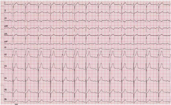
- Har QRS se pehle ek P wave hoti hai ✅
- Rhythm regular hoti hai ✅
- Heart rate: 60–100 bpm ✅
- ST segment seedha hota hai ✅
🏥 Common Diseases aur ECG Changes
1. 🔴 Heart Attack (STEMI — ST Elevation Myocardial Infarction)

- ST elevation — V1 se V6 mein (anterior wall attack → LAD artery block)
- Inferior leads (II, III, aVF) mein ST elevation → Right Coronary Artery block
- Reciprocal changes — opposite leads mein ST depression
- Yeh EMERGENCY hai — patient ko turant angioplasty chahiye!
2. 💜 Atrial Fibrillation (AF)

- ❌ Koi organized P wave nahi
- Baseline mein fibrillatory waves (titilte hue)
- R-R intervals — bilkul irregular ("irregularly irregular")
- Causes: hypertension, valve disease, thyroid, alcohol
3. 🟠 Ventricular Tachycardia (VT)

- Wide bizarre QRS (>0.12 sec)
- Rate: 150–250 bpm
- P waves nahi dikhte
- Life-threatening — cardioversion ki zaroorat
📐 ECG Padhne Ka Systematic Tarika (7 Steps)
1. RATE     → Kitni bpm? (300 ÷ R-R boxes)
2. RHYTHM   → Regular hai ya irregular?
3. P WAVES  → Har QRS se pehle P wave hai?
4. PR INTERVAL → 0.12-0.20 sec hai?
5. QRS WIDTH → Narrow (<0.12) ya wide (>0.12)?
6. ST SEGMENT → Elevated, depressed ya normal?
7. T WAVES  → Upright hain ya inverted?
🗺️ 12 Leads — Kaunsa Lead Dil Ka Kaunsa Hissa Dekhta Hai?
| Leads | Dil Ka Hissa |
|---|---|
| II, III, aVF | Inferior wall (neeche) → RCA |
| V1–V4 | Anterior wall (aage) → LAD |
| I, aVL, V5–V6 | Lateral wall (baaju) → LCx |
| V1–V2 | Posterior wall (peeche se dekhne ke liye) |
⚡ Quick Summary Table — Common ECG Abnormalities
| Bimari | ECG Finding |
|---|---|
| Heart Attack (STEMI) | ST elevation + reciprocal depression |
| Angina (NSTEMI) | ST depression + T wave inversion |
| Atrial Fibrillation | No P waves, irregular rhythm |
| Ventricular Tachycardia | Wide QRS, fast rate, no P waves |
| Complete Heart Block | P waves aur QRS ka koi relation nahi |
| Bundle Branch Block | QRS >0.12 sec (W pattern LBBB, M pattern RBBB) |
| Hyperkalemia | Peaked T waves → sine wave → cardiac arrest |
| Long QT | QT interval prolonged → Torsades de Pointes risk |
Achcha kiya tum mujhe cbc ko samjhna sikha sakte ho
CBC complete blood count normal values reference ranges
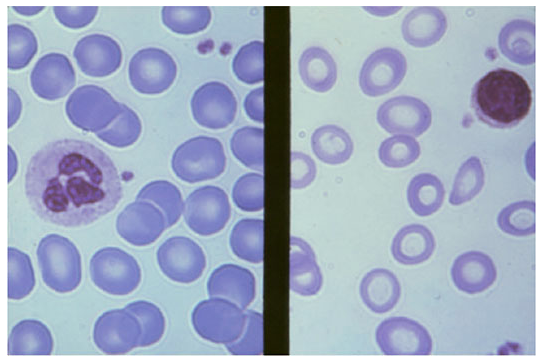
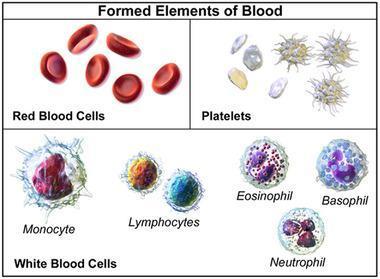
blood cells red white platelets microscopy smear
microcytic anemia iron deficiency blood smear small pale red cells
white blood cell types neutrophil lymphocyte monocyte eosinophil basophil diagram
🩸 CBC (Complete Blood Count) — Poori Guide Hindi Mein
CBC Kya Hota Hai?
- RBC — Red Blood Cells (lal khoon ke kankshaye)
- WBC — White Blood Cells (safed khoon ke kankshaye)
- Platelets — Khoon rokne waali cells
🔴 PART 1 — RBC (Red Blood Cells) — Lal Khoon
Normal Values (Adult):
| Parameter | Full Form | Normal Range | Kya Measure Karta Hai |
|---|---|---|---|
| Hb | Hemoglobin | M: 13–17 g/dL, F: 12–15 g/dL | Oxygen carry karne ki taakat |
| RBC | Red Blood Cell count | M: 4.5–5.5 million/µL, F: 4–5 million/µL | Lal cells ki ginti |
| Hct/PCV | Hematocrit | M: 40–52%, F: 36–48% | Khoon mein RBC ka hissa |
| MCV | Mean Corpuscular Volume | 80–100 fL | Ek RBC ka size |
| MCH | Mean Corpuscular Hemoglobin | 27–33 pg | Ek cell mein Hb kitna |
| MCHC | MCH Concentration | 32–36 g/dL | Cells kitni "bhuri" (colored) hain |
| RDW | RBC Distribution Width | 11.5–14.5% | Cells ka size kitna vary karta hai |
Anemia — Hb Kam Hona
🔵 Microcytic Anemia (MCV < 80 — Chhoti cells)
| Cause | Key Finding |
|---|---|
| Iron Deficiency (sabse common) | Serum ferritin ↓, TIBC ↑ |
| Thalassemia | Target cells, Hb electrophoresis abnormal |
| Chronic disease | Ferritin normal/high, TIBC ↓ |
🟡 Normocytic Anemia (MCV 80–100 — Normal size)
| Cause |
|---|
| Acute blood loss |
| Hemolytic anemia (cells toot rahe hain) |
| Aplastic anemia (bone marrow fail) |
| Kidney disease (EPO kam) |
🔴 Macrocytic Anemia (MCV > 100 — Badi cells)
| Cause | Key Finding |
|---|---|
| B12 deficiency | Neurological symptoms bhi |
| Folate deficiency | Pregnancy mein common |
| Liver disease / Alcohol | History se pata chalti hai |
🩺 PART 2 — WBC (White Blood Cells) — Safed Khoon
WBC ke Types (Differential Count):
| Cell | % (Normal) | Kaam | Increase (↑) Matlab |
|---|---|---|---|
| Neutrophil | 50–70% | Bacteria se ladta hai | Bacterial infection, stress |
| Lymphocyte | 20–40% | Virus se ladta hai, antibodies banata hai | Viral infection, leukemia |
| Monocyte | 2–8% | Bacteria khata hai (phagocytosis) | TB, fungal infection |
| Eosinophil | 1–4% | Allergy, parasites se ladta hai | Allergy, asthma, worms |
| Basophil | 0–1% | Allergic reaction mein | Allergic reaction, CML |
WBC Abnormalities:
| Finding | Normal Range | Kya Matlab |
|---|---|---|
| Leukocytosis (WBC > 11,000) | — | Infection, inflammation, leukemia |
| Leukopenia (WBC < 4,000) | — | Viral infection, chemotherapy, aplastic anemia |
| Neutrophilia (Neutrophil ↑) | — | Bacterial infection, steroids |
| Lymphocytosis (Lymphocyte ↑) | — | Viral infection (dengue, COVID), CLL |
| Eosinophilia (Eosinophil ↑ >4%) | — | Allergy, asthma, intestinal worms |
| Blast cells dikh rahe hain | Zero hone chahiye | Leukemia (EMERGENCY) |
🟣 PART 3 — Platelets (Thrombocytes) — Khoon Rokne Waali Cells
| Finding | Kya Matlab |
|---|---|
| Thrombocytopenia (< 1.5 lakh) | Dengue, ITP, leukemia, chemotherapy — bleeding ka risk |
| < 20,000 | Spontaneous bleeding ka danger! |
| Thrombocytosis (> 4 lakh) | Iron deficiency, infection, essential thrombocythemia |
📋 Ek Poori CBC Kaise Padhein — Step by Step
Step 1: Hb dekho → Anemia hai?
           ↓ Low → MCV dekho → Micro/Normo/Macro?

Step 2: WBC dekho →
           ↑ High → Differential dekho (Neutrophil ya Lymphocyte?)
           ↓ Low → Viral? Chemo? Aplastic?

Step 3: Platelets dekho →
           ↓ Low → Dengue? ITP? Bleeding risk?
           ↑ High → Infection? Thrombocythemia?
🧾 Quick Summary — Common CBC Patterns
| Pattern | Diagnosis |
|---|---|
| Hb↓, MCV↓, Ferritin↓ | Iron deficiency anemia |
| Hb↓, MCV↑, B12↓ | B12 / Folate deficiency anemia |
| WBC↑ (Neutrophil↑), Fever | Bacterial infection |
| WBC↑ (Lymphocyte↑), Fever | Viral infection (dengue, typhoid) |
| Platelets↓ + Fever | Dengue |
| Hb↓ + WBC↓ + Platelets↓ (Pancytopenia) | Aplastic anemia / Leukemia / Chemotherapy |
| Blast cells present | Acute Leukemia — EMERGENCY! |
Cbs lft lft mp in sabme aane wale problem or unke solution bhi btaao
liver function test jaundice hepatitis bilirubin elevated
malaria parasite blood smear Plasmodium ring form
dengue thrombocytopenia low platelet CBC findings
jaundice yellow skin scleral icterus liver disease