
Tinnitus maskers. Recent management modalities of tinnitus maskers in detail according to standard ENT textbook in simplified manner
tinnitus masking management
tinnitus treatment sound therapy
tinnitus masker sound therapy device diagram
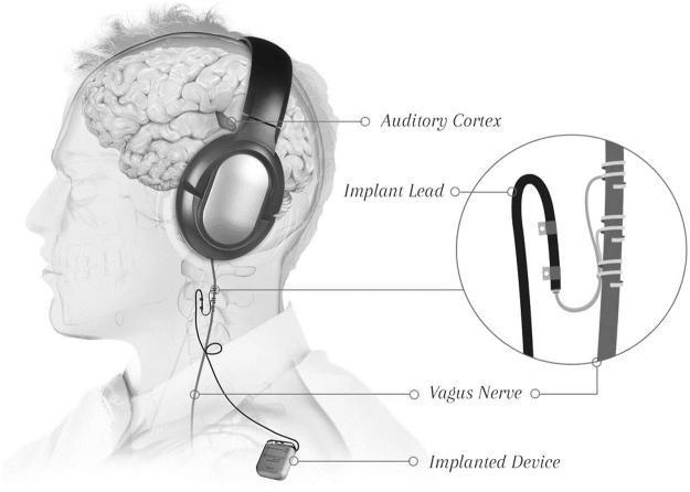
This medical illustration depicts the setup for a Vagus Nerve Stimulation (VNS) system paired with auditory tones, designed for neurological therapy such as tinnitus treatment. The diagram shows a lateral view of a human head and torso. An 'Implanted Device' (pulse generator) is situated under the chest wall, connected to an 'Implant Lead' that travels superiorly toward the neck. A magnified inset illustrates the electrode lead wrapped securely around the cervical Vagus Nerve using specialized helical anchors. Externally, the patient wears high-quality circumaural headphones, with a label pointing to the 'Auditory Cortex' to indicate the target of the sound therapy. The overall configuration demonstrates the integration of peripheral nerve stimulation with central auditory processing. This setup is used to facilitate neuroplasticity by timing electrical pulses to specific auditory stimuli, aiming to reorganize cortical maps.
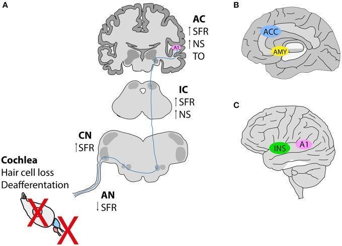
A multi-panel pathophysiology diagram illustrating the central auditory pathway and non-auditory brain networks involved in tinnitus pathogenesis. Panel A depicts a vertical ascending pathway starting from the cochlea (showing hair cell loss and deafferentation marked by red Xs) leading to the auditory nerve (AN), cochlear nucleus (CN), inferior colliculus (IC), and auditory cortex (AC). It highlights changes in spontaneous firing rate (SFR), neuronal synchrony (NS), and tonotopic organization (TO), showing a decrease in peripheral activity (AN) but central hyperactivity (CN, IC, AC). Panel B presents a medial/sagittal view of the brain highlighting the anterior cingulate cortex (ACC) and amygdala (AMY), representing the salience and emotional distress networks. Panel C shows a lateral view of the brain highlighting the insula (INS) and primary auditory cortex (A1). The illustration summarizes how peripheral auditory damage triggers neuroplastic changes and involves non-auditory cognitive and emotional centers in the perception of phantom sound.
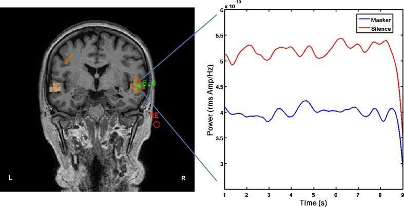
This composite image consists of a diagnostic brain scan and a corresponding line graph, illustrating neurophysiological activity related to auditory processing. On the left is a coronal T1-weighted MRI section overlayed with a Beamformer pseudo-Z image. The scan shows discrete orange-highlighted regions of interest (ROIs) in the bilateral auditory cortex, specifically concentrated in the superior temporal gyri. A green marker indicates the peak voxel in the right hemisphere. On the right, a line graph displays the time course of normalized oscillatory power in the delta band (1–4 Hz) for that right hemisphere peak voxel over a 9-second period. The y-axis measures power in rms Amp/Hz (x10^-10), and the x-axis represents time in seconds. Two distinct traces are shown: a red line representing the 'Silence' condition and a blue line representing the 'Masker' condition. The red trace shows consistently higher delta-band power compared to the blue trace, demonstrating the suppression of slow-wave cortical activity during auditory masking. This image is used in neuroscientific research to investigate the pathophysiology of tinnitus and the effects of sound masking on thalamocortical dysrhythmia.
Tinnitus Maskers: Management Modalities (ENT Textbook Reference)
What Is a Tinnitus Masker?
Classification of Tinnitus Masking Devices
| Type | Description |
|---|---|
| Pure Tinnitus Masker | Noise generator only; worn like a hearing aid; produces white/broadband noise |
| Hearing Aid as Masker | For patients with co-existing hearing loss; amplifies external sound which naturally suppresses tinnitus |
| Combination (Masker-Hearing Aid) | Combines amplification for hearing loss AND a built-in noise generator for tinnitus masking in one device |
| Sound Generators / White Noise Machines | Tabletop or bedside devices; emit pink noise, white noise, or nature sounds |
Mechanism: Residual Inhibition
Modern/Recent Management Modalities of Tinnitus Maskers
1. Sound Therapy (Broadband Noise / White Noise)
- Mechanism: Broadband or narrowband noise delivered at a low level to partially or completely mask tinnitus.
- Delivery: Ear-level devices (behind-the-ear or in-the-canal style), tabletop sound generators, smartphone apps.
- Current role: Used as a component within Tinnitus Retraining Therapy (TRT), not as standalone therapy.
- In TRT, sound generators deliver broadband noise at a sub-masking level (just below the level of tinnitus), rather than attempting total masking. This is intentional and mechanistically different from pure masking.
2. Tinnitus Retraining Therapy (TRT) - The Gold Standard Sound-Based Approach
- TRT Effectiveness: Several studies report significant improvement in 75-80% of patients. More severe cases tend to have superior results. Studies confirm that patients who use the recommended sound-generating instrumentation alongside counseling have better outcomes than counseling alone.
- Duration: Typically 12-18 months of treatment.
- TRT in hyperacusis: TRT has also shown superiority over tinnitus alone for hyperacusis (63% improvement vs 47%), improving loudness discomfort levels by ~12.5 dB and broadening dynamic range by ~11.3 dB.
3. Combination Hearing Aids with Maskers
- A combination device (hearing aid + sound generator) is the first-line device intervention.
- The amplification component restores auditory input, which itself reduces tinnitus by improving the signal-to-noise ratio in the brain.
- The noise generator provides additional masking when amplification alone is insufficient.
- Modern digital hearing aids with open-fit designs (thin tubing, open ear canal) are particularly useful as they amplify high-frequency sounds (where tinnitus is most common) while allowing ambient sound in naturally.
- Important: Hearing aids are not expected to work in patients whose tinnitus pitch is above the frequency capability of the device - but classical experiments by Feldman showed tinnitus can be suppressed by sounds across many frequencies, so this limitation is less than previously assumed.
4. Digital/Programmable Sound Masking Devices
- Adjust the frequency spectrum of the masking noise to match or bracket the patient's tinnitus pitch
- Set intensity precisely (especially sub-threshold for TRT protocols)
- Use multimemory settings - patient selects different environments (quiet room, noisy office, outdoors)
- Access datalogging - tracks how much time is spent in different environments and user preferences, enabling fine-tuning at follow-up appointments
5. Smartphone Apps and Internet-Based Sound Therapy
- Smartphone apps deliver customizable sound therapies (white noise, pink noise, notched music, nature sounds) at low cost.
- Notched music therapy - music with energy removed at the tinnitus frequency - is based on lateral inhibition; reduces tinnitus loudness by inhibiting the tinnitus-generating neurons in auditory cortex (emerging evidence).
- Internet-delivered CBT with sound therapy components has shown similar results to in-person therapy at lower cost and time burden.
6. Cognitive Behavioral Therapy (CBT) - Combined with Sound Therapy
- Cochrane review: Significant improvement in depression and QoL, though does not alter subjective tinnitus loudness.
- Meta-analysis of 15 RCTs: CBT effectively reduces annoyance and distress, with persistent long-term benefits.
- Sessions: 60-120 minutes weekly, for 8-24 weeks.
- Acceptance and Commitment Therapy (ACT) is a closely related approach that has shown comparable results to CBT for tinnitus distress and improves sleep compared to TRT alone.
7. Electrical Stimulation (Emerging/Experimental)
- Transcranial Magnetic Stimulation (TMS) and Transcranial Direct Current Stimulation (tDCS) target the hyperactive auditory cortex.
- Vagus Nerve Stimulation (VNS) paired with auditory tones - an implanted pulse generator delivers electrical impulses to the vagus nerve timed to specific auditory stimuli, aiming to reorganize cortical tonotopic maps via neuroplasticity (see illustration below).
- These remain adjunctive/experimental options and are not first-line.
8. Pharmacological Therapy (Limited Role)
- Antidepressants (TCAs, SSRIs, trazodone): Modest improvement seen in some studies, likely via modulation of comorbid anxiety/depression rather than a direct anti-tinnitus effect.
- Anticonvulsants (carbamazepine, gabapentin): All placebo-controlled trials negative.
- Anxiolytics (benzodiazepines - oxazepam, clonazepam): Some reduction in tinnitus severity on visual analog scale, but no significant THI difference; dependency risk limits use.
- Acamprosate (GABA/glutamate modulator): Promising in 2 small RCTs, but insufficient evidence to recommend routinely.
- Medications should only be used if comorbid psychiatric illness (anxiety, depression) is present and needs treatment in its own right.
9. Surgical Options (Rare and Targeted)
- Cochlear implantation - For patients with profound SNHL + tinnitus; restores auditory input and markedly reduces tinnitus in most cases.
- Microvascular decompression - If tinnitus is caused by vascular compression of CN VIII (rare).
- Removal of causative lesions (e.g., acoustic neuroma, glomus tumor, otosclerosis surgery, patulous eustachian tube repair) - Treats the underlying structural cause; tinnitus may or may not improve.
Simplified Summary Table
| Modality | Mechanism | Evidence Level | Role |
|---|---|---|---|
| Pure tinnitus masker | Drowns out tinnitus with noise | Moderate | Adjunct; less used alone now |
| Hearing aids (HL present) | Amplifies ambient sound, crowding out tinnitus | Good | First-line if SNHL present |
| Combination device | Amplification + built-in sound generator | Good | First-line for SNHL + tinnitus |
| TRT (counseling + sound generator) | Habituation via neuroplasticity | Good | Gold standard sound-based therapy |
| Smartphone/Internet sound apps | Accessible sound enrichment | Emerging | Cost-effective adjunct |
| CBT / ACT | Reduces distress; modifies maladaptive response | Excellent (Cochrane) | First-line for bothersome tinnitus |
| TMS / VNS | Cortical reorganization | Experimental | Adjunct, not routine |
| Pharmacotherapy | Modulates CNS activity | Poor (no FDA approval) | Comorbidities only |
| Surgery | Treats causative pathology | Limited to specific cases | Rare, targeted |
Key Clinical Points to Remember
- Tinnitus maskers do NOT cure tinnitus - they manage it by reducing perception, distress, and disability.
- The most effective modern approach combines sound therapy with counseling (TRT or CBT) - neither alone is as effective as the combination.
- Always treat the underlying hearing loss first - a hearing aid alone can resolve tinnitus in many patients with SNHL.
- Residual inhibition (post-masking quiet period) has theoretical appeal but is clinically impractical (lasts only seconds to minutes).
- Physician counseling and reassurance is the first and most important step - negative counseling (e.g., "nothing can be done") worsens outcomes significantly.
- Current AAO guidelines do not recommend routine medications, routine imaging, or intratympanic medications for uncomplicated subjective tinnitus.