
sarcoidosis
Reading File
Now let me search the medical library and PubMed in parallel for comprehensive sarcoidosis coverage.
Finding Sources
Searching PubMed
sarcoidosis
Good. Let me now search for the most comprehensive sarcoidosis sections - I'll also look in respiratory/internal medicine textbooks and get the main content blocks.
Finding Sources
Finding Sources
Excellent - found comprehensive sources. Let me now read the most detailed sections in parallel - from Goldman-Cecil Medicine (the main clinical chapter), the Rheumatology textbook (immunopathogenesis), and Fishman's Pulmonary Diseases.
Finding Sources
Finding Sources
Finding Sources
Now let me read the main content blocks in parallel - Goldman-Cecil chapter 83, Fishman's block 11, and Rheumatology immunopathogenesis.
Reading File
Reading File
Reading File
Excellent content. Let me now get more from Goldman-Cecil (clinical features, diagnosis, treatment) and the Fishman's detailed content, plus the granuloma formation diagram.
Reading File
Reading File
Reading File
Now let me get the treatment and radiology staging sections from Goldman-Cecil and Fishman's, plus fetch the granuloma diagram image.
Reading File
Reading File
I now have very rich content. Let me fetch the key images and get the treatment section from Fishman's and the Rheumatology immunopathogenesis content.
Loading Image
Reading File
Reading File
I now have comprehensive content from multiple authoritative textbooks. Let me compile the full, detailed response on sarcoidosis.
Sarcoidosis
Definition
Sarcoidosis is a systemic granulomatous disease of unknown cause characterized by a variable clinical presentation and course. More than 90% of patients exhibit thoracic involvement with mediastinal and hilar lymph node enlargement or parenchymal lung disease, but any organ may be involved. The presentation ranges from asymptomatic disease with spontaneous resolution to organ failure and death.
- Goldman-Cecil Medicine, Ch. 83
Epidemiology
-
Occurs worldwide; affects all racial and ethnic groups; usually presents before age 50, with peak incidence at 20-39 years
-
Highest incidence in Northern Europe: 5-40 per 100,000/year
-
In the United States, Black Americans have ~3.5x higher adjusted incidence than White Americans (35.5 vs. 10.9 per 100,000)
-
Black women have the highest lifetime risk: 2.7%
-
In Black patients: disease tends to occur later in life, peak in the 4th decade, and is more likely to be chronic and fatal
-
Women are affected more often across all racial/ethnic groups
-
Strong familial clustering: a Scandinavian registry study found an 80-fold increased risk in monozygotic twins
-
Low income and financial barriers to care are associated with more severe disease
-
Goldman-Cecil Medicine, Ch. 83
Immunopathogenesis
The noncaseating epithelioid granuloma is the histologic hallmark of sarcoidosis, forming via a stepwise process:
Step 1: Lymphocytic Alveolitis
A triggering antigen is taken up through Toll-like receptors and processed by antigen-presenting cells (type II alveolar epithelial cells, alveolar macrophages, dendritic cells) bearing MHC class II molecules. This activates alveolar macrophages to secrete chemoattractant cytokines (IL-15, IL-16) and chemokines (MCP1/CCL2, MIP-1/CCL3-4, RANTES/CCL5, IL-8/CXCL8, IP-10/CXCL10), coupled with TNF-α-, IL-1-, and IL-15-mediated upregulation of endothelial ICAM adhesion molecules. This drives marked accumulation and recruitment of CD4+ T cells from peripheral blood into the lung alveoli. An antigen-specific immune response is evidenced by oligoclonal expansion of αβ T cells with a restricted TCR repertoire (Vβ2, Vβ8, Vβ12, Vα2.3).
Candidate Antigens
Environmental substances, microbial remnants (Mycobacterium tuberculosis, Propionibacterium acnes proteins have been implicated), vimentin, and misfolded serum amyloid A.
Granuloma Formation
The noncaseating granuloma is formed by activated macrophages and CD4+ T cells. It is a protective response to isolate poorly degradable antigens. Granulomas are well-circumscribed, compact, noncaseating, of the epithelioid type, rimmed by hyaline collagen. Note: ~20% of sarcoidosis patients may show some minor necrosis on biopsy, which does not exclude the diagnosis.
- Rheumatology, 2-Volume Set (Elsevier), p. 1611
Clinical Features
Sarcoidosis is a multisystem disease. The table below summarizes organ involvement:
| Organ System | Prevalence (Cumulative) | Key Manifestations |
|---|---|---|
| Pulmonary | >90% | Restrictive/obstructive impairment, reduced DLCO, fibrocystic disease, pulmonary hypertension, bronchiectasis, cavitating nodules, aspergillomas, tracheal/bronchial stenosis |
| Constitutional | >50% | Fever, night sweats, malaise, fatigue, weight loss |
| Skin | 20-30% | Chronic nodules/plaques, lupus pernio, erythema nodosum, scar granulomas, tattoo granulomas, alopecia |
| Ocular | 20-30% | Anterior/posterior uveitis, chorioretinitis, optic neuritis, orbital inflammation |
| Hematologic | 20-30% | Lymphadenopathy, splenomegaly, anemia, lymphopenia, hypergammaglobulinemia |
| Cardiac | 5-20% | Arrhythmias, heart block, cardiomyopathy, sudden death, pericardial disease |
| Hepatic/Abdominal | 10-20% | Hepatomegaly, jaundice, cirrhosis with portal hypertension, splenomegaly, abdominal lymphadenopathy |
| Musculoskeletal | 10-20% | Arthralgia, polyarthritis, Achilles tendinitis, dactylitis, bone cysts, myopathy |
| Endocrine | 10-20% | Hypercalcemia, hypercalciuria, diabetes insipidus, hypopituitarism |
| Neurologic | 5-10% | Facial palsy (most common cranial neuropathy), basilar leptomeningitis, seizures, myelopathy, small fiber neuropathy |
| Upper respiratory tract | 5-10% | Nasal congestion, sinusitis, saddle nose deformity, hoarseness, laryngeal obstruction |
| Renal | <10% | Renal calculi, nephrocalcinosis, granulomatous interstitial nephritis, renal failure |
- Fishman's Pulmonary Diseases and Disorders, p. 271; Rheumatology 2-Vol Set, p. 1614
Special Clinical Syndromes
- Löfgren syndrome: Acute presentation with bilateral hilar adenopathy + erythema nodosum + arthritis (especially ankle periarthritis) + fever. Carries an excellent prognosis with spontaneous remission in most cases.
- Heerfordt syndrome (uveoparotid fever): Uveitis + parotid enlargement + fever + facial nerve palsy.
- Lupus pernio: Violaceous indurated plaques on nose, cheeks, ears, and lips - strongly associated with chronic fibrotic pulmonary disease and bone involvement.
Chest Radiology: Scadding Staging
The Scadding staging system classifies pulmonary sarcoidosis on plain chest X-ray:
| Stage | Findings | Spontaneous Resolution Rate |
|---|---|---|
| 0 | Normal CXR | - |
| I | Bilateral hilar lymphadenopathy (BHL) only | ~60-80% |
| II | BHL + pulmonary infiltrates | ~50-60% |
| III | Pulmonary infiltrates without BHL | ~30% |
| IV | Pulmonary fibrosis | Very low |
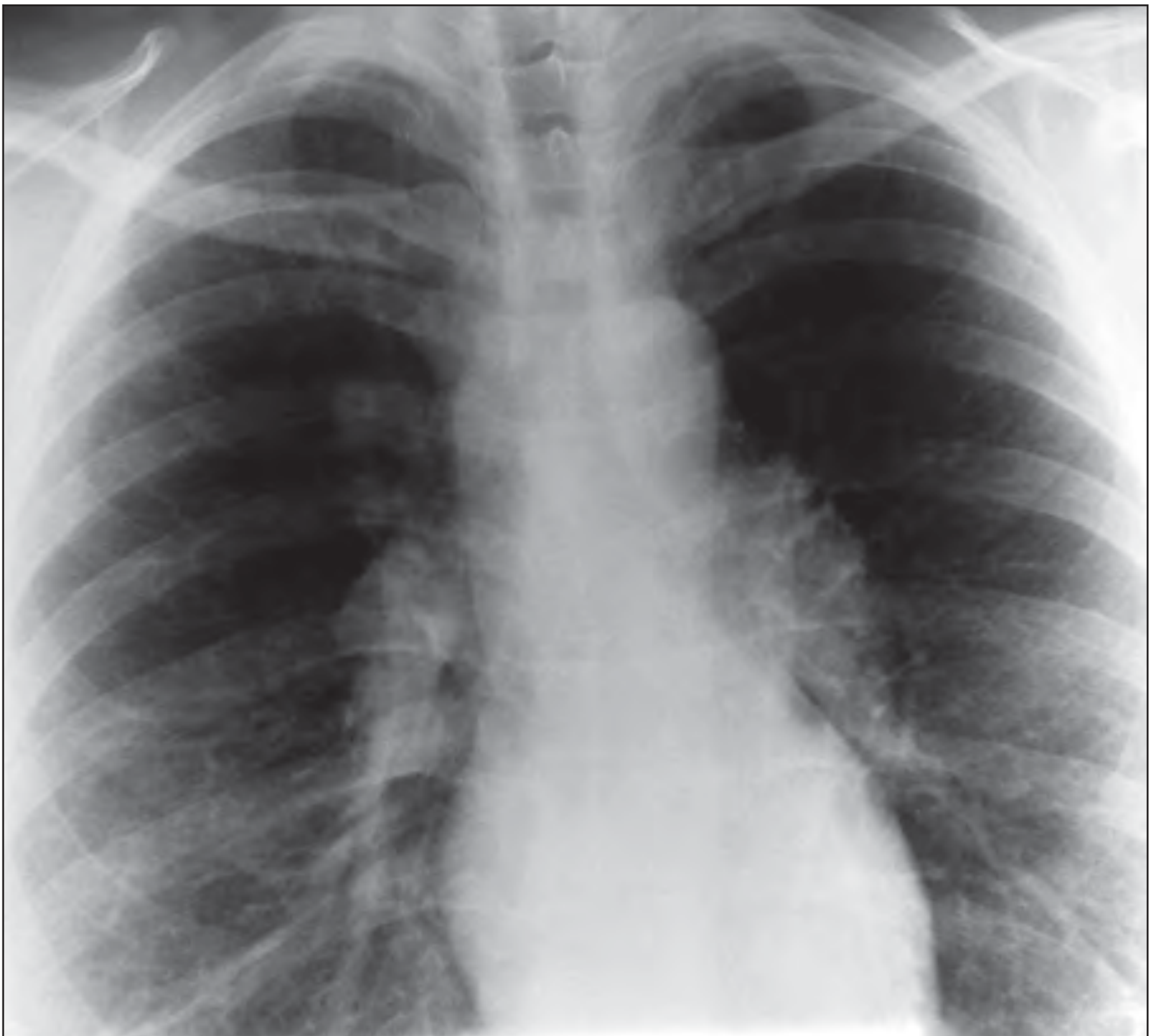
Airway disease (obstruction from endobronchial granulomas or bronchiolitis) occurs in ~50% and is a poor prognostic sign.
Diagnosis
Histology (Gold Standard)
Tissue biopsy showing noncaseating epithelioid granulomas after exclusion of other granulomatous diseases (TB, fungal infection, berylliosis, Crohn's, lymphoma).
Biopsy yield by site:
- Transbronchial biopsy: 40% (normal CXR) to >90% (abnormal CXR)
- Endobronchial/esophageal ultrasound-guided FNA of hilar/mediastinal nodes: 80-93%
- Skin and peripheral lymph node biopsies: high yield when involved
- BAL: not diagnostic alone, but CD4/CD8 ratio >3.5 is 94% specific, 52% sensitive
Key Labs and Tests
- Serum ACE (sACE): elevated in 30-80% of active disease - not specific (also elevated in TB, fungal disease, cirrhosis, diabetes); useful for monitoring
- Hypercalciuria (more common than hypercalcemia): due to macrophage/epithelioid cell conversion of 25(OH) vitamin D to 1,25(OH)₂ vitamin D
- Hypercalcemia: can cause fatigue, constipation, polyuria, altered mental status
- BAL: >30-60% lymphocytes; CD4/CD8 ratio >3.5
Recommended Initial Evaluation (all patients)
-
Chest radiograph + PFTs (spirometry, DLCO, lung volumes) + 6-minute walk
-
CBC, chemistry (calcium, LFTs, creatinine), urinalysis
-
25(OH) and 1,25(OH)₂ vitamin D levels
-
ECG (all patients, yearly)
-
Ophthalmologic exam (slit lamp + funduscopy + tonometry)
-
TB test (tuberculin skin test or IGRA)
-
Rheumatology, 2-Vol Set (ATS/ERS/WASOG guidelines)
Treatment
Indications
Not all patients require treatment. Observation for 3-6 months is appropriate for patients with:
- Good prognostic signs (Löfgren syndrome, Stage I, asymptomatic Stage II-III)
Treatment is indicated for:
- Progressive or symptomatic pulmonary disease (TLC decrease ≥10%, FVC decrease ≥15%, DLCO decrease ≥20%, or progressive radiographic changes)
- Cardiac, neurologic, ocular (non-anterior), or hypercalcemia
- Cosmetically disfiguring skin disease
Step 1: Corticosteroids (First-line)
- Prednisone: 40 mg/day (≤1 mg/kg/day) for 4-6 weeks, then taper by 5-10 mg every 4-8 weeks to maintenance of 10-15 mg/day
- Continue maintenance for 6-8 months (total ≥12 months before attempting to taper off)
-
70% respond favorably; however, relapse occurs in 25-50% after tapering or discontinuation
Step 2: Steroid-Sparing Agents (2nd line / chronic disease)
| Drug | Dose |
|---|---|
| Methotrexate | 15 mg/week |
| Mycophenolate mofetil | 1-1.5 g twice daily |
| Azathioprine | 2 mg/kg/day |
| Leflunomide | 10-20 mg/day |
| Infliximab (anti-TNF) | 3-5 mg/kg IV every 1-2 months |
| Rituximab | 1000 mg x2, 2 weeks apart |
Organ-Specific Considerations
- Cardiac sarcoidosis: Antiarrhythmics, diuretics, afterload reduction + corticosteroids (prednisone 10-25 mg/day maintenance). ICD placement for serious arrhythmias. Anti-TNF agents as steroid-sparing.
- Neurosarcoidosis/severe ocular: High-dose oral corticosteroids or IV pulse therapy, followed by maintenance. Steroid-sparing agents often needed for chronic disease. Anterior uveitis can be managed with topical steroid drops.
- Pulmonary hypertension: May respond to sildenafil, bosentan, or epoprostenol.
- Pregnancy: Corticosteroids only (other agents teratogenic). Spontaneous remission may occur during pregnancy, but exacerbation often follows delivery.
End-Stage Disease
-
Lung transplantation is an option; outcomes are comparable to other interstitial lung diseases.
-
Sarcoidosis recurs in the allograft in ~50% of cases, but this is usually asymptomatic and does not significantly affect outcomes.
-
Fishman's Pulmonary Diseases, p. 939; Rheumatology, 2-Vol Set, p. 3113-3121
Prognosis
- ~2/3 of patients experience spontaneous remission within 10 years
- Stage I: ~60-80% spontaneous resolution
- Stage IV: fibrosis is generally irreversible
- Chronic/progressive disease is more common in Black patients, older age at onset, multi-organ involvement, and Stage III-IV disease
- Mortality: ~1-5%; main causes are respiratory failure, cardiac arrhythmias/cardiomyopathy, and neurosarcoidosis
Recent Evidence (2025)
A 2025 systematic review and meta-analysis (PMID 40393718) published in Thorax evaluated the efficacy of biologic and targeted synthetic therapies in sarcoidosis, supporting the role of anti-TNF agents (especially infliximab) in refractory disease. This aligns with current guideline recommendations for steroid-sparing treatment.
Sources: Goldman-Cecil Medicine (26th ed., Ch. 83) | Fishman's Pulmonary Diseases and Disorders | Rheumatology, 2-Volume Set (Elsevier, 2022) | Firestein & Kelley's Textbook of Rheumatology
This is a shared conversation. Sign in to Orris to start your own chat.