
Anatomy of nose easy summary for revision dont leave any important points first year bds student
Reading File
Searching sources...
Finding Sources
Finding Sources
Reading File
Reading File
Reading File
Reading File
Finding Sources
Finding Sources
Reading File
Reading File
Reading File
Finding Sources
Reading File
Finding Sources
Reading File
Reading File
Reading File
Reading File
Reading File
Loading Image
Reading File
Loading Image
I now have comprehensive information from the textbooks to compile a thorough, well-structured revision summary. Let me put it all together.
Anatomy of the Nose - Complete Revision Summary (1st Year BDS)
1. OVERVIEW & PARTS
The nose has two parts:
- External nose - pyramidal structure projecting from the face
- Nasal cavity - extends from nares (nostrils) anteriorly to choanae posteriorly (where it becomes the nasopharynx)
- Divided into right and left cavities by the nasal septum
2. EXTERNAL NOSE
Shape & Surface Landmarks
- Pyramidal shape; apex points anteriorly
- Root (upper end, between orbits), Dorsum (bridge), Tip (apex), Alae (wings), Columella (between nostrils), Nares (external openings)
- Skin is thin and loosely adherent over dorsum; becomes thicker with many sebaceous glands at the tip and alae
Skeleton of External Nose
Bony part (upper 1/3):
- Nasal bones (paired)
- Parts of frontal bones
- Frontal processes of maxilla
Cartilaginous part (lower 2/3):
| Cartilage | Position/Notes |
|---|---|
| Septal (quadrilateral) cartilage | Forms anterior nasal septum in midline |
| Upper lateral (ULC) | Trapezoid shape; attaches to dorsal septum medially |
| Lower lateral (LLC) / Alar cartilage | Divided into medial, intermediate, lateral crura |
| Sesamoid cartilages (2-3) | Small, between ULC and LLC |
Key: ULC and septal cartilage together form the internal nasal valve (narrowest part of the nasal airway, ~10-15 degrees in Caucasians).
Muscles of External Nose
All supplied by facial nerve (CN VII)
| Action | Muscles |
|---|---|
| Elevators | Procerus, levator labii superioris alaeque nasi, anomalous nasi |
| Depressors | Alar nasalis, depressor septi nasi |
| Compressors | Transverse nasalis, compressor narium minor |
| Dilator | Dilator naris anterior |
Subcutaneous Layers (from superficial to deep)
- Superficial fatty layer
- Fibromuscular layer (SMAS)
- Deep fatty layer (contains neurovascular structures)
- Periosteum/perichondrium
3. NASAL VESTIBULE
- Anteriormost part of nasal cavity
- Lined by keratinizing stratified squamous epithelium
- Contains vibrissae (coarse hairs), sebaceous glands, sweat glands
- Demarcated by the limen nasi (at caudal border of lower lateral cartilage)
4. NASAL CAVITY
Boundaries
| Wall | Formed by |
|---|---|
| Floor | Palatine process of maxilla (anterior 3/4) + horizontal plate of palatine bone (posterior 1/4) |
| Roof | Skull base (sloping downward anteroposteriorly) - cribriform plate of ethmoid is its narrowest part |
| Medial wall | Nasal septum |
| Lateral wall | Turbinates, meatus, sinus openings |
Epithelium
- Nasal vestibule → Stratified squamous (keratinized)
- Superior part (superior septum, superior turbinate, upper middle turbinate) → Olfactory epithelium
- Remainder → Pseudostratified ciliated columnar (respiratory epithelium)
5. NASAL SEPTUM
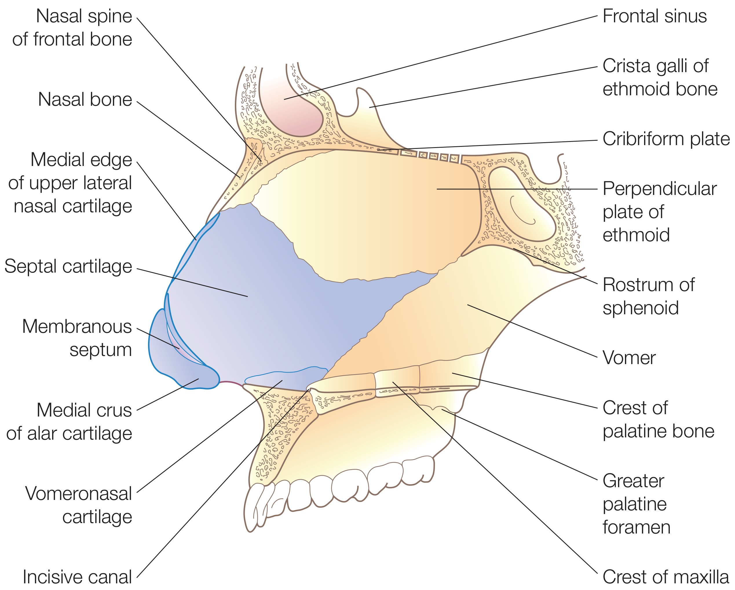
The septum has three parts:
Bony Part
| Bone | Location |
|---|---|
| Perpendicular plate of ethmoid | Upper 1/3; continuous superiorly with cribriform plate and crista galli |
| Vomer | Posterior and inferior septum; articulates with sphenoid rostrum above |
| Maxillary crest | Anteroinferior portion |
| Palatine bone (crest) | Posteroinferior portion |
Cartilaginous Part
- Quadrilateral (septal) cartilage - forms anterior septum; firmly attached to nasal bones, perpendicular plate of ethmoid, and vomer by collagenous fibers
- Its sphenoidal process (septal tail) extends posteriorly between vomer and perpendicular plate
Membranous Part
- Segment of connective tissue between caudal septal cartilage and columella
Exam point: Nasal septal deviation is the most common cause of nasal obstruction; the commonest site of septal perforation/epistaxis is Kiesselbach's plexus (Little's area)
6. LATERAL NASAL WALL & TURBINATES (Conchae)
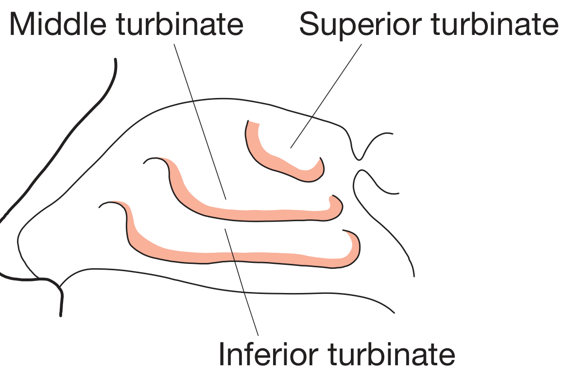
Turbinates
- 3 main turbinates: Superior, Middle, Inferior
- Middle and superior turbinates arise from ethmoid bone
- Inferior turbinate is an independent osseous structure (separate bone)
- Function: warm, humidify, and filter inspired air; modify nasal airflow resistance
Meatuses (spaces under each turbinate)
| Meatus | Opens into / Contains |
|---|---|
| Inferior meatus | Nasolacrimal duct opens here (at Hasner's valve, ~1 cm behind the head of inferior turbinate) |
| Middle meatus | Frontal sinus, maxillary sinus, anterior ethmoid cells all drain here (via the ostiomeatal complex) |
| Superior meatus | Posterior ethmoid air cells drain here |
| Sphenoethmoidal recess (above superior turbinate) | Sphenoid sinus drains here |
Memory tip: "FBI drains Middle" - Frontal, (anterior ethmoid,) maxillary drains into Middle meatus. Posterior ethmoid → Superior meatus. Sphenoid → Sphenoethmoidal recess.
Nasal Cycle
- Turbinates cyclically alternate congestion/decongestion every 0.5-3 hours under sympathetic control - this is normal physiology.
7. BLOOD SUPPLY
Two major sources: External + Internal carotid
External Carotid Artery (via Maxillary artery → Sphenopalatine artery)
- Sphenopalatine artery - main supply to turbinates and lateral nasal wall; enters via sphenopalatine foramen
- Posterior septal artery (branch) → posteroinferior septum
- Greater palatine artery → enters via incisive canal → anteroinferior septum
- Superior labial artery (branch of facial artery) → columella and caudal septum
- Angular/lateral nasal arteries (branch of facial artery) → ala and sidewall
Internal Carotid Artery (via Ophthalmic artery → Ethmoidal arteries)
- Anterior ethmoidal artery → anterosuperior septum + dorsum of external nose
- Posterior ethmoidal artery → posterosuperior septum
Kiesselbach's Plexus (Little's Area) ⭐ HIGH YIELD
- Located on the anterior nasal septum
- Anastomosis of: anterior ethmoidal artery + posterior septal artery + septal branch of superior labial artery
- Most common site of epistaxis (nosebleed) due to rich vascularity and susceptibility to trauma/turbulent airflow
Venous Drainage
- Posteriorly via sphenopalatine vessels → pterygoid plexus
- Anteriorly via facial vein
- Superiorly: ethmoidal veins → superior ophthalmic vein → cavernous sinus
⚠️ Danger area: Upper lip and nose infections can spread retrograde through the valveless facial vein to the cavernous sinus via the ophthalmic vein - potentially life-threatening!
8. NERVE SUPPLY
External Nose
| Nerve | Area Supplied |
|---|---|
| Supratrochlear & infratrochlear (from V1 - ophthalmic) | Nasal root, bridge, upper sidewall |
| External nasal branch of anterior ethmoidal nerve (V1) | Dorsum and nasal tip |
| Infraorbital nerve (V2 - maxillary) | Remaining skin of nasal sidewall and ala |
Nasal Cavity (Mucosa)
- Sensory: Ophthalmic (V1) and maxillary (V2) divisions of trigeminal nerve
- Lateral wall and turbinates: posterolateral nasal nerves from V2 (via sphenopalatine ganglion) + ethmoidal nerves from V1
- Anterior septum: nasopalatine nerve
- Parasympathetic: Via vidian nerve (greater superficial petrosal nerve) → synapse in sphenopalatine ganglion → regulates nasal secretions
- Sympathetic: Via vidian nerve (deep petrosal nerve) → passes through sphenopalatine ganglion (no synapse) → regulates vascular tone and turbinate congestion
9. LYMPHATIC DRAINAGE
- External nose: Submandibular, submental, and facial nodes; drainage may also reach parotid nodes
- Nasal cavity (anterior): Submandibular nodes
- Nasal cavity (posterior): Upper deep cervical and retropharyngeal nodes
10. PARANASAL SINUSES (Quick Summary)
| Sinus | Location | Drainage |
|---|---|---|
| Frontal sinus | Frontal bone | Middle meatus (via frontonasal duct) |
| Maxillary sinus | Maxillary bone | Middle meatus |
| Anterior ethmoid cells | Ethmoid bone | Middle meatus |
| Posterior ethmoid cells | Ethmoid bone | Superior meatus |
| Sphenoid sinus | Sphenoid bone | Sphenoethmoidal recess |
- All sinuses lined by respiratory (ciliated) mucosa and innervated by trigeminal nerve (CN V)
- The maxillary sinus ostium opens high on the medial wall - poor gravity drainage (clinically important!)
11. QUICK CLINICAL CORRELATIONS (BDS Favorites)
| Condition | Anatomical Basis |
|---|---|
| Epistaxis | Kiesselbach's plexus on anterior septum (Little's area) |
| Cavernous sinus thrombosis | Venous spread via valveless facial/ophthalmic vein from nasal/upper lip infections |
| Choanal atresia | Failure of bucconasal membrane to break down during embryological development |
| Nasal obstruction | Deviated nasal septum, turbinate hypertrophy, polyps |
| Referred dental pain to maxillary teeth | Maxillary sinus infection (roof of maxillary sinus = floor of orbit; floor overlies upper molar roots) |
| Nasolacrimal duct blockage | Presents as epiphora; duct opens in inferior meatus |
12. EMBRYOLOGY (Key Points)
- Nose develops in the 4th week of gestation from neural crest cell proliferation forming nasal placodes
- Medial nasal processes → philtrum, columella, premaxilla
- Lateral nasal processes → nasal bones and alar cartilages
- Nasal septum: downgrowth of frontonasal process; fuses with palate from 9th-12th week
- Vomeronasal (Jacobson's) organs: regress before birth; only vomeronasal cartilages remain in adults
Sources: Scott-Brown's Otorhinolaryngology Head & Neck Surgery Vol 1; Bailey and Love's Short Practice of Surgery 28th Ed; Gray's Anatomy for Students; Cummings Otolaryngology Head and Neck Surgery
Anatomy of triangle of neck easy summary for bds students dont leave any important points for exam points of view
Reading File
Searching sources...
Finding Sources
Finding Sources
Reading File
Reading File
Reading File
Reading File
Reading File
Reading File
Reading File
Reading File
Reading File
Reading File
Reading File
Loading Image
Reading File
Loading Image
Reading File
Reading File
Reading File
I now have all the essential information. Let me compile the complete, exam-focused revision summary.
Triangles of the Neck - Complete Revision Summary (1st Year BDS)
OVERVIEW
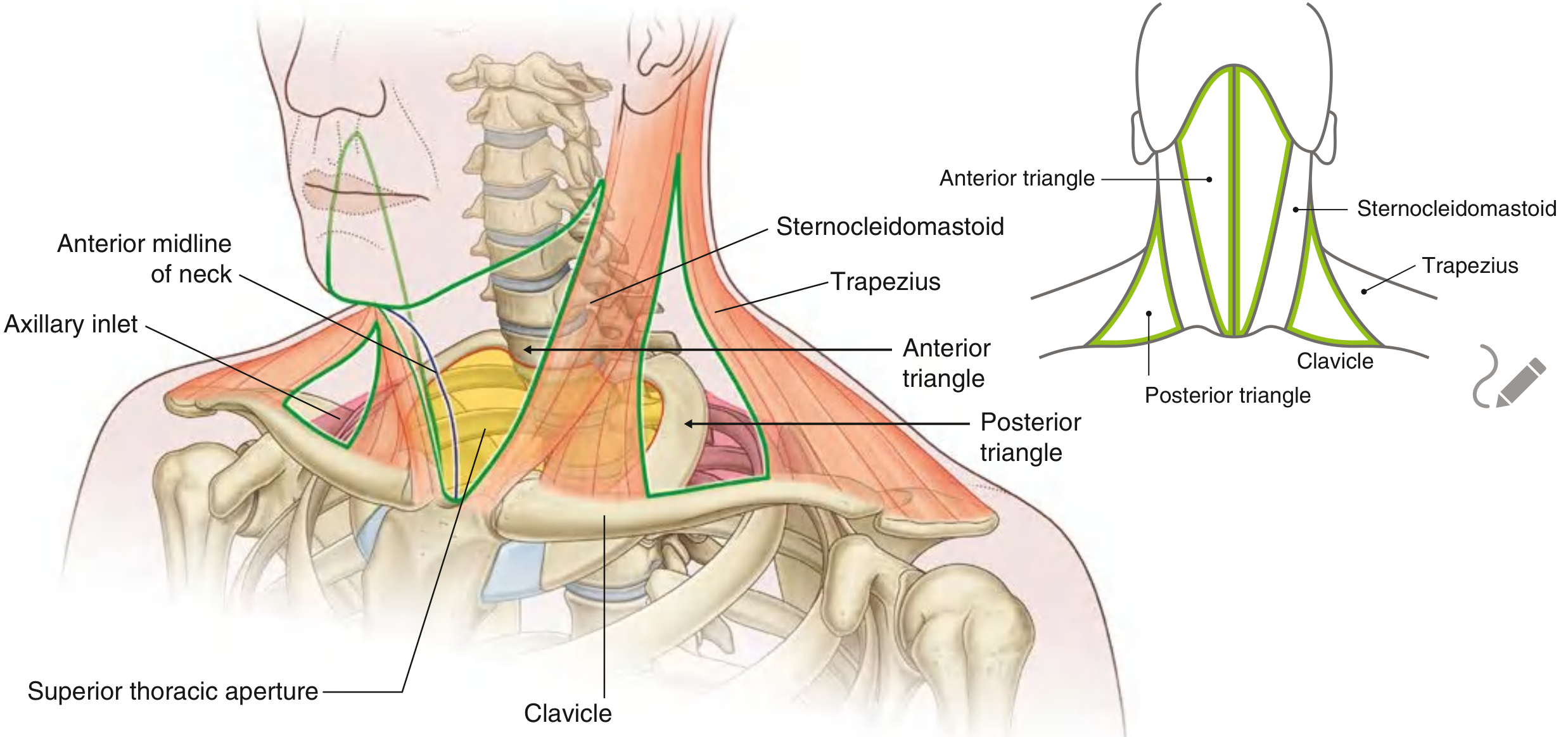
The neck is divided into two major triangles on each side by the sternocleidomastoid (SCM) muscle:
- Anterior triangle - medial to SCM
- Posterior triangle - lateral to SCM
THE STERNOCLEIDOMASTOID (SCM) MUSCLE - KEY LANDMARK
| Feature | Detail |
|---|---|
| Origin | Two heads: sternal head (manubrium) + clavicular head (medial 1/3 of clavicle) |
| Insertion | Mastoid process + lateral 1/2 of superior nuchal line |
| Nerve supply | Accessory nerve (CN XI) motor; C2, C3 proprioception |
| Action (unilateral) | Tilts head to same side, rotates face to opposite side |
| Action (bilateral) | Draws head forward (flexion) |
PART A: ANTERIOR TRIANGLE
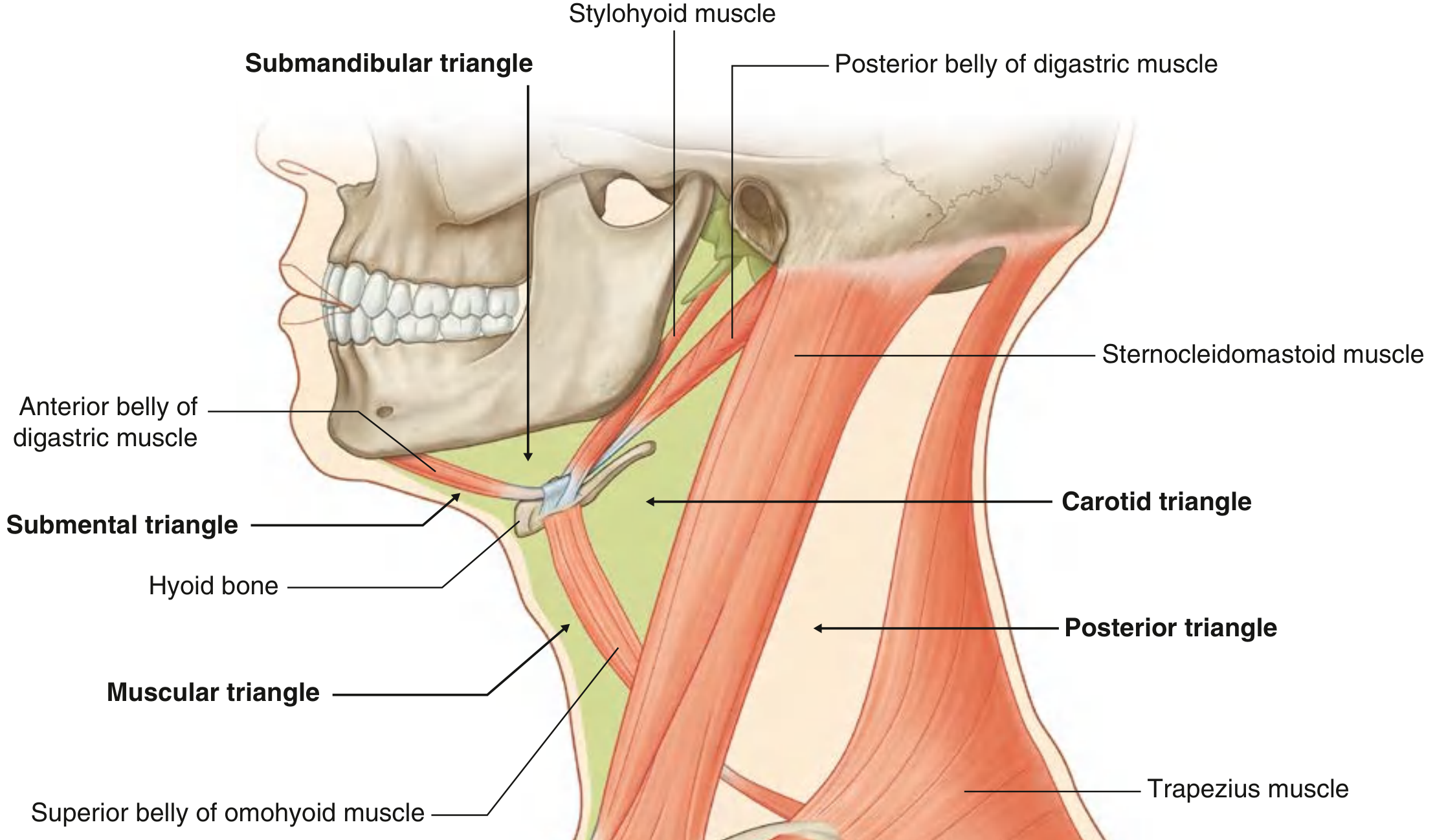
Boundaries
| Border | Structure |
|---|---|
| Lateral | Anterior border of SCM |
| Superior | Inferior border of mandible |
| Medial | Midline of neck |
| Roof | Investing layer of deep cervical fascia + platysma |
Subdivisions
The anterior triangle is divided into 4 smaller triangles by the digastric and omohyoid muscles:
1. SUBMENTAL TRIANGLE (unpaired/single)
| Boundaries | Mandibular symphysis (above), anterior belly of digastric (each side), body of hyoid (below) |
| Floor | Mylohyoid muscle |
| Contents | Submental lymph nodes, tributaries forming anterior jugular vein |
BDS point: Only unpaired triangle of the neck. Submental nodes drain tip of tongue, floor of mouth, lower incisors.
2. SUBMANDIBULAR (DIGASTRIC) TRIANGLE
| Boundaries | Lower border of mandible (above), anterior belly of digastric (anteroinferior), posterior belly of digastric (posteroinferior) |
| Floor (3 muscles, superficial to deep) | Mylohyoid → Hyoglossus → Superior constrictor |
| Roof | Investing fascia splits to enclose submandibular gland |
Contents:
| Structure | Notes |
|---|---|
| Submandibular gland | Main content; deep process extends between mylohyoid and hyoglossus |
| Wharton's duct (submandibular duct) | Opens on floor of mouth lateral to frenulum |
| Facial artery & vein | Artery grooves submandibular gland, appears on face at lower border of mandible |
| Hypoglossal nerve (CN XII) | Passes between mylohyoid and hyoglossus |
| Lingual nerve | Loops around Wharton's duct (lateral → inferior → medial to duct) |
| Submandibular ganglion | Hangs from lingual nerve; preganglionic PS fibers synapse here; supplies submandibular + sublingual glands |
| Submandibular lymph nodes | Important in head and neck infection/malignancy |
| Mylohyoid nerve | Branch of V3; motor to mylohyoid and anterior belly of digastric |
⭐ BDS High Yield: The lingual nerve spirals around Wharton's duct going lateral → inferior → medial - a classic exam question! The submandibular ganglion hangs from the lingual nerve like a "bag hanging from a nerve."
3. CAROTID TRIANGLE ⭐ MOST IMPORTANT
| Boundaries | Posterior belly of digastric (superior), superior belly of omohyoid (anteroinferior), anterior border of SCM (posterolateral) |
| Floor | Thyrohyoid + hyoglossus (anteriorly); middle and inferior pharyngeal constrictors + longus capitis (posteriorly) |
Contents:
Arteries
| Artery | Notes |
|---|---|
| Common carotid artery (CCA) | Right CCA from brachiocephalic; left CCA from aortic arch. NO branches in neck |
| Bifurcation of CCA | At level of upper border of thyroid cartilage (C3/C4) |
| Internal carotid artery (ICA) | No branches in neck; enters skull via carotid canal |
| External carotid artery (ECA) | Gives branches immediately; initially anteromedial to ICA |
Branches of ECA (mnemonic: Some Anatomists Like Feeling Other People's Soft Parts)
| Group | Branches |
|---|---|
| Anterior | Superior thyroid, Lingual, Facial |
| Posterior | Sternocleidomastoid, Occipital, Posterior auricular |
| Medial | Ascending pharyngeal |
| Terminal | Superficial temporal + Maxillary (at neck of mandible, inside parotid) |
Carotid sinus = dilation at origin of ICA → baroreceptor (monitors BP), innervated by CN IX (glossopharyngeal) Carotid body = ovoid body at bifurcation → chemoreceptor (monitors O2/CO2), innervated by CN IX + X
Veins
| Vein | Notes |
|---|---|
| Internal jugular vein (IJV) | Begins at jugular foramen; lies anterolateral to CCA in carotid sheath |
| Tributaries | Facial, lingual, superior thyroid veins (superior part); middle thyroid vein (inferior part) |
Carotid Sheath Contents (medial to lateral)
- Medial: Common/internal carotid artery
- Lateral: Internal jugular vein
- Posterior (between): Vagus nerve (CN X)
- Deep (on prevertebral fascia): Cervical sympathetic trunk (outside sheath)
Nerves in Carotid Triangle
| Nerve | Notes |
|---|---|
| Vagus (CN X) | In carotid sheath between artery and vein |
| Superior laryngeal nerve (branch of X) | Divides into: external laryngeal (motor to cricothyroid) + internal laryngeal (sensory to larynx above vocal folds - pierces thyrohyoid membrane) |
| Accessory nerve (CN XI) | Crosses superior part of triangle, then deep to SCM to reach posterior triangle |
| Hypoglossal nerve (CN XII) | Hooks around occipital artery; enters submandibular triangle |
| Ansa cervicalis | Loop on/in front of carotid sheath |
Ansa Cervicalis ⭐
- Superior root (descendens hypoglossi): C1 fibers carried by CN XII → innervates superior belly of omohyoid, upper sternohyoid, upper sternothyroid
- Inferior root (descendens cervicalis): C2 + C3 → innervates inferior belly of omohyoid, lower sternohyoid, lower sternothyroid
- Thyrohyoid is innervated by C1 fibers from hypoglossal nerve (not ansa itself)
Memory trick: Ansa cervicalis = "handle/loop" in Latin. It supplies all infrahyoid muscles EXCEPT thyrohyoid (which gets C1 via CN XII directly).
4. MUSCULAR (STRAP) TRIANGLE
| Boundaries | Midline (medial), superior belly of omohyoid (superolateral), anterior border of SCM (lateral) |
| Contents | Strap muscles (sternohyoid, omohyoid, sternothyroid, thyrohyoid), thyroid gland, parathyroid glands, trachea, esophagus, pharynx, larynx |
MUSCLES OF ANTERIOR TRIANGLE
Suprahyoid Muscles (above hyoid - raise hyoid during swallowing)
| Muscle | Origin | Insertion | Nerve Supply | Action |
|---|---|---|---|---|
| Digastric - anterior belly | Digastric fossa of mandible | Hyoid (via tendon) | V3 (nerve to mylohyoid) | Raises hyoid / opens mouth |
| Digastric - posterior belly | Mastoid notch | Hyoid (via tendon) | CN VII (facial) | Raises hyoid posterosuperiorly |
| Stylohyoid | Styloid process | Body of hyoid | CN VII (facial) | Pulls hyoid posterosuperiorly |
| Mylohyoid | Mylohyoid line of mandible | Hyoid + mylohyoid raphe | V3 (nerve to mylohyoid) | Forms floor of mouth; elevates hyoid/floor of mouth |
| Geniohyoid | Genial tubercle (mental spine) of mandible | Body of hyoid | C1 (via CN XII) | Elevates hyoid anteriorly |
⭐ Exam point: Digastric has dual innervation - posterior belly (CN VII), anterior belly (V3). This is because they have different embryological origins!
Infrahyoid Muscles (below hyoid - depress hyoid during swallowing)
| Muscle | Nerve Supply |
|---|---|
| Sternohyoid | Ansa cervicalis (C1-C3) |
| Sternothyroid | Ansa cervicalis (C1-C3) |
| Thyrohyoid | C1 via CN XII |
| Omohyoid (superior belly) | Ansa cervicalis (C1-C3) |
PART B: POSTERIOR TRIANGLE (Lateral Cervical Triangle)
Boundaries
| Border | Structure |
|---|---|
| Anterior | Posterior border of SCM |
| Posterior | Anterior border of trapezius |
| Inferior (base) | Middle 1/3 of clavicle |
| Apex | Occipital bone (where SCM and trapezius meet, posterior to mastoid) |
| Roof | Investing layer of deep cervical fascia |
| Floor | Prevertebral fascia covering scalene muscles |
Subdivisions
The inferior belly of omohyoid divides the posterior triangle into:
| Sub-triangle | Location | Key Contents |
|---|---|---|
| Occipital triangle | Larger, superior part | Accessory nerve, cervical plexus branches, brachial plexus roots/trunks |
| Omoclavicular (Subclavian/Supraclavicular) triangle | Smaller, inferior part | Subclavian artery (3rd part), external jugular vein (pierces fascia here) |
Floor Muscles (superficial to deep, anterior to posterior)
- Splenius capitis (at apex)
- Levator scapulae
- Posterior scalene
- Middle scalene
- Anterior scalene
⭐ Brachial plexus roots emerge between anterior and middle scalene muscles
Contents of Posterior Triangle
Nerves
Accessory Nerve (CN XI) ⭐
- Exits jugular foramen → passes through/deep to SCM (innervates it) → crosses posterior triangle within investing fascia → reaches trapezius (innervates it)
- Lies superficial to prevertebral fascia (unlike most other motor nerves here)
- Most superior nerve in the posterior triangle
- Vulnerable to injury during lymph node biopsy → causes drooping shoulder (trapezius paralysis)
Cervical Plexus (C1-C4) - Cutaneous branches:
All emerge at nerve point of the neck (posterior border of SCM, at its midpoint):
| Nerve | Spinal Level | Area Supplied |
|---|---|---|
| Lesser occipital | C2 | Skin behind ear, posterior scalp |
| Great auricular | C2, C3 | Skin over parotid, angle of mandible, auricle |
| Transverse cervical | C2, C3 | Anterior neck skin |
| Supraclavicular (medial, intermediate, lateral) | C3, C4 | Skin over shoulder, clavicle, upper chest |
Mnemonic for cutaneous branches: "My Great Aunt Talks Softly" → Medial/Lesser occipital, Great auricular, Anterior (transverse cervical), Two (C3,C4) Supraclavicular
Phrenic nerve (C3,4,5):
- Formed on surface of anterior scalene muscle
- "C3,4,5 keeps the diaphragm alive"
- Descends across anterior scalene within prevertebral fascia
Brachial Plexus (roots + trunks):
- Roots emerge between anterior and middle scalene muscles
- C5 + C6 → Superior trunk; C7 → Middle trunk; C8 + T1 → Inferior trunk
Vessels
| Vessel | Notes |
|---|---|
| External jugular vein | Crosses SCM superficially → enters posterior triangle → pierces investing fascia to drain into subclavian vein |
| Subclavian artery (3rd part) | Only part appearing in omoclavicular triangle |
| Transverse cervical artery | Branch of thyrocervical trunk; crosses posterior triangle |
| Suprascapular artery | Branch of thyrocervical trunk; crosses posterior triangle |
| Occipital artery | Enters apex of posterior triangle |
DEEP CERVICAL FASCIA - QUICK SUMMARY (BDS Favorite!)
| Layer | Encloses | Clinical Importance |
|---|---|---|
| Investing layer | SCM, trapezius, parotid, submandibular gland | Roof of both triangles; splits to enclose glands |
| Pretracheal layer | Trachea, thyroid, oesophagus, infrahyoid muscles | Limits spread of thyroid infections to anterior mediastinum |
| Prevertebral layer | Vertebral column + prevertebral muscles | Floor of posterior triangle; retropharyngeal abscess |
| Carotid sheath | CCA/ICA, IJV, vagus nerve | Blends with all three layers |
QUICK CLINICAL CORRELATIONS (⭐ BDS Exam Favorites)
| Scenario | Anatomical Basis |
|---|---|
| Carotid sinus massage (treats SVT) | Stimulates baroreceptors → reflex bradycardia via CN IX |
| "Carotid blow out" in neck dissection | Common carotid artery injury |
| Drooping shoulder after neck biopsy | CN XI (accessory nerve) injury in posterior triangle |
| Winging of scapula | Long thoracic nerve injury (C5,6,7) in posterior triangle |
| Phrenic nerve palsy (raised hemidiaphragm) | Injury to nerve crossing anterior scalene in posterior triangle |
| Submandibular abscess spreading | Can track along deep process of gland into floor of mouth (Ludwig's angina) |
| Horner's syndrome in neck | Cervical sympathetic trunk injury (posterior to carotid sheath) → ptosis, miosis, anhidrosis, enophthalmos |
| Lingual nerve injury in submandibular gland surgery | Loss of general sensation from anterior 2/3 tongue + loss of taste (chorda tympani fibers) |
MEMORY AIDS
ECA branches: "Some Anatomists Like Feeling Other People's Soft Parts"
Superior thyroid | Ascending pharyngeal | Lingual | Facial | Occipital | Posterior auricular | Superficial temporal | (maxillary) - terminal branches
Posterior triangle cutaneous nerves emerge at "Erb's point" (nerve point) - behind midpoint of SCM
Ansa cervicalis: "C1 superior, C2+C3 inferior; supplies all strap muscles EXCEPT thyrohyoid"
Carotid sheath contents (medial to lateral): "I Am Very Nervous" - Internal carotid Artery | (i)nternAl jugular Vein | Vagus Nerve
Sources: Gray's Anatomy for Students; Fischer's Mastery of Surgery 8th Ed; Scott-Brown's Otorhinolaryngology Head & Neck Surgery
Fascia of neck bds students first year exam points of view important points revision and summary dont skip imp points for exam points of view
Searching sources...
Finding Sources
Finding Sources
Reading File
Reading File
Reading File
Reading File
Reading File
Reading File
Finding Sources
Finding Sources
Finding Sources
Finding Sources
Finding Sources
Reading File
Reading File
Reading File
Reading File
Loading Image
Loading Image
I now have all the essential content. Let me compile the comprehensive revision summary.
Fascia of the Neck - Complete Revision Summary (1st Year BDS)
OVERVIEW
The fascia of the neck is organised into two broad categories:
- Superficial cervical fascia
- Deep cervical fascia (most important for exams - has 3 main layers + carotid sheath)
The fascial layers create compartments and potential spaces that control how infections, tumours, and surgical dissection planes behave in the neck.
PART 1: SUPERFICIAL CERVICAL FASCIA
- A layer of loose areolar connective tissue lying directly beneath the skin
- Contains:
- Platysma muscle (most important content)
- Superficial veins (external jugular, anterior jugular veins)
- Cutaneous nerves (branches of cervical plexus)
- Superficial lymph nodes
- Fat
Platysma ⭐
| Feature | Detail |
|---|---|
| Type | Broad, flat, thin sheet of muscle of facial expression |
| Origin | Superficial fascia of upper chest/shoulder (below clavicle) |
| Insertion | Skin + muscles of lower face/lip (crosses mandible) |
| Nerve supply | Cervical branch of facial nerve (CN VII) |
| Action | Depresses mandible; tenses skin of neck; draws lip/angle of mouth downward |
| Surgical note | Plane immediately deep to platysma is avascular - best surgical plane |
⭐ BDS Exam point: Platysma is the only muscle in the superficial cervical fascia. It is supplied by the cervical branch of CN VII (not a muscle of mastication nerve). Damage during surgery → loss of lower facial/lip tone.
PART 2: DEEP CERVICAL FASCIA
The deep cervical fascia has 3 main layers:
- Investing layer (superficial)
- Pretracheal layer / middle layer (visceral)
- Prevertebral layer (deep)
- Carotid sheath (a separate condensation formed from all 3 layers)
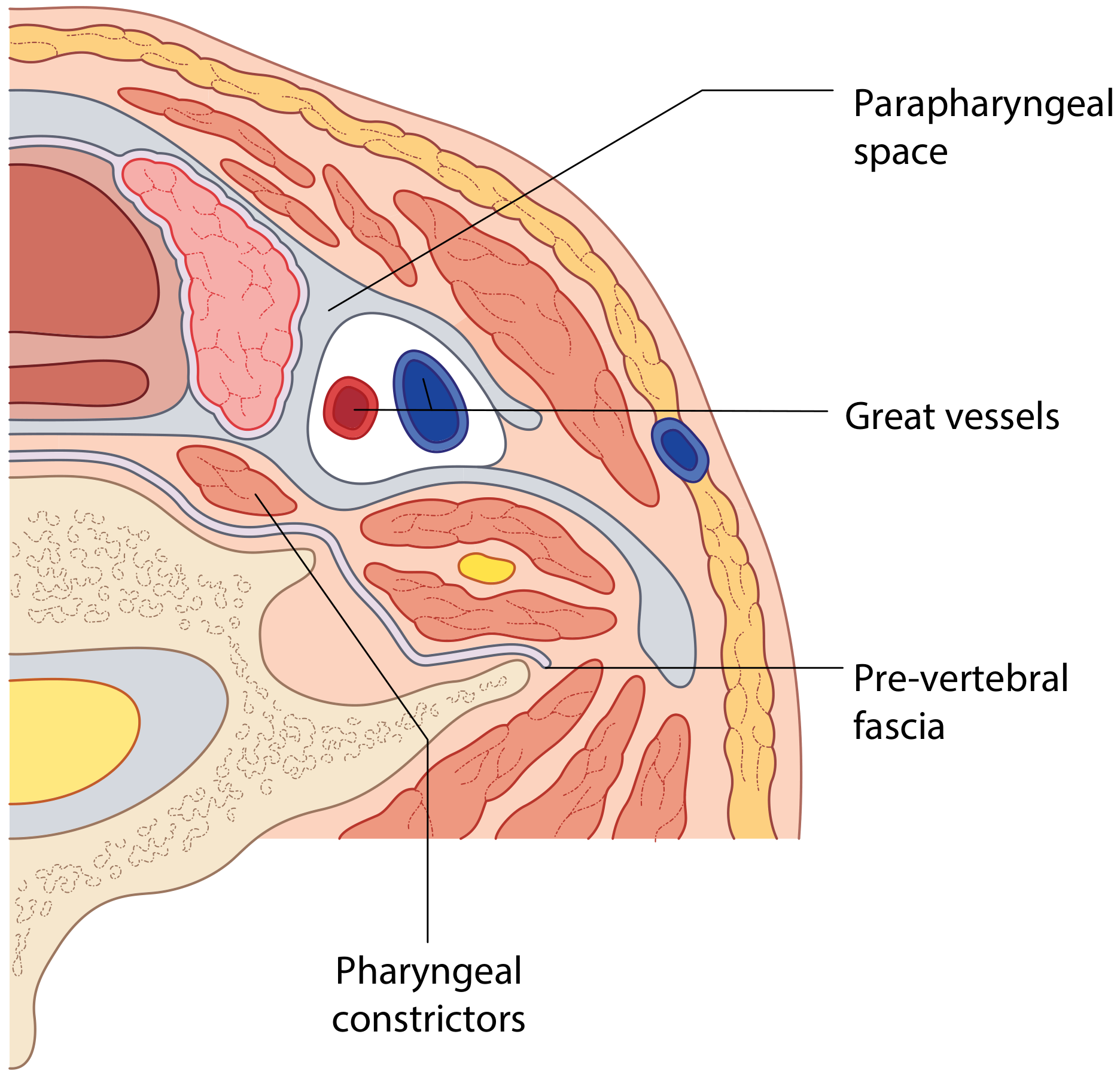
LAYER 1: INVESTING (SUPERFICIAL) LAYER OF DEEP CERVICAL FASCIA ⭐
Also called: Superficial layer of deep cervical fascia
Attachments
| Superior | Inferior |
|---|---|
| Superior nuchal line of occipital bone | Manubrium sterni |
| Mastoid process | Clavicle |
| Zygomatic arch | Acromion and spine of scapula |
| Inferior border of mandible | |
| Hyoid bone (anteriorly) | |
| Spinous processes of cervical vertebrae (posteriorly) |
What it encloses/does
- Envelops the entire neck like a cylinder
- Splits to enclose: SCM, trapezius, omohyoid (with a fascial sling to clavicle), infrahyoid muscles
- Splits to enclose glands:
- Parotid gland → forms parotid capsule/fascia (deep layer fuses with ICA fascia)
- Submandibular gland → splits to form a tough supporting membrane
- Forms roof of both the anterior and posterior triangles of neck
- Contains all cervical lymph nodes within its boundary
- Forms the stylomandibular ligament posteriorly (separates parapharyngeal and submandibular spaces)
- Splits inferiorly at manubrium to form the suprasternal space of Burns
Suprasternal Space of Burns ⭐
- A potential space between the two leaves of the investing fascia above the manubrium
- Contains: anterior jugular veins + jugular venous arch, lymph nodes, fat
- Can collect blood/pus after thyroid surgery or neck infections
⭐ Clinical: Because the investing layer forms a tough cylinder, deep abscesses build up under it and cause airway compression before they point superficially. This is why deep neck infections are dangerous.
LAYER 2: PRETRACHEAL (MIDDLE/VISCERAL) LAYER
Also called: Middle layer of deep cervical fascia
Limited to the anterior neck only.
Has two divisions:
| Division | Encloses | Notes |
|---|---|---|
| Muscular division | Infrahyoid strap muscles (sternohyoid, sternothyroid, thyrohyoid, omohyoid) | Also called muscular fascia |
| Visceral division | Trachea, thyroid + parathyroid glands, pharynx, larynx, upper oesophagus | Also called pretracheal or visceral fascia |
Attachments
- Superior: Hyoid bone + oblique line of thyroid cartilage
- Inferior: Descends into superior mediastinum → fuses with fibrous pericardium
- Lateral: Blends/connects to carotid sheath
Buccopharyngeal Fascia ⭐
- Part of the visceral division that overlies the posterior surface of the pharynx and buccinator
- Forms the anterior wall of the retropharyngeal space
- Forms the midline pharyngeal raphe (posterior midline attachment of constrictors)
- Forms the pterygomandibular raphe laterally
⭐ Clinical: Because the pretracheal layer descends into the mediastinum, thyroid infections and pretracheal abscesses can spread directly down into the anterior mediastinum - a life-threatening complication.
LAYER 3: PREVERTEBRAL (DEEP) LAYER
Also called: Deep layer of deep cervical fascia
What it covers
- Cervical vertebrae and their bodies
- Prevertebral muscles (longus colli, longus capitis, scalene muscles)
- Posterior neck muscles (paraspinous)
Attachments
- Superior: Base of skull
- Inferior: Fuses with anterior longitudinal ligament at T3 in the posterior mediastinum
- Lateral: Extends as the axillary sheath (encloses brachial plexus + axillary vessels)
Significance
- Forms the floor of the posterior triangle of neck
- The phrenic nerve (C3,4,5) and brachial plexus run deep to this fascia - protected during neck dissection
- Deep limit/margin of surgical neck dissection - should not be breached unless tumour invades it
- Allows the pharynx to glide freely over vertebral column during swallowing
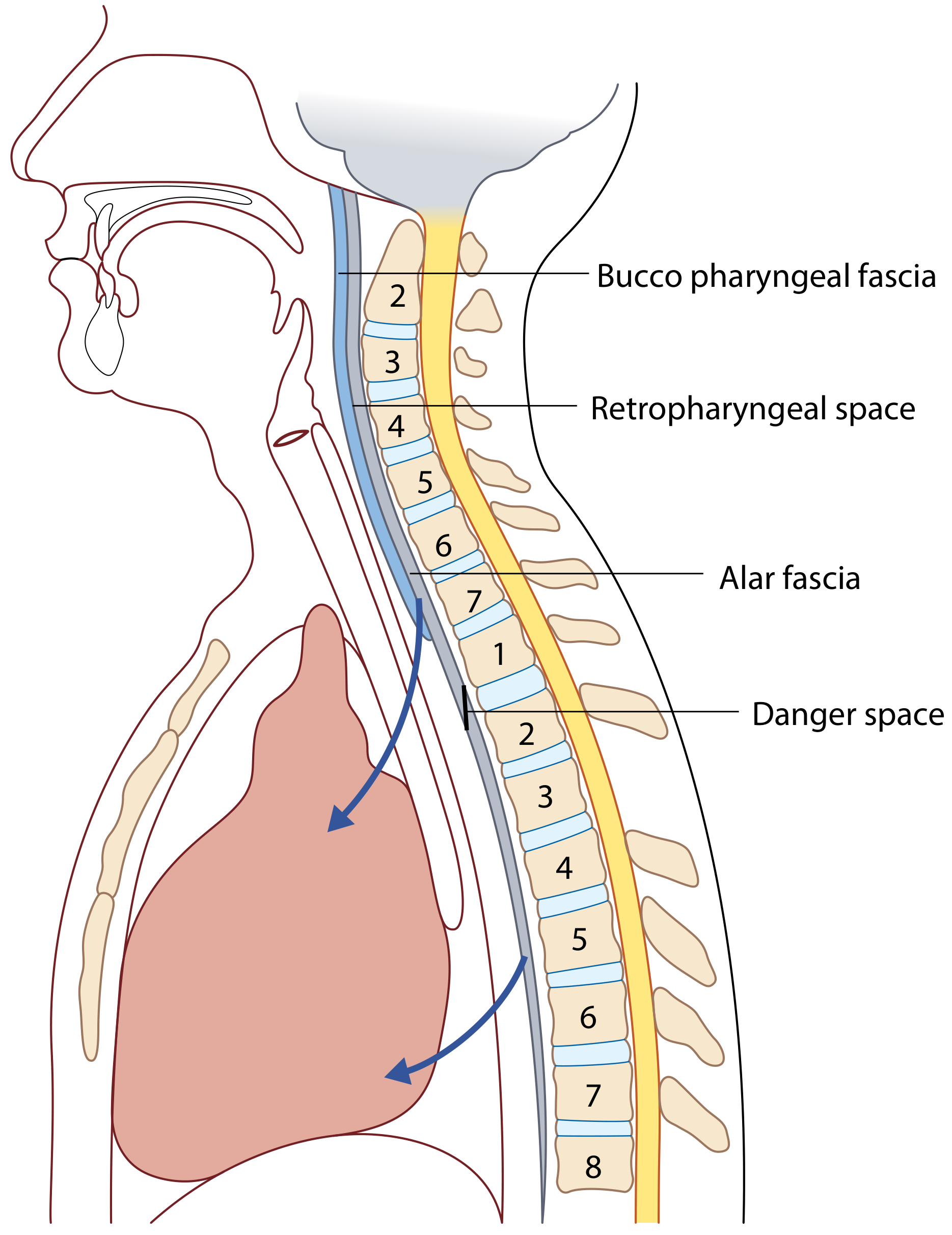
ALAR FASCIA ⭐ (often asked separately)
- A supplementary sheet anterior to the prevertebral fascia
- Lies between the prevertebral layer (behind) and the middle/visceral layer (in front)
- Extends from skull base to T2 vertebra (where it fuses with prevertebral fascia)
- Creates the critical "Danger Space" posterior to it
LAYER 4: CAROTID SHEATH ⭐⭐
Formation
- Formed from contributions of all three layers of the deep cervical fascia
- A tubular condensation running from skull base to thorax
Contents (medial to lateral)
| Contents | Position |
|---|---|
| Common carotid artery (CCA) / Internal carotid artery (ICA) | Medial |
| Internal jugular vein (IJV) | Anterolateral |
| Vagus nerve (CN X) | Posterior (between artery and vein) |
| Sympathetic nerve fibers (periarterial plexus) | On arterial wall |
| Ansa cervicalis | Anterior surface |
| Deep cervical lymph nodes | Within sheath |
Note: The sheath is thicker around the arteries but loose around the IJV to allow expansion during increased venous flow.
⭐ Classic exam fact: The carotid sheath is called the "Lincoln Highway of the Neck" because it provides a fast route for infection to spread from the skull base to the thorax.
PART 3: FASCIAL SPACES OF THE NECK ⭐⭐ (Very High Yield for BDS)
Spaces between fascial layers contain only loose areolar tissue normally, but are highways for spread of infection.
1. Submental Space
| Boundaries | Corresponds to submental triangle: mandibular symphysis, anterior bellies of digastric, hyoid |
| Floor | Mylohyoid + geniohyoid |
| Roof | Superficial cervical fascia |
| Contents | Submental lymph nodes (Level Ia) |
| Clinical | Spread from mandibular incisors, floor of mouth |
2. Submandibular Space ⭐
| Superior | Mucosa of floor of mouth |
| Inferior | Investing layer (between mandible and hyoid) |
| Anteroinferior | Anterior belly of digastric |
| Posteroinferior | Posterior belly of digastric |
| Divided by | Mylohyoid muscle → into sublingual (superior) and submandibular (inferior) compartments |
| Communication | Both compartments connect around free posterior edge of mylohyoid |
| Clinical importance | Ludwig's angina - rapidly spreading infection of submandibular space; starts in inferior compartment, spreads to sublingual, then to parapharyngeal, retropharyngeal → airway compromise |
3. Parapharyngeal Space (Lateral Pharyngeal Space) ⭐⭐
Shape: Inverted cone (base at skull, apex at hyoid)
| Superior | Petrous temporal bone (skull base) |
| Inferior | Hyoid bone |
| Medial | Pharyngobasilar fascia + superior constrictor |
| Lateral | Deep lobe of parotid, mandible, medial pterygoid |
| Anterior | Pterygomandibular raphe |
| Posterior | Prevertebral fascia |
Divided by styloid process + its attachments into 2 compartments:
| Compartment | Contents | Mnemonic |
|---|---|---|
| Pre-styloid (anterior) | Fat, connective tissue, maxillary artery, inferior alveolar nerve, lingual nerve, auriculotemporal nerve | "FAT" - Fat And Trigeminal branches |
| Post-styloid (posterior) | Carotid sheath (ICA, IJV, CN X), CN IX, CN XII, sympathetic chain, lymph nodes | "VANISH" - Vagus, Accessory, Nerve IX, IX+X, Sympathetic, Hypoglossal |
⭐ BDS point: Tonsil abscess/peritonsillar abscess can spread to the parapharyngeal space. Parotid tumours can bulge medially into this space (causing medial pharyngeal wall displacement). Parapharyngeal space infections can spread to all adjacent spaces.
4. Retropharyngeal Space ⭐⭐
| Anterior wall | Buccopharyngeal fascia (middle/visceral layer) |
| Posterior wall | Alar fascia |
| Extends | Skull base → Tracheal bifurcation (T4) |
| Contents | Loose areolar tissue + retropharyngeal lymph nodes (Nodes of Rouvière - drain nasopharynx, posterior nasal cavity) |
| Clinical | Retropharyngeal abscess; more common in children (nodes regress by age 5) |
5. "Danger Space" (Space 4) ⭐⭐ MOST CLINICALLY DANGEROUS
| Anterior wall | Alar fascia |
| Posterior wall | Prevertebral fascia |
| Extends | Skull base ALL THE WAY to the DIAPHRAGM |
⭐ The MOST DANGEROUS space - because it has no natural barriers, infection can spread from the neck all the way to the diaphragm, causing descending necrotizing mediastinitis - a potentially fatal complication with mortality up to 40%.
6. Prevertebral Space
| Between | Prevertebral fascia (anterior) and vertebral bodies |
| Extends | Skull base to coccyx (along anterior longitudinal ligament) |
| Clinical | Pott's disease (TB of vertebral bodies) → psoas abscess tracking down into thigh; cold abscess |
7. Masticator Space
| Contents | Muscles of mastication (masseter, pterygoids), ramus of mandible, inferior alveolar nerve + artery, mandibular division of V (V3) |
| Communication | With pterygopalatine fossa via pterygomaxillary fissure |
| Clinical | Odontogenic infections (molar teeth) can track here → trismus (inability to open mouth) |
SUMMARY TABLE: LAYERS OF DEEP CERVICAL FASCIA
| Layer | Also Called | Encloses | Attachments (Inf.) | Clinical Significance |
|---|---|---|---|---|
| Investing | Superficial layer | SCM, trapezius, parotid, submandibular gland, all nodes | Manubrium, clavicle | Roof of triangles; contains all nodes; suprasternal space of Burns |
| Pretracheal | Middle/visceral | Strap muscles, thyroid, trachea, oesophagus, pharynx | Fibrous pericardium | Infection → anterior mediastinum; buccopharyngeal fascia |
| Prevertebral | Deep layer | Vertebrae, prevertebral + scalene muscles | T3 ant. long. lig. | Floor of posterior triangle; axillary sheath; limits neck dissection |
| Alar | (part of deep layer) | Between middle and deep layers | T2 (fuses with prevertebral) | Forms danger space posteriorly |
| Carotid sheath | - | CCA/ICA, IJV, CN X, lymph nodes | Thorax | "Lincoln Highway"; formed by all 3 layers |
SUMMARY TABLE: FASCIAL SPACES
| Space | Between | Extends To | Key Clinical Condition |
|---|---|---|---|
| Submental | Mylohyoid and skin | Limited | Dental abscess (incisors) |
| Submandibular | Mucosa/mandible and investing fascia | Parapharyngeal | Ludwig's angina |
| Parapharyngeal | Pharynx medially, mandible/parotid laterally | Skull base to hyoid | Peritonsillar abscess, parotid tumour |
| Retropharyngeal | Buccopharyngeal fascia to alar fascia | Skull base to T4 (bifurcation) | Retropharyngeal abscess (children) |
| Danger space | Alar fascia to prevertebral fascia | Skull base to diaphragm | Descending necrotizing mediastinitis |
| Prevertebral | Prevertebral fascia to vertebrae | Skull base to coccyx | Pott's disease, TB cold abscess |
| Masticator | Investing fascia around ramus | Skull base to mandible | Odontogenic infection, trismus |
TOP EXAM POINTS - QUICK RECALL ⭐
| Fact | Answer |
|---|---|
| Muscle in superficial fascia of neck | Platysma (supplied by cervical branch CN VII) |
| Number of layers of deep cervical fascia | 3 (investing, pretracheal, prevertebral) + carotid sheath |
| Roof of both triangles of neck | Investing layer of deep cervical fascia |
| Space above manubrium in investing fascia | Suprasternal space of Burns |
| Carotid sheath contents | CCA/ICA, IJV, Vagus (CN X), ansa cervicalis, lymph nodes |
| "Lincoln Highway of the Neck" | Carotid sheath |
| Infection following pretracheal layer | Anterior mediastinum |
| Infection following danger space | From skull base to diaphragm (most dangerous) |
| Anterior wall of retropharyngeal space | Buccopharyngeal fascia |
| Most dangerous fascial space | Danger space (skull base to diaphragm) |
| Axillary sheath is extension of | Prevertebral fascia |
| Deep limit of neck dissection | Prevertebral fascia |
| Styloid process divides | Parapharyngeal space into pre- and post-styloid |
| Ludwig's angina space | Submandibular space |
| Space divided by mylohyoid | Submandibular space (sublingual above, submandibular below) |
| Investing fascia splits to enclose | Parotid gland, submandibular gland |
CLINICAL MNEMONICS
Contents of carotid sheath: "I AM Very Nervous"
- Internal carotid Artery - Medially
- Vagus Nerve - between the two
Layers of deep cervical fascia (superficial to deep): "I Prefer Pizza"
- Investing → Pretracheal → Prevertebral
Danger space = most dangerous because it has no inferior barrier; connects neck directly to diaphragm.
Sources: Scott-Brown's Otorhinolaryngology Head & Neck Surgery; K.J. Lee's Essential Otolaryngology; Fischer's Mastery of Surgery 8th Ed
Fascia of neck bds students first hear exam points of visw imp points revision and summary dont skip imp points for exams
You already have a complete, detailed revision summary for Fascia of the Neck from the previous response! Here it is reorganised and presented as a clean final revision sheet - no need to search again since all the content is already in our session:
Fascia of the Neck - BDS 1st Year Revision Sheet
BASIC CONCEPT
The neck has two fascial layers:
- Superficial cervical fascia - under the skin
- Deep cervical fascia - deeper, most important for exams (3 layers + carotid sheath)
Fascial layers create potential spaces - these control how infections spread in the neck (extremely important clinically and in exams).
PART 1: SUPERFICIAL CERVICAL FASCIA
- Lies directly beneath the skin, above the deep fascia
- Contains loose areolar tissue + fat
Contents:
- Platysma muscle ⭐ (most important content)
- External jugular vein + anterior jugular vein
- Cutaneous branches of cervical plexus
- Superficial lymph nodes
PLATYSMA ⭐⭐
| Feature | Detail |
|---|---|
| Type | Broad, flat, thin sheet of muscle of facial expression |
| Origin | Superficial fascia of upper chest/shoulder (below clavicle) |
| Insertion | Skin and muscles of lower face and lower lip (crosses the mandible) |
| Nerve supply | Cervical branch of facial nerve (CN VII) |
| Actions | Depresses mandible; wrinkles skin of neck; draws corner of mouth/lip downward |
| Surgical importance | Plane immediately deep to platysma is avascular - the best surgical dissection plane in the neck |
⭐ Exam point: Platysma is the only muscle in the superficial fascia of the neck. It is a muscle of facial expression, NOT mastication, so it is supplied by CN VII (not V3). Injury during surgery causes loss of lower lip tone.
PART 2: DEEP CERVICAL FASCIA
THE 3 MAIN LAYERS + CAROTID SHEATH:
LAYER 1: INVESTING (SUPERFICIAL) LAYER ⭐⭐
Most superficial layer of deep fascia.
Attachments:
| Superior | Inferior |
|---|---|
| Superior nuchal line (occipital bone) | Manubrium sterni |
| Mastoid process | Clavicle |
| Zygomatic arch | Acromion + spine of scapula |
| Inferior border of mandible | |
| Hyoid bone (anteriorly) | |
| Spinous processes of cervical vertebrae (posteriorly) |
What it does:
- Wraps the neck like a tight stocking/cylinder
- Splits to enclose muscles: SCM, trapezius, omohyoid (sling to clavicle), infrahyoid strap muscles
- Splits to enclose glands:
- Parotid gland → forms parotid capsule
- Submandibular gland → forms tough supporting membrane
- Forms roof of both anterior and posterior triangles
- Contains ALL cervical lymph nodes within it
- Forms the stylomandibular ligament (separates parapharyngeal and submandibular spaces)
- Splits above manubrium → forms Suprasternal Space of Burns
Suprasternal Space of Burns ⭐
- Space between 2 leaves of investing fascia, just above manubrium
- Contains: anterior jugular veins + jugular venous arch, fat, lymph nodes
- Can collect pus/blood after thyroid surgery
⭐ Clinical: Investing layer forms a rigid cylinder around the neck. Deep abscesses are trapped inside it and can compress the airway before pointing superficially - reason deep neck infections are life-threatening.
LAYER 2: PRETRACHEAL (MIDDLE/VISCERAL) LAYER ⭐⭐
Only in the anterior neck.
Two divisions:
| Division | Encloses |
|---|---|
| Muscular (pretracheal) | Infrahyoid strap muscles (sternohyoid, sternothyroid, thyrohyoid, omohyoid) |
| Visceral | Thyroid + parathyroid glands, trachea, oesophagus, pharynx, larynx |
Attachments:
- Superior: Hyoid bone + thyroid cartilage (oblique line)
- Inferior: Descends into superior mediastinum → fuses with fibrous pericardium
- Lateral: Connects to carotid sheath
Buccopharyngeal Fascia ⭐
- Part of the visceral division
- Covers the posterior surface of pharynx + buccinator
- Forms anterior wall of retropharyngeal space
- Forms pharyngeal raphe (posterior midline)
- Forms pterygomandibular raphe (lateral - between buccinator and superior constrictor)
⭐ Clinical: Pretracheal layer tracks directly into the anterior mediastinum - thyroid infections and pretracheal abscesses can spread to cause anterior mediastinitis (life-threatening).
LAYER 3: PREVERTEBRAL (DEEP) LAYER ⭐
Deepest layer of deep fascia.
What it covers:
- Cervical vertebrae + vertebral bodies
- Prevertebral muscles (longus colli, longus capitis)
- Scalene muscles
- Posterior neck (paraspinous) muscles
Attachments:
- Superior: Base of skull
- Inferior: Fuses with anterior longitudinal ligament at T3 (posterior mediastinum)
- Lateral: Extends as axillary sheath (covering brachial plexus + axillary vessels)
Significance:
- Forms the floor of the posterior triangle of the neck
- Phrenic nerve + brachial plexus run DEEP to this fascia - protected during neck surgery
- Deep limit/surgical margin of neck dissection - not breached unless tumour invades it
- Allows pharynx to glide freely over vertebrae during swallowing
ALAR FASCIA ⭐ (a sub-layer of deep layer)
- An extra fascial sheet that lies anterior to the prevertebral fascia
- Between: prevertebral layer (behind) and buccopharyngeal fascia/middle layer (in front)
- Extends from skull base to T2 (fuses with prevertebral fascia at T2)
- Creates the critical "Danger Space" between itself and the prevertebral fascia
LAYER 4: CAROTID SHEATH ⭐⭐
Formation:
- Formed from contributions of ALL THREE layers of deep cervical fascia
- A tough tube running from skull base to thorax
Contents (know in order!):
| Structure | Position in sheath |
|---|---|
| Common + internal carotid artery | Medial |
| Internal jugular vein (IJV) | Anterolateral |
| Vagus nerve (CN X) | Posterior (between artery and vein) |
| Ansa cervicalis | Anterior surface |
| Sympathetic periarterial plexus | On arterial wall |
| Deep cervical lymph nodes | Within sheath |
Note: Sheath is thick around arteries, loose around IJV (allows expansion with increased venous flow). Cervical sympathetic trunk lies outside/posterior to the carotid sheath on the prevertebral fascia.
⭐ Classic exam: Carotid sheath = "Lincoln Highway of the Neck" - rapid spread of infection from skull base to thorax along this structure.
MASTER SUMMARY TABLE
| Layer | Also Known As | Encloses | Inferior Attachment | Key Clinical Point |
|---|---|---|---|---|
| Investing | Superficial layer | SCM, trapezius, parotid, submandibular gland, ALL nodes | Manubrium, clavicle | Roof of triangles; suprasternal space of Burns |
| Pretracheal | Middle/visceral | Strap muscles, thyroid, trachea, pharynx | Fibrous pericardium | Infection → anterior mediastinum |
| Prevertebral | Deep layer | Vertebrae, scalene + prevertebral muscles | T3 (ant. long. lig.) | Floor of post. triangle; axillary sheath |
| Alar | Part of deep layer | Space between middle and deep layers | T2 | Creates danger space |
| Carotid sheath | - | CCA/ICA, IJV, CN X, ansa cervicalis | Thorax | "Lincoln Highway"; all 3 layers contribute |
PART 3: FASCIAL SPACES OF THE NECK ⭐⭐⭐ (Highest Yield!)
Normally contain only loose areolar tissue, but become pathways for spread of infection.
1. Submental Space
| Walls | Detail |
|---|---|
| Roof | Superficial cervical fascia |
| Floor | Mylohyoid + geniohyoid |
| Sides | Anterior bellies of digastric |
| Contents | Submental lymph nodes (Level Ia) |
| Infection source | Lower incisor teeth |
2. Submandibular Space ⭐⭐ (Ludwig's Angina)
| Wall | Structure |
|---|---|
| Superior | Mucosa of floor of mouth |
| Inferior + lateral | Investing fascia (between mandible and hyoid) |
| Anteroinferior | Anterior belly of digastric |
| Posteroinferior | Posterior belly of digastric |
- Divided by mylohyoid into:
- Sublingual compartment (superior) - contains sublingual gland
- Submandibular compartment (inferior) - contains submandibular gland + nodes
- Both communicate around free posterior edge of mylohyoid
⭐ Ludwig's Angina = rapidly spreading bilateral cellulitis of submandibular space, usually from 2nd/3rd molar infection. Starts inferiorly, spreads to sublingual space → parapharyngeal → retropharyngeal space → airway compromise. Life-threatening. No pus/abscess formation - it is a brawny, woody induration.
3. Parapharyngeal Space ⭐⭐ (Lateral Pharyngeal Space)
Shape: Inverted cone - base at skull, apex at hyoid
| Wall | Structure |
|---|---|
| Superior | Petrous temporal bone (skull base) |
| Inferior | Hyoid bone |
| Medial | Pharyngobasilar fascia + superior constrictor |
| Lateral | Deep lobe of parotid, ramus of mandible, medial pterygoid |
| Anterior | Pterygomandibular raphe |
| Posterior | Prevertebral fascia |
Divided by styloid process into 2 compartments:
| Compartment | Contents |
|---|---|
| Pre-styloid (anterior) | Fat, maxillary artery, inferior alveolar nerve, lingual nerve, auriculotemporal nerve |
| Post-styloid (posterior) | Carotid sheath (ICA, IJV, CN X), CN IX, CN XII, sympathetic chain, lymph nodes |
⭐ BDS exam: Peritonsillar abscess spreads to parapharyngeal space. Parotid tumours bulge medially into this space → medial pharyngeal wall displaced. Parapharyngeal space is a communicating hub - connects with almost every other space.
4. Retropharyngeal Space ⭐⭐
| Anterior wall | Buccopharyngeal fascia (middle layer) |
| Posterior wall | Alar fascia |
| Extends | Skull base → tracheal bifurcation (T4) |
| Contents | Loose areolar tissue + retropharyngeal lymph nodes (Nodes of Rouvière) |
| Note | Nodes drain nasopharynx, posterior nasal cavity, middle ear - regress after age 5 |
⭐ Retropharyngeal abscess is common in children under 5 (nodes still present). Presents with neck stiffness, dysphagia, muffled voice, drooling. X-ray lateral neck shows widening of retropharyngeal space.
5. DANGER SPACE ⭐⭐⭐ (THE MOST IMPORTANT SPACE)
| Anterior wall | Alar fascia |
| Posterior wall | Prevertebral fascia |
| Extends | Skull base ALL THE WAY to the DIAPHRAGM |
| Significance | No inferior barrier - infection travels freely from neck to chest |
⭐ THE MOST DANGEROUS SPACE in the neck - infection spreads from neck to diaphragm causing Descending Necrotizing Mediastinitis (DNM) - mortality can reach 40%. That's why it's called the "danger space."
6. Prevertebral Space
| Boundaries | Between prevertebral fascia and vertebral bodies |
| Extends | Skull base to coccyx |
| Clinical | Pott's disease (TB of vertebral bodies) → cold abscess tracks down to psoas muscle and into thigh |
7. Masticator Space
| Contents | Ramus of mandible, masseter, pterygoid muscles, inferior alveolar nerve + artery, V3 |
| Communication | With pterygopalatine fossa via pterygomaxillary fissure |
| Clinical | Odontogenic infection (molar teeth) → trismus (inability to open mouth) |
FASCIAL SPACES SUMMARY TABLE ⭐⭐
| Space | Boundaries | Extends To | Key Condition |
|---|---|---|---|
| Submental | Mylohyoid + investing fascia | Limited | Incisor abscess |
| Submandibular | Mucosa/mandible above, investing fascia below | Parapharyngeal | Ludwig's angina |
| Parapharyngeal | Pharynx to parotid/mandible | Skull base to hyoid | Peritonsillar abscess |
| Retropharyngeal | Buccopharyngeal to alar fascia | Skull base to T4 | Retropharyngeal abscess (children <5) |
| Danger space | Alar to prevertebral fascia | Skull base to DIAPHRAGM | Descending necrotizing mediastinitis |
| Prevertebral | Prevertebral fascia to vertebrae | Skull base to coccyx | Pott's disease, TB cold abscess |
| Masticator | Investing fascia (ramus of mandible) | Skull base to mandible | Trismus, odontogenic infection |
QUICK-FIRE EXAM FACTS TABLE
| Question | Answer |
|---|---|
| Muscle in superficial fascia of neck | Platysma |
| Nerve to platysma | Cervical branch of CN VII |
| Layers of deep cervical fascia | 3: Investing, Pretracheal, Prevertebral (+ carotid sheath) |
| Roof of both neck triangles | Investing layer |
| Space above manubrium in investing fascia | Suprasternal space of Burns |
| What does investing fascia enclose (glands) | Parotid + submandibular glands |
| Carotid sheath contents | CCA/ICA, IJV, CN X, ansa cervicalis, lymph nodes |
| What structure is the "Lincoln Highway" | Carotid sheath |
| What forms the axillary sheath | Extension of prevertebral fascia |
| Deep limit of neck dissection | Prevertebral fascia |
| Anterior wall of retropharyngeal space | Buccopharyngeal fascia |
| Most dangerous neck space | Danger space (skull base to diaphragm) |
| Danger space is between | Alar fascia (ant.) and prevertebral fascia (post.) |
| Ludwig's angina = infection of | Submandibular space |
| Mylohyoid divides | Submandibular space into sublingual (above) and submandibular (below) |
| Pretracheal layer descends to | Fibrous pericardium (→ anterior mediastinitis) |
| Styloid process divides | Parapharyngeal space into pre- and post-styloid |
| Retropharyngeal nodes regress by | Age 5 |
| Where carotid sheath formed from | All 3 layers of deep cervical fascia |
| Cervical sympathetic trunk is | Outside the carotid sheath (on prevertebral fascia) |
MNEMONICS
Layers of deep cervical fascia (superficial to deep):
"I Prefer Pizza" → Investing → Pretracheal → Prevertebral
Carotid sheath contents:
"I AM Very Nervous" → Internal carotid Artery Medially, Vagus Nerve posterior
Pre-styloid compartment contents:
"FAT" → Fat + Arterial (maxillary) + Trigeminal branches (lingual, inf. alveolar, auriculotemporal)
Post-styloid compartment contents:
"CASH" → Carotid sheath + Accessory lymphatics + Sympathetic chain + Hypoglossal (CN XII) + CN IX
Infection spread order in Ludwig's angina:
Submandibular → Sublingual → Parapharyngeal → Retropharyngeal → Mediastinum
Sources: Scott-Brown's Otorhinolaryngology Head & Neck Surgery; K.J. Lee's Essential Otolaryngology; Fischer's Mastery of Surgery 8th Ed; Gray's Anatomy for Students
This is a shared conversation. Sign in to Orris to start your own chat.