
TENSION PNEUMOTHORAX
Reading File
Finding Sources
Finding Sources
Searching PubMed
"pneumothorax"[MeSH Terms] AND management
Searching the Web
tension pneumothorax emergency management guideline needle decompression official
Searching Images
tension pneumothorax chest x-ray mediastinal shift diagram
Reading File
Reading File
Reading File
query_images_db
Tension pneumothorax
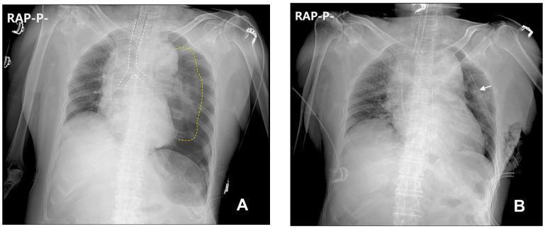
A tension pneumothorax is a rapidly fatal form of pneumothorax in which air enters the pleural space but cannot escape. Rising intrapleural pressure collapses the affected lung, shifts the mediastinum, obstructs venous return to the heart, and causes obstructive shock. Roberts and Hedges’ Clinical Procedures in Emergency, p. 264
Causes
- Chest trauma, especially penetrating trauma
- Positive-pressure ventilation or barotrauma
- CPR, especially with rib fractures
- Iatrogenic causes, such as central venous access
- Rarely, rupture of a bulla in asthma or emphysema
Clinical features
Diagnose it clinically. Do not delay treatment to obtain a chest x-ray.
- Acute severe dyspnea, tachypnea, hypoxia, agitation
- Unilateral absent or markedly reduced breath sounds
- Hyperresonance on the affected side
- Hypotension, tachycardia, signs of shock
- Raised jugular venous pressure, though it may be absent with hypovolemia
- Tracheal deviation away from the affected side is a late and unreliable sign
- In ventilated patients: suddenly increased airway pressures or resistance to ventilation, falling oxygen saturation, and hypotension
The mechanism is progressive lung collapse plus mediastinal shift, which compresses the venae cavae, reduces preload, and lowers cardiac output. Current Surgical Therapy 14e, p. 1310.
Emergency management
This requires immediate emergency treatment.
- Give high-flow oxygen and assess airway/breathing/circulation.
- Immediately decompress the affected pleural space if tension pneumothorax is suspected in an unstable patient.
- Needle thoracostomy using a large-bore catheter, or
- Finger thoracostomy where trained personnel and equipment permit.
- Preferred practical needle site: 4th or 5th intercostal space at the anterior to mid-axillary line. An anterior 2nd intercostal space mid-clavicular approach is an alternative in some protocols.
- Follow with definitive tube thoracostomy on the affected side.
Needle decompression is a temporary measure. A chest tube is needed immediately afterward to prevent recurrent tension. ROSEN's Emergency Medicine, p. 1156.
Key exam point
Suspected tension pneumothorax + hemodynamic or respiratory instability = decompress first, investigate later.
Chest radiography can show a hyperlucent hemithorax, collapsed lung, depressed diaphragm, and mediastinal shift, but imaging must not delay treatment. The MSD emergency guidance likewise recommends immediate decompression followed by tube thoracostomy.
Recent pneumothorax evidence reviews mainly address stable or non-tension pneumothorax, so they do not change this immediate clinical-decompression approach.
This is a shared conversation. Sign in to Orris to start your own chat.