
Radiological aspects of heart failure
Reading File
Finding Sources
Finding Sources
Searching Images
Chest radiograph features of congestive heart failure: cardiomegaly, pulmonary venous congestion, Kerley B lines, bat-wing edema, pleural effusions
Searching PubMed
heart failure AND chest radiography
Searching the Web
heart failure chest x ray radiographic signs pulmonary edema cardiomegaly review
Finding Sources
Finding Sources
Finding Sources
query_images_db
Searching PubMed
"Heart Failure"[MeSH Terms] AND "Radiography, Thoracic"[MeSH Terms]
Reading Web Page
https://radiopaedia.org/articles/congestive-cardiac-failure
Radiological aspects of heart failure
Chest radiography is the first-line imaging test in suspected acute or chronic heart failure. It helps identify pulmonary congestion and edema, cardiac enlargement, pleural effusion, and alternative diagnoses such as pneumonia or pneumothorax. However, a normal chest radiograph does not exclude heart failure, especially early acute decompensation.
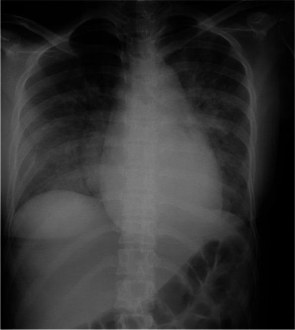
1. Chest X-ray findings in left-sided heart failure
The radiographic progression reflects increasing pulmonary venous pressure:
| Stage | Main X-ray signs | Meaning |
|---|---|---|
| Pulmonary venous hypertension | Upper-lobe venous diversion or cephalization, enlarged hilar vessels | Redistribution of pulmonary blood flow to upper lobes |
| Interstitial pulmonary edema | Kerley B lines, peribronchial cuffing, indistinct vessel margins, diffuse reticular haziness | Fluid in interlobular septa and peribronchial interstitium |
| Alveolar pulmonary edema | Bilateral perihilar fluffy air-space opacities, often in a bat-wing distribution | Alveolar fluid accumulation |
| Pleural effusion | Blunted costophrenic angles, meniscus sign, fluid in fissures | Common accompanying finding, often bilateral or right-predominant |
A useful mnemonic is ABCDE:
- A: Alveolar edema, classically bat-wing opacity
- B: Kerley B lines
- C: Cardiomegaly
- D: Dilated upper-lobe pulmonary vessels
- E: Pleural effusions
This sequence is summarized in the Radiopaedia heart-failure review.
2. Cardiac silhouette and cardiomegaly
- Cardiothoracic ratio (CTR) greater than 0.5 on an erect posteroanterior radiograph suggests cardiomegaly.
- CTR is unreliable on portable AP films because magnification can make the heart appear enlarged.
- Cardiomegaly is more typical of chronic congestive failure, dilated cardiomyopathy, and valvular regurgitation.
- Acute pulmonary edema may occur with a normal-sized heart, for example in acute myocardial infarction, acute mitral regurgitation, or acute hypertensive heart failure.
3. Radiographic features of right-sided heart failure
Chest radiography may show:
- Right atrial and right ventricular enlargement
- Prominent central pulmonary arteries, if pulmonary hypertension is present
- Pleural effusions
- Occasionally reduced peripheral pulmonary vascularity in advanced pulmonary arterial hypertension
Systemic venous congestion, hepatomegaly, ascites, and peripheral edema are clinically important but are not directly assessed on a routine chest radiograph.
4. Important patterns and diagnostic caveats
- Cardiogenic edema is usually bilateral, symmetric, central or perihilar, and associated with vascular congestion, cardiomegaly, and/or pleural effusions.
- Non-cardiogenic edema such as ARDS often has diffuse bilateral opacities but typically lacks cardiomegaly and vascular redistribution.
- Unilateral pulmonary edema can occur in acute severe mitral regurgitation, commonly affecting the right upper lobe, and can mimic pneumonia.
- In chronic mitral stenosis, pulmonary venous hypertension may produce upper-lobe diversion, septal lines, and sometimes pulmonary hemosiderosis.
The textbook evidence describes cephalization, Kerley B lines, and pulmonary edema as supportive signs of acute heart failure, but emphasizes that chest X-ray sensitivity is limited. Clinical assessment, natriuretic peptides, echocardiography, and bedside ultrasound must be integrated with the film.
5. Role of other imaging modalities
Echocardiography
- Main test for confirming structural and functional cardiac disease.
- Assesses ejection fraction, chamber size, wall motion, valve disease, pulmonary pressures, and pericardial effusion.
- Differentiates HFrEF from HFpEF and identifies causes such as severe mitral or aortic valve disease.
Lung ultrasound
- Demonstrates diffuse bilateral B-lines, indicating increased extravascular lung water.
- Also detects pleural effusions and can assess the inferior vena cava.
- It is particularly useful when chest X-ray is equivocal or when serial assessment of congestion is needed. A recent systematic review found lung-ultrasound-guided decongestion beneficial in heart-failure care, though this does not replace comprehensive cardiac assessment (PMID 41643462).
CT chest
- Not routinely required for uncomplicated heart failure.
- Useful when the diagnosis is unclear or when pulmonary embolism, pneumonia, malignancy, interstitial lung disease, or another alternative pathology is suspected.
- CT shows smooth interlobular septal thickening, ground-glass opacity, dependent consolidation, pleural effusions, and cardiomegaly with high sensitivity.
Cardiac MRI
- Best for detailed myocardial characterization.
- Helps identify myocarditis, infiltrative disease such as amyloidosis, ischemic scar, cardiomyopathy, ventricular volumes, and myocardial fibrosis.
Exam-style summary
Heart failure on chest X-ray:
- Cardiomegaly
- Pulmonary venous congestion and cephalization
- Interstitial edema: Kerley B lines and peribronchial cuffing
- Alveolar edema: bilateral perihilar bat-wing opacities
- Pleural effusions
The expected early-to-late radiographic sequence is pulmonary venous congestion, interstitial edema, alveolar edema, then pleural effusion, as outlined in this radiographic review.
This is a shared conversation. Sign in to Orris to start your own chat.