
Lump on thighs
lump thigh soft tissue tumor lipoma
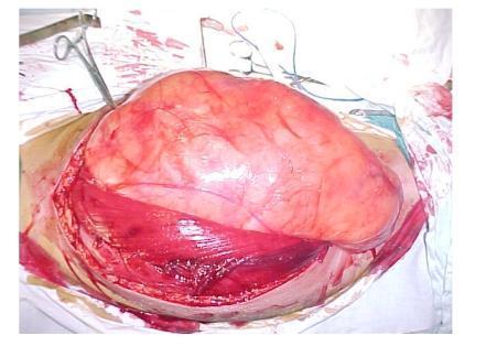
Intraoperative clinical photograph showing a large, encapsulated soft tissue tumor within the right thigh of a 44-year-old male. The surgical field displays a massive, well-circumscribed, lobulated mass with a predominantly reddish-pink and yellowish-pale surface, characteristic of a liposarcoma or a giant lipoma. The tumor, measuring approximately 27 cm by 25 cm, is seen displacing and stretching the adjacent thigh musculature, specifically the adductor and quadriceps muscle groups. The overlying skin and subcutaneous tissues are retracted, revealing the deep muscular planes and the substantial size of the neoplasm relative to the surrounding anatomy. Surgical instruments, including clamps, are visible at the periphery of the incision site. This image demonstrates the clinical presentation of a giant soft tissue tumor and the necessary surgical exposure for marginal excision.
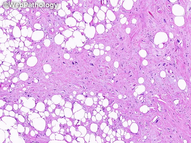
Histopathology image of a soft tissue tumor from a thigh shows mature adipocytes with variable size and scattered lipoblast-like cells embedded in variably dense fibrous septa. The specimen demonstrates a mixed morphology with lipoma-like adipose tissue interlaced by sclerosing areas containing thick, hyalinized collagen and sparse spindle cells. Adipocytic atypia is mild, with slightly enlarged, hyperchromatic nuclei, but there is no overt pleomorphism or high-grade mitotic activity. The fibrous stroma often blends with focal myxoid zones, yielding a mosaic pattern characteristic of the ALT/WDL spectrum. Overall, regions rich in adipose elements alternate with dense, fibrous septa and sclerotic foci, reflecting the combining lipoma-like and sclerosing components described in well-differentiated liposarcoma. The image does not demonstrate definitive dedifferentiation within the sampled fields. Clinically this correlates with a large, deep-seated thigh lipomatous tumor in an older adult. Distinguishing ALT/WDL from benign lipoma typically requires molecular confirmation of MDM2/CDK4 amplification or fluorescence in situ hybridization, in addition to histologic assessment of atypia and lipoblasts. Surgical excision with clear margins is recommended due to local recurrence risk. This image emphasizes the diagnostic spectrum and supports integrated histopathology and molecular testing in soft tissue sarcoma workup. Correlation with clinical history strengthens diagnostic confidence and management.
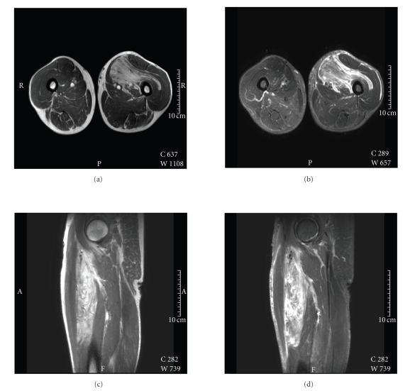
A multi-panel MRI series of the left thigh demonstrating a large, lipomatous soft tissue mass. The collection includes (a) axial T1-weighted, (b) axial fat-saturated STIR, (c) coronal T1-weighted, and (d) coronal T2-weighted images. The mass measures approximately 19 x 8 x 4 cm and is located within the soft tissues of the left thigh. On T1-weighted images, the lesion displays high signal intensity, though slightly lower than the surrounding subcutaneous fat. In the STIR and T2-weighted images, the mass maintains high signal intensity with internal foci of hyperintensity, suggesting a heterogeneous internal structure. The lesion is characterized by a lobulated morphology and irregular, poorly defined boundaries that show intramuscular and subfascial extensions. The lack of a clear capsule and the infiltrative margins are key diagnostic features for assessing soft tissue lipomatous tumors such as pleomorphic lipoma or atypical lipomatous tumor/well-differentiated liposarcoma.
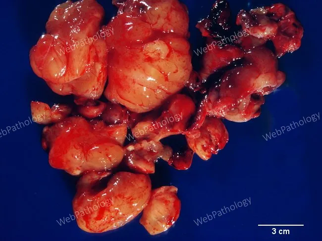
This is a macroscopic, ex vivo photograph of a thigh soft tissue liposarcoma resection specimen from a 68-year-old man. The tumor is composed of multiple lobulated, adipose-dense nodules with a yellow-tan, greasy appearance and glistening surfaces, separated by pale fibrous septa. The nodules vary from a few centimeters to several centimeters in greatest dimension; a 3 cm scale bar allows size estimation. The surface is largely smooth, with occasional gentle indentations and minor hemorrhagic streaks, consistent with a large, deep-seated lipomatous tumor. Although gross features mimic benign lipomas, histologic correlation (not shown in this image) demonstrates a well-differentiated liposarcoma characterized by mature adipocytes interspersed with scattered lipoblasts and fibrous septa containing atypical stromal cells. This ex vivo image illustrates the gross-pathology correlation used in surgical planning: complete excision with negative margins, assessment of tumor size and nodularity, and integration with genetic/immunohistochemical testing (e.g., MDM2, CDK4) to differentiate from lipoma and guide prognosis. Clinically, thigh liposarcomas occur in older adults and are typically large and deep to muscle; they require long-term follow-up due to local recurrence risk, potential for regional spread, and the need for adjuvant therapy decisions based on histologic subtype and margin status.
Lump on the Thigh - A Clinical Overview
Common Causes - Benign (Most Likely)
1. Lipoma
- Soft, mobile, painless, lobulated, compressible
- Grows slowly and usually stops after reaching a certain size
- Located in the subcutis of the proximal extremities (thigh is very common)
- Skin over the lump is normal but may dimple on traction
- Histology: well-encapsulated mass of mature adipocytes
- Most have chromosomal rearrangements involving chromosome 12q (dysregulating HMGA2)
- Treatment: observation if small/asymptomatic; simple excision is curative
2. Sebaceous/Epidermoid Cyst
- Smooth, round, firm, attached to skin
- Central punctum often visible
- Can become inflamed/infected
3. Lymph Node (inguinal/femoral)
- Lumps near the groin/upper thigh may be reactive or pathological lymph nodes
- Look for underlying infection, skin lesion, or systemic illness
4. Hematoma / Seroma
- History of trauma or surgery
- Fluctuant, may be tender
- Resolves spontaneously in most cases
5. Ganglion / Bursa
- Around the knee or hip joint extension
- Transilluminates; fluid-filled
6. Myositis Ossificans
- Painful firm mass following muscular trauma
- Shows calcification on X-ray after 4-6 weeks
Worrying Causes - Malignant
Red Flags ("Rule of 5s" - suspect malignancy if):
- Size >5 cm
- Deep to the deep fascia
- Increasing in size
- Recurrence after previous excision
- Firmness or fixation to underlying structures
Malignant Tumors of the Thigh
| Tumor | Who | Presentation | Imaging |
|---|---|---|---|
| Undifferentiated Pleomorphic Sarcoma (MFH) | Older adults | Large, deep, painless mass | Well-circumscribed, dark T1 / bright T2, may have central necrosis |
| Well-differentiated Liposarcoma (ATL) | Middle-aged adults | Slow-growing, non-painful | MRI: near-isointense to fat, hard to distinguish from lipoma |
| Myxoid Liposarcoma | Adults | Large, deep, painless | Heterogeneous MRI signal |
| Pleomorphic Liposarcoma | Adults (male slight predominance) | Rapid growth, large, 25% subcutaneous | Hypointense T1, hyperintense T2 |
| Synovial Sarcoma | Young adults (male predominance) | Juxtaarticular; slow-growing, may be painful | May have calcifications on X-ray |
| Rhabdomyosarcoma | Children/young adults | Rapidly growing | MRI shows muscle involvement |
Key Imaging
- Lipoma: bright on T1, suppressed on fat-saturation sequences, well-circumscribed
- Liposarcoma/Sarcoma: heterogeneous signal, may not suppress on fat-sat, irregular margins, deep to fascia
- CT chest/abdomen/pelvis is needed for staging if sarcoma is suspected
Evaluation Approach
- History: duration, growth rate, pain, trauma, prior similar lesions, family history, weight loss/systemic symptoms
- Examination: site (superficial vs deep to fascia), size, consistency, mobility, skin changes, lymph nodes
- X-ray: calcifications (myositis ossificans, synovial sarcoma), bony involvement
- MRI: best for characterization - always get if deep, >5 cm, or uncertain
- Biopsy: core needle biopsy (image-guided) for any suspicious mass before resection
- Do not excise a potentially malignant mass without pre-operative biopsy and specialist referral - it compromises subsequent limb-salvage surgery
Management Summary
| Lesion | Treatment |
|---|---|
| Lipoma (confirmed) | Observe, or excise if symptomatic/large |
| Soft tissue sarcoma | Wide excision + radiation therapy (± chemotherapy) by specialist team |
| ALT/Well-diff liposarcoma | Wide excision; no radiation or chemo usually needed; long follow-up |
| Hematoma | Conservative; drain if persistent |
| Infected cyst | Incision and drainage ± antibiotics |
When to Refer Urgently
- Deep to fascia
- Larger than 5 cm
- Growing rapidly
- Associated with pain or neurological symptoms
- Hard/fixed consistency
Disclaimer: This is general medical information. A clinical examination and imaging by a qualified healthcare professional are essential for any new lump. Do not attempt biopsy or excision of a thigh mass without specialist assessment.